
Today’s NCLEX-RN® question of the day focuses on the physiology of vision in infants. Do you know the answer? Let’s find out!
The labor and delivery (L&D) nurse explains the physiology of vision in infants to a newly graduated nurse. The nurse knows the teaching has been successful when the newly graduated nurse identifies which receptors are responsible for detecting light within the retina?
A. Nociceptors
B. Baroreceptors
C. Photoreceptors
D. Chemoreceptors
Scroll down for the correct answer!
The correct answer to today’s NCLEX® Question is…
C. Photoreceptors
Rationale: Photoreceptors are located in the retina and convert light into electrical impulses. These impulses travel through the optic nerve to the brain’s visual cortex, where visual information is processed.
Main Takeaway
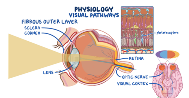
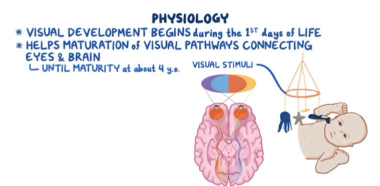
Visual development begins during the first days of life when the newborn starts receiving visual stimuli from the external world. This helps maturation of the visual pathways connecting the eyes and brain until they reach maturity at about four years of age. The visual pathways begin in the eye. In the wall of the eye, there is an outer fibrous layer, which contains the cornea and sclera, and helps control and focus the entry of light. The light that passes through the cornea is directed to the lens, which collects light arrays and focuses them onto the retina at the back of the eye. The retina houses photoreceptors that translate light into electrical impulses, which are then carried by the optic nerve to the brain’s visual cortex. Finally, the visual cortex processes the impulses from both eyes and fuses them into a single visual image.
Incorrect Answer Explanations
A. Nociceptors
Rationale: Nociceptors are pain receptors responsible for sending the sensory stimulus of pain to the brain. These receptors are not involved in the detection of visual signals.
B. Baroreceptors
Rationale: Baroreceptors detect changes in blood pressure within blood vessels and help regulate cardiovascular function. They are not involved in vision.
D. Chemoreceptors
Rationale: Chemoreceptors are located throughout the body and are responsible for detecting changes in pH levels, carbon dioxide, and oxygen. They are not involved in detecting visual signals.
Want to learn more about this topic?
Watch the Osmosis video: Amblyopia: Nursing

Try Osmosis from Elsevier today! Access your free trial to find out why future clinicians and caregivers love learning by Osmosis.
Leave a Reply