Aortic aneurysm: Nursing process (ADPIE)
Notes
| AORTIC ANEURYSM | ||
| KEY POINTS | NOTES | |
| PATIENT REPORT |
| |
| PATHOPHYSIOLOGY |
| |
| DIAGNOSIS AND TREATMENT |
| |
| ASSESSMENT |
| |
| NURSING DIAGNOSES |
| |
| PLANNING |
| |
| IMPLEMENTATION |
| |
| EVALUATION |
| |
Transcript
David Carter is a 65-year-old male client who arrives at the Primary Care Clinic for his annual follow-up appointment.
He is a current smoker and has a history of hypertension and atherosclerosis.
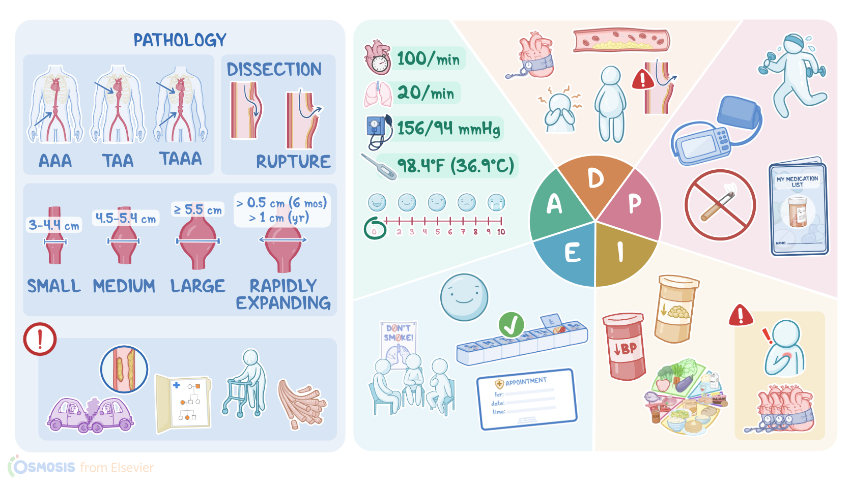
He was diagnosed with 4.0 cm asymptomatic abdominal aortic aneurysm last year.
Mr. Carter brought in his blood pressure machine from home, and he tells the front desk staff he is feeling anxious about his appointment today.
The aorta is a large elastic artery that carries blood from the left ventricle of the heart, down through the thorax and abdomen.
The artery wall consists of three layers: the tunica intima, tunica media, and the tunica adventitia, which are composed of smooth muscle, elastic fibers and collagen which give the artery strength and elasticity.
If an area of weakness develops along the aorta, a dilation or bulge forms, called an aneurysm.
Aortic aneurysms are described according to their location, shape, size, and whether they involve all or part of the artery wall.
So, if the aneurysm forms within the abdominal cavity, it’s called an abdominal aortic aneurysm, or Triple-A.
Likewise, if the aneurysm is found within the thorax, or chest, it’s called a thoracic aortic aneurysm, or T double-A.
Finally, a thoracoabdominal aortic aneurysm, or T triple-A involves both the thoracic and abdominal aorta.
Aortic aneurysms come in two basic shapes.
A circular dilation that involves the entire circumference of the aorta is called a fusiform aneurysm.
In contrast, a saccular aneurysm is formed when there’s only a localized outpouching, like a bubble on the side of the aorta.
Both fusiform and saccular aneurysms are classified as true aneurysms because they involve all three layers of the aortic wall.
In cases where there’s only a partial disruption of the artery wall, it’s called a false, or pseudoaneurysm.
In general, an aneurysm that measures between 3 and 4.4 centimeters is considered a small aneurysm; medium aneurysms have a diameter between 4.5 and 5.4 centimeters, and large aneurysms have a diameter 5.5 centimeters or more.
A rapidly expanding aneurysm is defined as one that grows more than 0.5 centimeters in 6 months or more than 1 centimeter per year.
An aortic aneurysm can form due to a number of factors that impair the integrity of the arterial wall.
These factors can be mechanical, inflammatory or congenital.
Mechanical causes such as blunt or penetrating trauma can result in immediate damage to the aorta, whereas the chronic stress from uncontrolled hypertension can weaken the aorta over time.
Hypertension also promotes the formation of atherosclorotic plaque, which results in inflammation and subsequent breakdown of collagen and elastin, two of the most important building blocks of the aortic wall.
Likewise, other risk factors for atherosclerosis such as smoking increase the risk of an aortic aneurysm.
Inherited connective tissue disorders like Marfan syndrome and Ehler-Danlos syndrome are associated with abnormal elastin and collagen and a weak aortic wall.
Other risk factors for aortic aneurysms include biological male sex, a family history of aneurysms, and increasing age, where the natural process of aging results in decreased elasticity of the aorta.
A TripIe-A is often asymptomatic and only detected incidentally during a routine examination or screening.
The weakened aortic wall can produce signs like a prominent pulsating mass in the abdomen, usually felt at or above the umbilicus.
Turbulent blood flow through the aneurysm produces a systolic bruit which can be auscultated over the aorta.
Sometimes the turbulence produces microthrombi, which can travel down and occlude the lower extremities, producing cool, painful cyanotic toes, a condition known as blue toe syndrome.
If the aneurysm presses on nearby structures, such as the intestines, it can cause altered bowel elimination.
A major complication of aortic aneurysm is dissection and rupture, which happens when the aneurysm enlarges and the layers of the artery wall split, allowing blood to leak in between them.
As the layers begin to tear and eventually rupture, hemorrhage and hypovolemic shock can result.
In cases where the hemorrhage occurs within the retroperitoneal space, the bleeding can be slowed by surrounding structures.
As blood continues to leak into the retroperitoneal space, ecchymoses often develop on the back or flank, producing the Grey Turner sign.
In any case, a dissecting aortic aneurysm classically presents with chest pain that radiates to the upper back, between the scapula.
A rupture is a medical emergency and requires immediate stabilization and surgery.
Ultrasound is used to detect the presence, location, and size of the aneurysm, and to monitor it’s growth over time.
Other useful diagnostic studies include computed tomography scan, or CT scan, which can provide a more accurate measurement of the aneurysm’s size and shape.
Treatment for abdominal aneurysm depends on its size and location and if there are symptoms.
Sources
- "Ackley and Ladwig’s Nursing Diagnosis Handbook: An Evidence-Based Guide to Planning Care, 13th edition" Mosby (2022)
- "Aortic Coarctation: Basic Imaging Findings and Management" Journal of Radiology Nursing (2020)
- "A Systematic Review of Total Endovascular Aortic Arch Repair: A Promising Technology" Can J Cardiol (2023)
- "Abdominal Aortic Aneurysm: A Case Report and Literature Review" Perm J (2019)
- "Updates of Recent Aortic Aneurysm Research" Arterioscler Thromb Vasc Biol (2019)
- "Unveiling the Hidden Landscape of Arterial Diseases at Single-Cell Resolution" Can J Cardiol (2023)
- "Critical Care Nursing: Diagnosis and Management, 9th edition" Elsevier (2021)