Aortic aneurysm: Nursing process (ADPIE)
Client Report0:00–0:31
David Carter is a 65-year-old male client who arrives at the primary care clinic for his annual follow-up appointment. He is a current smoker and has a history of hypertension and atherosclerosis.
He was diagnosed with 4.0 centimeter asymptomatic abdominal aortic aneurysm last year. Mr.
Carter brought in his BP machine from home, and he tells the front desk staff he is feeling anxious about his appointment today.
The aorta is a large elastic artery that carries blood from the left ventricle of the heart down through the thorax and abdomen.
Pathology0:31–5:58
The artery wall consists of three layers the tunica intima, tunica media, and the tunica adventitia, which are composed of smooth muscle, elastic fibers, and collagen which give the artery strength and elasticity.
So if the aneurysm forms within the abdominal cavity, it's called an abdominal aortic aneurysm or AAA. Likewise, if the aneurysm is found within the thorax or chest, it's called a thoracic aortic aneurysm or TAA.
Finally, a thoracoabdominal aortic aneurysm or TAAA involves both the thoracic and abdominal aorta. Aortic aneurysms come in two basic shapes.
A circular dilation that involves the entire circumference of the aorta is called a fusiform aneurysm. In contrast, a saccular aneurysm is formed when there's only a localized outpouching like a bubble on the side of the aorta.
Both fusiform and saccular aneurysms are classified as true aneurysms because they involve all three layers of the aortic wall.
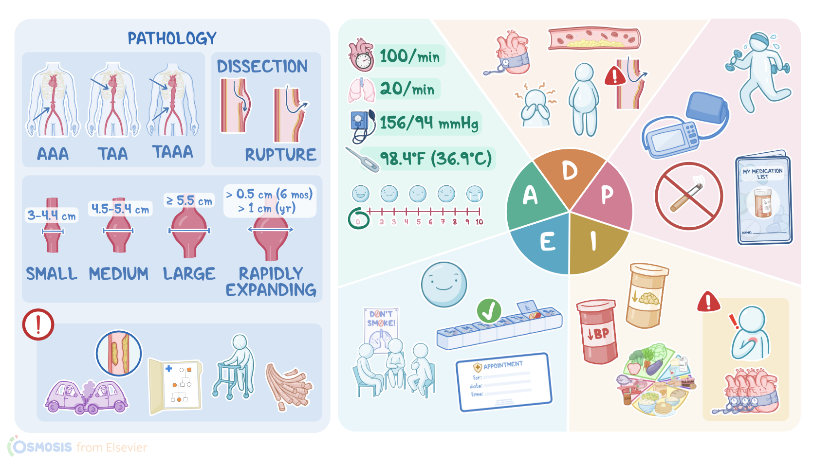
In cases where there's only a partial disruption of the artery wall, it's called a false or pseudoaneurysm. In general, an aneurysm that measures between 3 and 4.4 centimeters is considered a small aneurysm.
Medium aneurysms have a diameter between 4.5 and 5.4 centimeters, and large aneurysms have a diameter of 5.5 centimeters or more.
A rapidly expanding aneurysm is defined as one that grows more than 0.5 centimeters in six months or more than 1 centimeter per year.
An aortic aneurysm can form due to a number of factors that impair the integrity of the arterial wall. These factors can be mechanical, inflammatory, or congenital.
Mechanical causes such as blunt or penetrating trauma can result in immediate damage to the aorta, whereas the chronic stress from uncontrolled hypertension can weaken the aorta over time.
Hypertension also promotes the formation of atherosclerotic plaque, which results in inflammation and subsequent breakdown of collagen and elastin, two of the most important building blocks of the aortic wall.
Likewise, other risk factors for atherosclerosis, such as smoking, increase the risk of an aortic aneurysm. Inherited connective tissue disorders like Marfan syndrome and Ehler-Danlos syndrome are associated with abnormal elastin and collagen and a weak aortic wall.
Other risk factors for aortic aneurysms include biological male sex, a family history of aneurysms, and increasing age, where the natural process of aging results in decreased elasticity of the aorta.
Turbulent blood flow through the aneurysm produces a systolic brute which can be auscultated over the aorta. Sometimes the turbulence produces microthrombi, which can travel down and occlude the lower extremities, producing cool, painful, cyanotic toes, a condition known as blue toe syndrome.
If the aneurysm presses on nearby structures such as the intestines, it can cause altered bowel elimination. A major complication of aortic aneurysm is dissection and rupture, which happens when the aneurysm enlarges and the layers of the artery wall split, allowing blood to leak in between them.
As the layers begin to tear and eventually rupture, hemorrhage and hypovolemic shock can result. In cases where the hemorrhage occurs within the retroperitoneal space, the bleeding can be slowed by surrounding structures.
As blood continues to leak into the retroperitoneal space, ecchymoses often develop on the back or flank, producing the gray turner sign.
In any case, a dissecting aortic aneurysm classically presents with chest pain that radiates to the upper back between the scapula.
A rupture is a medical emergency and requires immediate stabilization and surgery. Ultrasound is used to detect the presence, location, and size of the aneurysm and to monitor its growth over time.
Other useful diagnostic studies include computed tomography scan or CT scan, which can provide a more accurate measurement of the aneurysm's size and shape.
Treatment for abdominal aneurysm depends on its size and location and if there are symptoms. For a small asymptomatic aortic aneurysm, the treatment consists of conservative management like smoking cessation, lipid lowering medications, anti-hypertensive medications, and diet.
And lifestyle changes. There should be regular follow-ups to monitor the size and growth of the aneurysm.
Surgical repair is the treatment of choice when the aneurysm size exceeds 5.0 to 5.5 centimeters, is rapidly expanding, or if symptoms develop.
Assessment5:58–7:57
So after you greet Mister Carter and introduce yourself as his nurse, you wash your hands, confirm his identity, and begin your assessment by asking how he's feeling today.
Your initial assessment findings are height 5 ft 10 inches, weight 212 lbs or 96 kg, heart rate 100 per minute and regular, respiratory rate 20 per minute with clear breath sounds bilaterally, BP 156/94 millimeters of mercury, temperature 98.4 °F or 36.9 °C, pain 0 out of 10.
Before leaving the room, you check his recent diagnostic tests and note the following hemoglobin 14 g per deciliter, hematocrit 40%, BUN 20 mg per deciliter, creatinine 1.0 mg per deciliter, cholesterol 240 mg per deciliter, HDL 30 mg per deciliter, LDL 150 mg per deciliter, triglycerides 190 mg per deciliter.
His medical record lists lisinopril, an angiotensin converting enzyme inhibitor or ACE inhibitor, as his only current medication.
After documenting your assessment findings, you inform Mister Carter that the nurse practitioner, or APRN will be in shortly to examine him and discuss his plan of care.
After the examination, you note the additional findings assessed by the APRN including 3+ pedal pulses, a visible pulsation, and a systolic brute auscultated at the level of the umbilicus.
You compare the abdominal ultrasound from 1 year ago, which showed a 4.0 centimeter AAA with his ultrasound results from today, which shows the aneurysm is now 4.3 centimeters.
Diagnosis7:57–8:21
Now that you've completed your nursing assessment, you've put together your nursing diagnoses for Mr. Carter.
Anxiety related to his verbalized stress about his aneurysm, ineffective health maintenance related to current smoking status, hypertension, dyslipidemia, and increased body mass index, or BMI, and risk for ineffective tissue perfusion related to potential aneurysm rupture.
Now that you've gathered the information and have formulated your nursing diagnoses, it's time to collaborate with the APRN and Mr.
Planning8:21–9:14
Carter to develop a holistic plan of care. By the end of his visit today, your goals are that Mr.
Carter will be able to demonstrate how to check his BP and identify when a reading is not within the desired range, verbalize understanding of the importance of smoking cessation, and enroll in a smoking cessation program.
Identify necessary lifestyle changes to address his hypertension, dyslipidemia, and weight. He will state the importance of taking his BP medication and any new prescriptions as prescribed, and he will experience less anxiety as a result of his increased knowledge of how to maintain his health.
Finally, you establish long-term goals that he will have a normal BP measurement by his next follow-up appointment and that this aneurysm will remain stable.
After collaborating with the APRN you implement the plan of care. The APRN orders include an increased dose of lisinopril and a new prescription of atorvastatin, an HMGCOA reductase inhibitor, also known as a statin.
Implementation9:14–10:16
You give him the prescriptions and explain the use, possible side effects, and importance of taking the medications as prescribed.
You teach Mr. Carter the importance of checking his BP daily at the same time each day and to keep a log of his BP readings to bring to his next appointment.
Mr. Carter brought in his BP machine from home, and you review how to correctly measure his BP.
You discuss lifestyle and dietary changes to promote a normal BP and maintain a healthy weight. Go over smoking cessation resources and sign him up for a smoking cessation support group.
He will be returning for a follow-up in 6 months for a BP check, lipid lab draw, and ultrasound. Finally, you tell him that if he experiences pain or persistent high BP, he should notify his APRN immediately.
Evaluation10:16–10:48
All right, let's check back in on Mister Carter now that he is ready to go home. Mr.
He goes on to tell you that he downloaded a smoking cessation app on his phone, has picked a stop date, and will be attending his first support group meeting next week.
He describes his plan for a healthier lifestyle and adherence to his medication regimen. Mr.
Carter has already scheduled his next follow-up appointments. OK, as a quick recap, your client, Mr.
Summary10:48–12:00
Carter, has a history of smoking, hypertension, atherosclerosis, and an asymptomatic abdominal aortic aneurysm. He presented to the primary care clinic today for his annual follow-up.
An aortic aneurysm is an abnormal dilation of the arterial wall, usually resulting from localized weakness. Risk factors include hypertension, advanced age, biological male sex, smoking, atherosclerosis, connective tissue disorders, and having a family history of aneurysms.
Your nursing diagnoses included anxiety, ineffective health maintenance, and risk for ineffective tissue perfusion. The planning goals were to decrease Mr.
Carter's anxiety, increase his knowledge of health maintenance, and decrease the risk of disease progression. Along with the APRN, you work to implement these actions to achieve the goals of the plan of care.
Throughout the appointment and up until Mr. Carter goes home, you will continue to evaluate if those goals have been met.
| AORTIC ANEURYSM | ||
| KEY POINTS | NOTES | |
| PATIENT REPORT |
| |
| PATHOPHYSIOLOGY |
| |
| DIAGNOSIS AND TREATMENT |
| |
| ASSESSMENT |
| |
| NURSING DIAGNOSES |
| |
| PLANNING |
| |
| IMPLEMENTATION |
| |
| EVALUATION |
| |
- "Ackley and Ladwig’s Nursing Diagnosis Handbook: An Evidence-Based Guide to Planning Care, 13th edition" Mosby (2022)
- "Aortic Coarctation: Basic Imaging Findings and Management" Journal of Radiology Nursing (2020)
- "A Systematic Review of Total Endovascular Aortic Arch Repair: A Promising Technology" Can J Cardiol (2023)
- "Abdominal Aortic Aneurysm: A Case Report and Literature Review" Perm J (2019)
- "Updates of Recent Aortic Aneurysm Research" Arterioscler Thromb Vasc Biol (2019)
- "Unveiling the Hidden Landscape of Arterial Diseases at Single-Cell Resolution" Can J Cardiol (2023)
- "Critical Care Nursing: Diagnosis and Management, 9th edition" Elsevier (2021)
No notes for this video yet
Try adding a note below