Appendicitis: Nursing process (ADPIE)

2,030views

Appendicitis: Nursing process (ADPIE)

FINAL

FINAL

ACE inhibitors, ARBs and direct renin inhibitors
Thiazide and thiazide-like diuretics
Calcium channel blockers
Adrenergic antagonists: Beta blockers
Acyanotic congenital heart defects: Pathology review
Atherosclerosis and arteriosclerosis: Pathology review
Coronary artery disease: Pathology review
Peripheral artery disease: Pathology review
Valvular heart disease: Pathology review
Cardiomyopathies: Pathology review
Heart failure: Pathology review
Supraventricular arrhythmias: Pathology review
Ventricular arrhythmias: Pathology review
Heart blocks: Pathology review
Aortic dissections and aneurysms: Pathology review
Pericardial disease: Pathology review
Endocarditis: Pathology review
Hypertension: Pathology review
Shock: Pathology review
Vasculitis: Pathology review
Cardiac and vascular tumors: Pathology review
Dyslipidemias: Pathology review
Cardiac tamponade
Endocarditis
Myocarditis
Rheumatic heart disease
Heart failure
Cor pulmonale
Long QT syndrome and Torsade de pointes
Ventricular tachycardia
Premature ventricular contraction
Ventricular fibrillation
Atrial flutter
Premature atrial contraction
Atrial fibrillation
Atrioventricular nodal reentrant tachycardia (AVNRT)
Deep vein thrombosis
Hypotension
Orthostatic hypotension
Polycystic kidney disease
Pheochromocytoma
Cushing syndrome
Renal artery stenosis
Hypertension
Aneurysms
Aortic dissection
Peripheral artery disease
Angina pectoris
Unstable angina
Prinzmetal angina
Myocardial infarction
Stable angina
Arterial disease
ECG normal sinus rhythm
ECG cardiac hypertrophy and enlargement
ECG cardiac infarction and ischemia
ECG basics
ECG intervals
ECG axis
ECG QRS transition
ECG rate and rhythm
Cardiac conduction system
Cardiac conduction velocity
Normal heart sounds
Abnormal heart sounds
Cardiovascular changes during postural change
Cardiovascular changes during hemorrhage
Cardiac preload
Cardiac contractility
Cardiac afterload
Measuring cardiac output (Fick principle)
Thrombocytopenia: Clinical
Heparin-induced thrombocytopenia
Immune thrombocytopenia
Gout
Chronic kidney disease: Clinical
Traumatic brain injury: Pathology review
Traumatic brain injury: Clinical
Concussion and traumatic brain injury
Blood groups and transfusions
Blood products and transfusion: Clinical
HIV (AIDS)
Hodgkin lymphoma
Acromegaly
Musculoskeletal injuries: Nursing process (ADPIE)
Hemophilia: Nursing process (ADPIE)
Diabetes insipidus
Diabetes mellitus
Diabetes mellitus: Clinical
Diabetes mellitus: Pathology review
Diabetes mellitus (DM): Nursing process (ADPIE)
Diabetes insipidus: Nursing process (ADPIE)
Managing diabetes during the holidays: Information for patients and families
Hypoglycemics: Insulin secretagogues
Insulins
Epistaxis: Nursing process (ADPIE)
Appendicitis
Appendicitis: Clinical
Appendicitis: Pathology review
Appendicitis: Nursing process (ADPIE)
Hypothyroidism medications
Hyperosmolar hyperglycemic state (HHS): Nursing process (ADPIE)
Sympathomimetics: Direct agonists
Cushing syndrome and Cushing disease: Pathology review
Cushing syndrome: Clinical
Metabolic and respiratory alkalosis: Clinical
Metabolic and respiratory acidosis: Clinical
Conjunctivitis: Nursing process (ADPIE)
Stroke: Clinical
Stroke: Nursing process (ADPIE)
Peptic ulcer
Peptic ulcer disease (PUD): Nursing process (ADPIE)
Peptic ulcers and stomach cancer: Clinical
Gallbladder histology
Gallbladder disorders: Clinical
Acute cholecystitis
Oral cancer
Hepatitis A and Hepatitis E virus
Viral hepatitis: Clinical
Hepatitis medications
Seizures: Pathology review
Seizures: Clinical
Seizures and epilepsy
Febrile seizure
Seizure disorder: Nursing process (ADPIE)
Non-urothelial bladder cancers
Inflammatory bowel disease: Clinical
Inflammatory bowel disease: Pathology review
Anticoagulants: Heparin
Postoperative evaluation: Clinical
Trigeminal neuralgia
Trigeminal neuralgia: Nursing process (ADPIE)
Hypoparathyroidism
Pancreatitis: Pathology review
Pancreatitis: Clinical
Acute pancreatitis
Pancreatitis: Nursing process (ADPIE)
Chronic pancreatitis
Sickle cell disease (NORD)
Sickle cell disease: Clinical
Sickle cell disease: Nursing process (ADPIE)
Class IV antiarrhythmics: Calcium channel blockers and others
Hypertension: Clinical
Pulmonary hypertension
Hypertension: Nursing process (ADPIE)
Osteoarthritis
Joint pain: Clinical
Hyperthyroidism: Pathology review
Hyperthyroidism: Clinical
Deep vein thrombosis and pulmonary embolism: Pathology review
Hyperthyroidism
Hyperthyroidism medications
Hyperthyroidism: Nursing process (ADPIE)

Notes

APPENDICITIS

KEY POINTS
NOTES
PATIENT REPORT
  • 16-year-old boy
  • Diagnosis: appendicitis
  • Scheduled for appendectomy

PATHOPHYSIOLOGY
  • Appendectomy
    • Inflammation and obstruction of appendix
    • Mucosal secretions and bacteria build up
    • Parietal membrane becomes irritated
    • Causes
      • Lymphoid hyperplaisa
      • Fecalith
      • Tumor
      • Parasites
    • Signs and symptoms
      • Abdominal pain
      • Fever
      • Anorexia
      • Nausea
      • Vomiting
  • Complications
    • Necrosis
    • Abscess 
    • Peritonitis
  • Diagnosis
    • History
    • Physical assessment
    • Ultrasound
    • CT
    • MRI
    • Laboratory tests
  • Treatment
    • Surgery

ASSESSMENT
  • Lying on side with knees drawn up
  • Blood pressure: 112/78 mmHg
  • Heart rate: 108
  • Respiratory rate: 22
  • Oxygen saturation: 99% room air
  • Temperature: 100.4 F (38.0 C)
  • 5'9'' (175 cm)
  • 148 lbs (67 kg) 
  • Right-sided abdominal pain
  • Pain: 7/10
  • Nauseated
  • White blood count: 13,500 cells/mm(13.5 x 109 cells/L)

NURSING DIAGNOSES
  • Acute pain related to inflammation of the appendix
  • Nausea related to inflammation of appendix
  • Risk for infection related to possible rupture
  • Risk for delayed surgical recovery

PLANNING
  • Manageable pain
  • Absence of nausea
  • Tolerate oral intake
  • No infection
  • No postoperative complications

IMPLEMENTATION
  • Administer medications as prescribed
  • Maintain NPO status
  • Assist to semi-Fowler's position
  • Place ice pack on abdomen
  • Explain surgery
  • Provide teaching on preventing postoperative complications

EVALUATION
  • Blood pressure: 110/80 mmHg
  • Heart rate: 88
  • Respiratory rate: 16
  • Clear lungs
  • Oxygen saturation: 98% 2L nasal cannula
  • Pain: 2/10
  • Temperature: 98.8 F (37.1 C)
  • Active bowel sounds
  • No nausea
  • Incisions clean, dry, and intact

Transcript

Watch video only

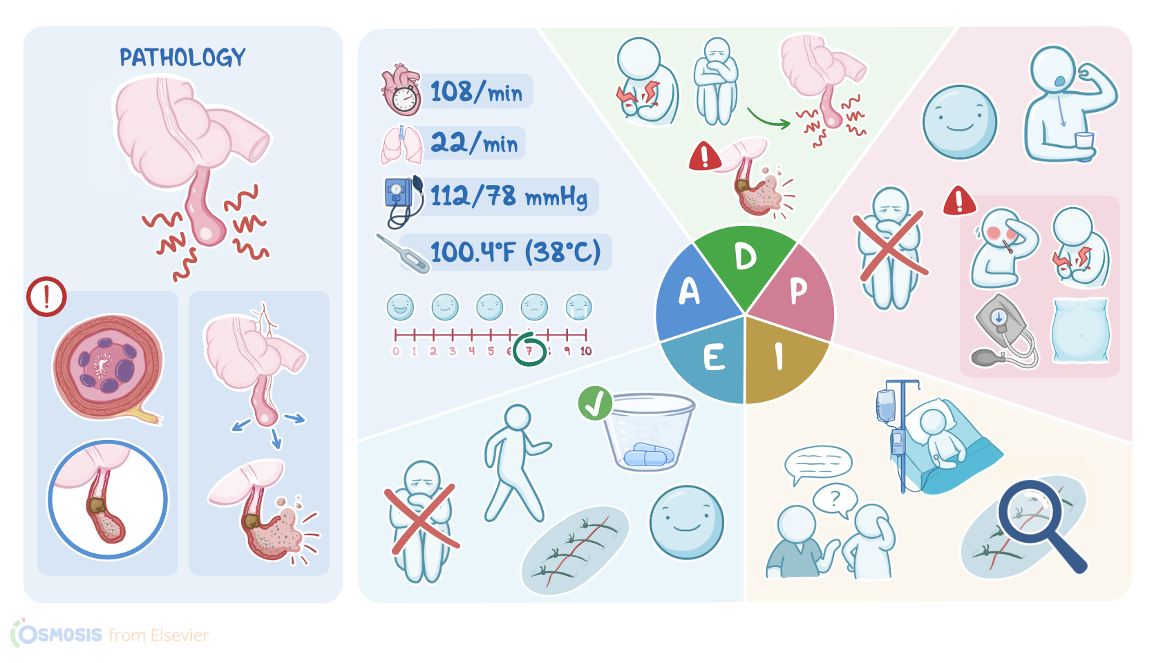
16-year-old Manny Correa is admitted to the pediatric inpatient unit from the urgent care clinic.

Manny’s father George brought him to the clinic after Manny experienced pain around his belly button for the past 24 hours.

An abdominal ultrasound confirmed an inflamed appendix. Manny is scheduled for an appendectomy today.

Appendicitis is a condition where the appendix, a finger-like projection that hangs off of the cecum of the large intestine, becomes obstructed and inflamed.

Appendicitis can be caused by lymphoid hyperplasia, where overgrown lymphoid follicles obstruct the appendix, or when a hard lump of stool called a fecalith, a tumor, or even parasites like pinworms cause an obstruction, resulting in appendicitis.

Appendicitis is more common in the second and third decades of life, with the highest incidence between the ages of 10 and 19.

It is more common in men than in women.

Now, when the appendix becomes obstructed, mucosal secretions and the bacteria that normally live in the appendix build up inside, causing the appendix to expand and press against the visceral nerve fibers, resulting in pain that is often felt in the periumbilical area.

Multiplication of bacteria inside the appendix leads to inflammation, which is accompanied by fever, anorexia, nausea, and sometimes vomiting.

Eventually, the appendix starts to irritate the nearby parietal membrane lining the walls of the abdominal cavity, causing the pain to intensify and migrate to the right lower quadrant, an area known as McBurney’s point, which is located 1.5 to 2 inches from the navel to the anterior superior iliac spine.

Palpating McBurney’s point and quickly releasing pressure will demonstrate rebound tenderness, and the person may show guarding, where their abdominal muscles tense up when pressed in an attempt to avoid pain.

Rovsing's sign is elicited if pressure over the person’s left lower abdominal quadrant causes pain in the right lower abdominal quadrant.

The psoas sign is when pain occurs with passive extension of the right hip.

Lastly, the obturator sign is elicited by flexing the right knee to a 90 degree angle while internally rotating the hip.

Complications can develop as the appendix continues to swell, compress the venous and arterial blood flow.

This leads to edema, ischemia and eventual necrosis of the appendiceal wall.

As cells die, the wall becomes weaker and weaker, until it eventually ruptures, allowing bacteria, pus, and fluid to escape into the peritoneum.

The result is the formation of abscesses around the appendix, or peritoneal inflammation, referred to as peritonitis.

At first, the person may notice a sudden decrease in pain as the pressure inside the appendix is relieved.

However, as infection and inflammation continues to develop inside the peritoneal cavity, the person can develop severe abdominal pain and rigidity, vomiting, tachycardia, and hypotension.

If untreated, peritonitis can progress to shock or septicemia.

After a client history and physical examination is complete, laboratory and other diagnostic tests can help confirm a diagnosis of appendicitis.

Abdominal ultrasound can be used to visualize inflammatory changes.

Abdominal CT scans are more sensitive, and can show a more precise view of these findings.

An MRI is useful in diagnosing appendicitis in pregnancy or when contrast must be avoided.

A CBC will often show leukocytosis with a left shift, or an increased number of immature neutrophils.

A C-reactive protein, which is an indication of inflammation, is often elevated.

Sometimes, uncomplicated appendicitis is treated with a course of antibiotics, but the definitive treatment for appendicitis is the surgical removal of the appendix, referred to as an appendectomy.

If an abscess has formed and there is no evidence of peritonitis, treatment may involve draining the abscess with a needle.

Sometimes a tube may be left in place to allow ongoing drainage.

Manny and George have arrived on the pediatric inpatient unit, and it’s time for you to begin your assessment.

After introducing yourself, confirming Manny’s identity, and performing hand hygiene, you note that Manny is laying on his right side with his knees drawn up towards his chest.

Sources

  1. "Ackley and Ladwig’s Nursing Diagnosis Handbook: An Evidence-Based Guide to Planning Care, 13th edition" Mosby (2022)
  2. "Post operative pediatric appendicitis nurse-driven discharge: Patient outcomes and nursing perspectives" Am J Surg (2021)
  3. "Appendicitis in a ventral hernia" Visual Journal of Emergency Medicine (2023)
  4. "Medical-Surgical Nursing: Concepts for Interprofessional Collaborative Care, 10th Edition" Elsevier (2020)
  5. "Harrison’s Principles of Internal Medicine, 21st edition" McGraw Hill / Medical (2022)
  6. "Simultaneous occurrence of acute appendicitis and appendicular band syndrome in a patient with intestinal obstruction" Visual Journal of Emergency Medicine (2023)
  7. "Routine post-operative labs and healthcare system burden in acute appendicitis" Am J Surg (2023)
  8. "Health Assessment for Nursing Practice, 7th edition" Elsevier (2021)