Chest tube care: Nursing

2,513views

Chest tube care: Nursing

Adult Nursing 1 AAS

Adult Nursing 1 AAS

Charge nurse as a leader: Nursing
Health history: Nursing
Health literacy: Nursing
Health and illness models: Nursing
Health promotion and illness prevention: Nursing
Chronic disease: Nursing
Palliative and hospice care: Nursing
Body fluid compartments
Complete metabolic panel (CMP) - Chloride: Nursing
Complete metabolic panel (CMP) - Sodium: Nursing
Complete metabolic panel (CMP) - Potassium: Nursing
Complete metabolic panel (CMP) - Calcium: Nursing
Renin-angiotensin-aldosterone system
Arterial blood gas (ABG) - Metabolic acidosis: Nursing
Arterial blood gas (ABG) - Respiratory acidosis: Nursing
Arterial blood gas (ABG) - Metabolic alkalosis: Nursing
Arterial blood gas (ABG) - Respiratory alkalosis: Nursing
Arterial blood gas (ABG) - Overview: Nursing
Preoperative care: Nursing
Postoperative care: Nursing
Biology of cancer: Nursing
Physiology of pain: Nursing
Respiratory system anatomy and physiology
Geriatric considerations - Respiratory: Nursing
Ventilation
Foreign body aspiration and upper airway obstruction: Nursing process (ADPIE)
Pharyngitis: Nursing
Anatomy of the larynx and trachea
Laryngeal cancer: Nursing
Care of an intubated client: Nursing skills
Acute respiratory distress syndrome (ARDS): Nursing
Tuberculosis (TB): Nursing
Atelectasis: Nursing
Chronic obstructive pulmonary disease (COPD): Nursing process (ADPIE)
Lung cancer: Nursing
Asthma: Nursing process (ADPIE)
Pleural effusion: Nursing
Chronic bronchitis
Emphysema
Pulmonary embolism
Pneumonia
Chest tube care: Nursing
Oxygenation - Oxygen therapy: Nursing skills
Pulmonary edema: Nursing
Pulmonary hypertension
Pleurisy: Nursing
Flail chest: Nursing
Pulmonary contusion: Nursing
Rib fracture: Nursing
Pneumothorax and hemothorax: Nursing
Bacterial pneumonia: Nursing process (ADPIE)
Geriatric considerations - Cardiac: Nursing
Cardiac work
Stroke volume, ejection fraction, and cardiac output
Cardiac cycle
Arrhythmias - Heart blocks: Nursing
Shock - Cardiogenic: Nursing
Arrhythmias - Premature atrial contractions (PACs): Nursing
Cardiac afterload
Cardiac preload
Abnormal heart sounds
Heart failure: Pathology review
Heart failure
Normal heart sounds
Cardiovascular system anatomy and physiology
Pericardial effusion and cardiac tamponade: Nursing process (ADPIE)
Cardiac contractility
Arrhythmias - Supraventricular tachycardia (SVT): Nursing
Anatomy of the heart
Vital signs - Pulse: Nursing skills
Cardiac biomarkers - Troponin: Nursing
Cardiac conduction system
Arrhythmias - Sinus tachycardia and sinus bradycardia: Nursing
ECG rate and rhythm
Arrhythmias - Ventricular tachycardia (Vtach): Nursing
Cardiac biomarkers - Creatine kinase (CK): Nursing
Valvular heart disease: Nursing
ECG basics
Pericarditis: Nursing
Rheumatic heart disease: Nursing process (ADPIE)
Aortic valve disease
Cardiomyopathy: Nursing
Arrhythmias - Premature ventricular contractions (PVCs): Nursing
Physical assessment - Heart and neck vessels: Nursing
Vital signs - Blood pressure (BP): Nursing skills
Hypertension: Nursing process (ADPIE)
Atherosclerosis and arteriosclerosis: Pathology review
Coronary artery disease (CAD) and angina pectoris: Nursing process (ADPIE)
Myocardial infarction (MI): Nursing process (ADPIE)
Myocarditis: Nursing
Left-sided heart failure: Nursing process (ADPIE)
Arrhythmias - Atrial flutter (Aflutter): Nursing
Arrhythmias - Asystole: Nursing
Arrhythmias - Ventricular fibrillation (Vfib): Nursing
Mitral valve disease
Endocarditis: Nursing
ECG axis
ECG intervals
Electrocardiogram (ECG) - Normal sinus rhythm (NSR): Nursing
Arrhythmias - Atrial fibrillation (Afib): Nursing
Action potentials in pacemaker cells
Peripheral arterial disease (PAD): Nursing process (ADPIE)
Peripheral venous disease (PVD): Nursing process (ADPIE)
Arterial embolism: Nursing
Physical assessment - Peripheral vascular system: Nursing
Raynaud phenomenon: Nursing
Aortic aneurysm: Nursing process (ADPIE)
Aneurysms
Venous thromboembolism (VTE): Nursing process (ADPIE)
Blood components
Platelet plug formation (primary hemostasis)
Coagulation (secondary hemostasis)
Complete blood count (CBC) - Red blood cells (RBC): Nursing
Complete blood count (CBC) - White blood cells (WBC) and differential: Nursing
Complete blood count (CBC) - Hemoglobin and hematocrit: Nursing
Complete blood count (CBC) - Platelets: Nursing
Anemia - Macrocytic: Nursing
Anemia - Aplastic: Nursing
Anemia - Iron-deficiency: Nursing
Non-hemolytic normocytic anemia: Pathology review
Anemia of chronic disease: Year of the Zebra
Pernicious anemia: Year of the Zebra
Thalassemia: Nursing
Vitamin B12 deficiency
Polycythemia: Nursing
Platelet disorders: Pathology review
Essential thrombocythemia (NORD)
Coagulation studies - Prothrombin time (PT) and international normalized ratio (INR): Nursing
Coagulation studies - Partial thromboplastin time (PTT): Nursing
Disseminated intravascular coagulation (DIC): Nursing
Thrombocytopenia: Nursing
Leukemia: Nursing process (ADPIE)
Lymphoma - Hodgkin and non-Hodgkin: Nursing
Multiple myeloma: Nursing
Blood groups and transfusions
Blood transfusion reactions and transplant rejection: Pathology review
Gastrointestinal system anatomy and physiology
Geriatric considerations - Gastrointestinal: Nursing
Physical assessment - Abdomen: Nursing
Physical assessment - Anus, rectum, and prostate: Nursing
Liver anatomy and physiology
Complete metabolic panel (CMP) - Liver function tests (LFT): Nursing
Jaundice: Nursing
Achalasia: Year of the Zebra
Hiatal hernia: Nursing process (ADPIE)
Gastroesophageal reflux disease (GERD): Nursing process (ADPIE)
Esophageal disorders: Pathology review
Esophageal cancer: Nursing
Nutrition - Enteral: Nursing skills
Peptic ulcer disease (PUD): Nursing process (ADPIE)
Gastric cancer: Nursing
Diarrhea: Nursing
Irritable bowel syndrome (IBS): Nursing
Malabsorption syndromes: Pathology review
Appendicitis: Nursing process (ADPIE)
Diverticular disease: Nursing
Diverticulosis and diverticulitis
Inflammatory bowel disease - Crohn disease and ulcerative colitis: Nursing
Hygiene - Ostomy care: Nursing skills
Colorectal cancer: Nursing
Intestinal obstruction: Nursing
Gastrointestinal bleeding: Pathology review
Medications for hepatic encephalopathy: Nursing pharmacology
Hepatitis: Nursing
Hepatitis A and Hepatitis E virus
Hepatitis C virus
Cirrhosis: Nursing process (ADPIE)
Liver cancer: Nursing
Cholelithiasis: Nursing
Cholecystitis: Nursing
Pancreatitis: Nursing process (ADPIE)
Pancreatic cancer: Nursing
Renal system anatomy and physiology
Anatomy of the urinary organs of the pelvis
Glomerulonephritis: Nursing
Poststreptococcal glomerulonephritis
Nephrotic syndrome: Nursing
Acute kidney injury (AKI): Nursing process (ADPIE)
Chronic kidney disease (CKD): Nursing
The role of the kidney in acid-base balance
Complete metabolic panel (CMP) - Estimated glomerular filtration rate (eGFR): Nursing
Erythropoietin
Geriatric considerations - Urinary: Nursing
Dialysis care: Nursing
Renal cancer: Nursing
Urinary tract infections (UTIs): Nursing process (ADPIE)
Pyelonephritis: Nursing
Urinary incontinence - Stress: Nursing process (ADPIE)
Urinary retention: Nursing
Renal and urinary calculi: Nursing
Bladder tumors: Nursing
Hygiene - Urinary catheter care: Nursing skills
Renal clearance

Notes

CHEST TUBE CARE

KEY POINTS
NOTES
DEFINITION
  • Chest tube
    • Remove air, fluid, or blood from pleural space
    • Prevent air or fluid from reentering pleural space
    • Reestablish intra pleural and intrapulmonary pressures
    • Drain fluid from pericardial sac

PHYSIOLOGY
  • Pleura
    • Tow-layered serous membrane
    • Covers the lungs
    • Visceral pleura covers lungs
    • Parietal pleura lines chest wall
    • Pleural space between two layers filled with serous fluid
  • Breathing
    • Inspiration
      • Increased intrathoracic volume
      • Decreases intrathoracic pressure
    • Expiration
      • Decreased intrathoracic volume
      • Increases intrathoracic pressure

CHEST TUBES AND DRAINAGE SYSTEMS
  • Three-compartment drainage system
    • Collection chamber
      • Collects air and fluid from pleural cavity
    • Water seal chamber
      • One-way valve
      • Allows air to flow into it during exhalation and prevents air moving back into pleural space during inhalation
      • Tidaling
    • Suction control chamber
      • Regulates prescribed amount of suction
      • Dry or wet
  • Other
    • Digital drainage systems
    • Hemlich chest drain

PROCEDURE
  • Indications
    • Pneumothorax
    • Hemothorax
    • Hydrothorax
  • Setting
    • Emergency department
    • Operating room
    • Bedside
  • Procedure
    • Obtain informed consent
    • X-ray
    • Gather equipment
    • Position on healthy side with arm raise
    • Elevate head
    • Cleanse site of entry
    • Incision made over top of inferior rib
    • Clamped chest tube inserted and sutured into place
    • Site covered with occlusive dressing
    • Tube placement confirmed with X-ray

MANAGEMENT OF CARE
  • Goals of care
    • Assist with insertion
    • Promote lung expansion
    • Monitor for complications
  • Confirm informed consent obtained
  • Provide premedication
  • Set up drainage system
  • Assist with positioning 
  • Secure all connections
  • Institute pulse oximetry
  • Place hemostats at bedside
  • Ensure proper drainage system function
  • Avoid dependent loops of chest tubing
  • Avoid stripping the tube
  • Assist with turning, coughing, and deep breathing
  • Manage pain
  • Check for tidaling and intermittent bubbling
  • Check connections
  • Notify HCP
    • No bubbling
    • Subcutaneous emphysema
    • Excessive or bloody drainage 
    • Chest pain
    • Dyspnea
    • Oxygen saturation decrease
    • Neck vein distention
    • Diminished or absent breath sounds
    • Tracheal shift
  • Palpate insertion site
  • Apply occlusive dressing to dislodged chest tube site
    • Notify HCP
  • Place dislodged tubing in sterile water below chest level if dislodged
    • Notify HCP

PATIENT AND FAMILY TEACHING
  • Explain condition, plan of care, and how to safely self-administer medications
  • Explain coughing and deep breathing
  • Teach range of motion exercises
  • Avoid submerging sutures
  • Keep follow-up appointments
  • Notify HCP
    • Shortness of breath
    • Fever
    • Redness
    • Swelling
    • Bleeding

Transcript

Watch video only

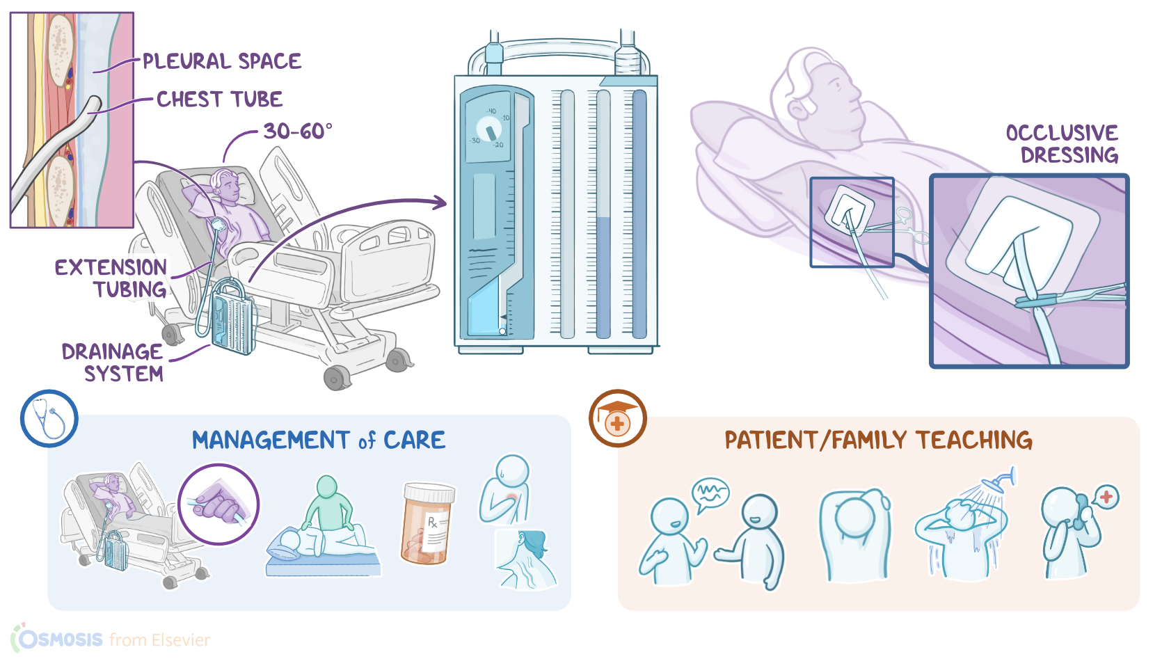
A chest tube can be used to remove air, fluids, or blood from the pleural space; to prevent air or fluid from reentering the pleural space; and to reestablish intrapleural and intrapulmonary pressures after surgery or trauma, in order to re-expand the lungs. A chest tube can also be inserted into the mediastinum after open heart surgery to drain fluid from the pericardial sac. Now, let’s quickly review the physiology of the pleura, which is a two-layered serous membrane that covers the lungs. The first layer of the pleura adheres to the surface of the lungs, called visceral pleura, whereas the second layer lines the chest wall, and it’s called the parietal pleura. Between these two layers there’s the pleural space, which is typically filled with a small amount of serous fluid that prevents friction between the two layers while breathing.

Moreover, let’s look at the physiology of breathing, which consists of two stages: inspiration and expiration. Inspiration is an active process, meaning that it requires muscles such as the diaphragm and the intercostal muscles to contract, which increases the intrathoracic volume and lowers the intrapulmonary pressure below the atmospheric pressure. This pressure gradient makes air flow from a high pressure area, meaning the atmospheric air, to a lower pressure area; so air is pulled inside the lungs. On the other hand, expiration is a passive process, meaning that it doesn’t require muscle contraction. That’s because the lungs have a special feature called elastic recoil, meaning they have a tendency to return to their normal size after being expanded. This allows the lungs to deflate after inspiration, decreasing the intrathoracic volume and increasing the intrathoracic pressure.

This process passively pushes the air from the area of higher pressure, which is the lungs, to the area of lower pressure, which is the atmosphere.Now, one end of the chest tube is typically inserted through the rib cage into the pleural space, while the other end is connected to extension tubing that drains into a drainage system. The most commonly used drainage system is the three-compartment drainage system. This system consists of three interconnected chambers: First, the collection chamber connects directly to the chest tube, and it collects air and fluid from the pleural cavity. Fluid stays in this chamber, while air flows to the next chamber, called the water seal chamber. This chamber acts as a one-way valve that allows air to flow into it during exhalation, and prevents air from moving back into the pleural space during inhalation. It is kept filled with 2 cm of sterile water that rises and falls during inhalation and exhalation, a process called tidaling.

When tidaling is present, we can be sure the chest tube is present. The suction control chamber is used to regulate the prescribed amount of suction, and it can be dry or wet. In wet suction devices, the amount of suction is regulated by the level of sterile water in the chamber, which is typically at -20 cm of pressure. In dry suction devices, a mechanical regulator is used to adjust the suction level. Other types of drainage systems include digital drainage systems, as well as a drainage system called the Hemlich chest drain. It is used to remove air from the pleural space in cases of a small pneumothorax where drainage or suction are not needed. It consists of a one-way flutter valve contained within a plastic tube. This device has an inlet nozzle, which allows air to pass from the pleural space to the valve; and an outlet nozzle, which allows air to flow to the outside.

All right, now the indications of chest tube placement include air in the pleural space, or pneumothorax, blood in the pleural space, called a hemothorax, or serous fluid from the pleural space, called a hydrothorax. Now, placing a chest tube can be done in the emergency room, the operating room, or at the bedside, and a couple of measures should be taken before inserting it. First is to make sure that the client is aware of the procedure and to obtain informed consent. Next is to perform an X-ray to confirm the affected side of the chest. Finally, gather the equipment needed for the procedure. Now, once everything is set up, the procedure begins by placing the client on the healthy side of the chest, with the arm on the affected side raised above the head to expose the midaxillary line.

The client’s head should also be elevated 30 to 60 degrees to minimize the risk of injury to the diaphragm. To insert the chest tube, first an antiseptic solution is used to cleanse the point of chest tube entry, which varies depending on the procedure. For draining fluid from the pleural space, the needle is typically inserted between the fifth or sixth intercostal space. On the other hand, when draining air, the needle is inserted between the second and third intercostal space, because air tends to pool at the top of the lungs. Now, after cleansing the point of entry, remember that the incision is made over the top of the inferior rib to avoid injury to intercostal blood vessels and nerves, which lie behind and below each rib. Next, a clamped chest tube is inserted into the incision and sutured into place; and the insertion site is covered with an occlusive dressing to prevent air from entering the pleural cavity.

Finally, the tube placement should be confirmed with an X-ray. Okay, let’s look at the nursing care you’ll provide to a client with a wet-suction chest tube. Your priority goals of care are to assist with chest tube insertion, promote lung expansion, and monitor for complications. Now, you can assist with the chest tube insertion by confirming informed consent has been obtained and premedicating your client with the prescribed analgesic. Set up the CDU by filling the water seal chamber with sterile water to the 2 cm mark and then filling the suction control chamber with sterile water to the -20 cm level. After the CDU is ready, assist with positioning your client for the procedure and providing reassurance during the insertion of the chest tube. Once the chest tube is in place, connect the drainage tube to the chest tube, and remove the clamp. Secure all of the connections with occlusive tape to prevent air leaks or disconnections.

Then, connect the suction control chamber to the suction unit, and increase the pressure until you see gentle, steady bubbling in the chamber. Assist your client into a semi-Fowler position and institute pulse oximetry so their oxygen saturation can be monitored. Lastly, place two soft-tipped hemostats by the bedside so they can be retrieved quickly. To promote lung expansion, ensure the CDU is functioning properly. Begin by ensuring the CDU is kept in an upright position and at a level lower than the client’s chest. Be sure to watch the water level in the water seal chamber, because it can evaporate over time; refill it if the water level drops below the 2 cm water mark. Likewise, because the continuous bubbling in the suction control chamber promotes evaporation, check this chamber often and keep the water in this chamber filled to the prescribed level.