Chronic kidney disease (CKD): Nursing

3,723views

Chronic kidney disease (CKD): Nursing

Adult Nursing 1 AAS

Adult Nursing 1 AAS

Charge nurse as a leader: Nursing
Health history: Nursing
Health literacy: Nursing
Health and illness models: Nursing
Health promotion and illness prevention: Nursing
Chronic disease: Nursing
Palliative and hospice care: Nursing
Body fluid compartments
Complete metabolic panel (CMP) - Chloride: Nursing
Complete metabolic panel (CMP) - Sodium: Nursing
Complete metabolic panel (CMP) - Potassium: Nursing
Complete metabolic panel (CMP) - Calcium: Nursing
Renin-angiotensin-aldosterone system
Arterial blood gas (ABG) - Metabolic acidosis: Nursing
Arterial blood gas (ABG) - Respiratory acidosis: Nursing
Arterial blood gas (ABG) - Metabolic alkalosis: Nursing
Arterial blood gas (ABG) - Respiratory alkalosis: Nursing
Arterial blood gas (ABG) - Overview: Nursing
Preoperative care: Nursing
Postoperative care: Nursing
Biology of cancer: Nursing
Physiology of pain: Nursing
Respiratory system anatomy and physiology
Geriatric considerations - Respiratory: Nursing
Ventilation
Foreign body aspiration and upper airway obstruction: Nursing process (ADPIE)
Pharyngitis: Nursing
Anatomy of the larynx and trachea
Laryngeal cancer: Nursing
Care of an intubated client: Nursing skills
Acute respiratory distress syndrome (ARDS): Nursing
Tuberculosis (TB): Nursing
Atelectasis: Nursing
Chronic obstructive pulmonary disease (COPD): Nursing process (ADPIE)
Lung cancer: Nursing
Asthma: Nursing process (ADPIE)
Pleural effusion: Nursing
Chronic bronchitis
Emphysema
Pulmonary embolism
Pneumonia
Chest tube care: Nursing
Oxygenation - Oxygen therapy: Nursing skills
Pulmonary edema: Nursing
Pulmonary hypertension
Pleurisy: Nursing
Flail chest: Nursing
Pulmonary contusion: Nursing
Rib fracture: Nursing
Pneumothorax and hemothorax: Nursing
Bacterial pneumonia: Nursing process (ADPIE)
Geriatric considerations - Cardiac: Nursing
Cardiac work
Stroke volume, ejection fraction, and cardiac output
Cardiac cycle
Arrhythmias - Heart blocks: Nursing
Shock - Cardiogenic: Nursing
Arrhythmias - Premature atrial contractions (PACs): Nursing
Cardiac afterload
Cardiac preload
Abnormal heart sounds
Heart failure: Pathology review
Heart failure
Normal heart sounds
Cardiovascular system anatomy and physiology
Pericardial effusion and cardiac tamponade: Nursing process (ADPIE)
Cardiac contractility
Arrhythmias - Supraventricular tachycardia (SVT): Nursing
Anatomy of the heart
Vital signs - Pulse: Nursing skills
Cardiac biomarkers - Troponin: Nursing
Cardiac conduction system
Arrhythmias - Sinus tachycardia and sinus bradycardia: Nursing
ECG rate and rhythm
Arrhythmias - Ventricular tachycardia (Vtach): Nursing
Cardiac biomarkers - Creatine kinase (CK): Nursing
Valvular heart disease: Nursing
ECG basics
Pericarditis: Nursing
Rheumatic heart disease: Nursing process (ADPIE)
Aortic valve disease
Cardiomyopathy: Nursing
Arrhythmias - Premature ventricular contractions (PVCs): Nursing
Physical assessment - Heart and neck vessels: Nursing
Vital signs - Blood pressure (BP): Nursing skills
Hypertension: Nursing process (ADPIE)
Atherosclerosis and arteriosclerosis: Pathology review
Coronary artery disease (CAD) and angina pectoris: Nursing process (ADPIE)
Myocardial infarction (MI): Nursing process (ADPIE)
Myocarditis: Nursing
Left-sided heart failure: Nursing process (ADPIE)
Arrhythmias - Atrial flutter (Aflutter): Nursing
Arrhythmias - Asystole: Nursing
Arrhythmias - Ventricular fibrillation (Vfib): Nursing
Mitral valve disease
Endocarditis: Nursing
ECG axis
ECG intervals
Electrocardiogram (ECG) - Normal sinus rhythm (NSR): Nursing
Arrhythmias - Atrial fibrillation (Afib): Nursing
Action potentials in pacemaker cells
Peripheral arterial disease (PAD): Nursing process (ADPIE)
Peripheral venous disease (PVD): Nursing process (ADPIE)
Arterial embolism: Nursing
Physical assessment - Peripheral vascular system: Nursing
Raynaud phenomenon: Nursing
Aortic aneurysm: Nursing process (ADPIE)
Aneurysms
Venous thromboembolism (VTE): Nursing process (ADPIE)
Blood components
Platelet plug formation (primary hemostasis)
Coagulation (secondary hemostasis)
Complete blood count (CBC) - Red blood cells (RBC): Nursing
Complete blood count (CBC) - White blood cells (WBC) and differential: Nursing
Complete blood count (CBC) - Hemoglobin and hematocrit: Nursing
Complete blood count (CBC) - Platelets: Nursing
Anemia - Macrocytic: Nursing
Anemia - Aplastic: Nursing
Anemia - Iron-deficiency: Nursing
Non-hemolytic normocytic anemia: Pathology review
Anemia of chronic disease: Year of the Zebra
Pernicious anemia: Year of the Zebra
Thalassemia: Nursing
Vitamin B12 deficiency
Polycythemia: Nursing
Platelet disorders: Pathology review
Essential thrombocythemia (NORD)
Coagulation studies - Prothrombin time (PT) and international normalized ratio (INR): Nursing
Coagulation studies - Partial thromboplastin time (PTT): Nursing
Disseminated intravascular coagulation (DIC): Nursing
Thrombocytopenia: Nursing
Leukemia: Nursing process (ADPIE)
Lymphoma - Hodgkin and non-Hodgkin: Nursing
Multiple myeloma: Nursing
Blood groups and transfusions
Blood transfusion reactions and transplant rejection: Pathology review
Gastrointestinal system anatomy and physiology
Geriatric considerations - Gastrointestinal: Nursing
Physical assessment - Abdomen: Nursing
Physical assessment - Anus, rectum, and prostate: Nursing
Liver anatomy and physiology
Complete metabolic panel (CMP) - Liver function tests (LFT): Nursing
Jaundice: Nursing
Achalasia: Year of the Zebra
Hiatal hernia: Nursing process (ADPIE)
Gastroesophageal reflux disease (GERD): Nursing process (ADPIE)
Esophageal disorders: Pathology review
Esophageal cancer: Nursing
Nutrition - Enteral: Nursing skills
Peptic ulcer disease (PUD): Nursing process (ADPIE)
Gastric cancer: Nursing
Diarrhea: Nursing
Irritable bowel syndrome (IBS): Nursing
Malabsorption syndromes: Pathology review
Appendicitis: Nursing process (ADPIE)
Diverticular disease: Nursing
Diverticulosis and diverticulitis
Inflammatory bowel disease - Crohn disease and ulcerative colitis: Nursing
Hygiene - Ostomy care: Nursing skills
Colorectal cancer: Nursing
Intestinal obstruction: Nursing
Gastrointestinal bleeding: Pathology review
Medications for hepatic encephalopathy: Nursing pharmacology
Hepatitis: Nursing
Hepatitis A and Hepatitis E virus
Hepatitis C virus
Cirrhosis: Nursing process (ADPIE)
Liver cancer: Nursing
Cholelithiasis: Nursing
Cholecystitis: Nursing
Pancreatitis: Nursing process (ADPIE)
Pancreatic cancer: Nursing
Renal system anatomy and physiology
Anatomy of the urinary organs of the pelvis
Glomerulonephritis: Nursing
Poststreptococcal glomerulonephritis
Nephrotic syndrome: Nursing
Acute kidney injury (AKI): Nursing process (ADPIE)
Chronic kidney disease (CKD): Nursing
The role of the kidney in acid-base balance
Complete metabolic panel (CMP) - Estimated glomerular filtration rate (eGFR): Nursing
Erythropoietin
Geriatric considerations - Urinary: Nursing
Dialysis care: Nursing
Renal cancer: Nursing
Urinary tract infections (UTIs): Nursing process (ADPIE)
Pyelonephritis: Nursing
Urinary incontinence - Stress: Nursing process (ADPIE)
Urinary retention: Nursing
Renal and urinary calculi: Nursing
Bladder tumors: Nursing
Hygiene - Urinary catheter care: Nursing skills
Renal clearance

Notes

CHRONIC KIDNEY DISEASE (CKD)

KEY POINTS
NOTES
DEFINITION
  • Structural or functional kidney abnormality that lasts more than 3 months

PHYSIOLOGY
  • Kidney
    • Body's natural blood filter
    • Clears blood of metabolic wastes and toxins
  • Secrete hormones
  • Regulate acid-base balance, pH, BP, and electrolyte levels
  • Nephron
    • Renal corpuscle
      • Where filtration occurs
      • Glomerulus
      • Bowman's capsule
        • Ultrafiltrate of blood
    • Renal tubules
      • Urine produced and modified
  • Glomerular filtration rate (GFR)
    • Rate that renal filtration takes place
    • Main measure of kidney function

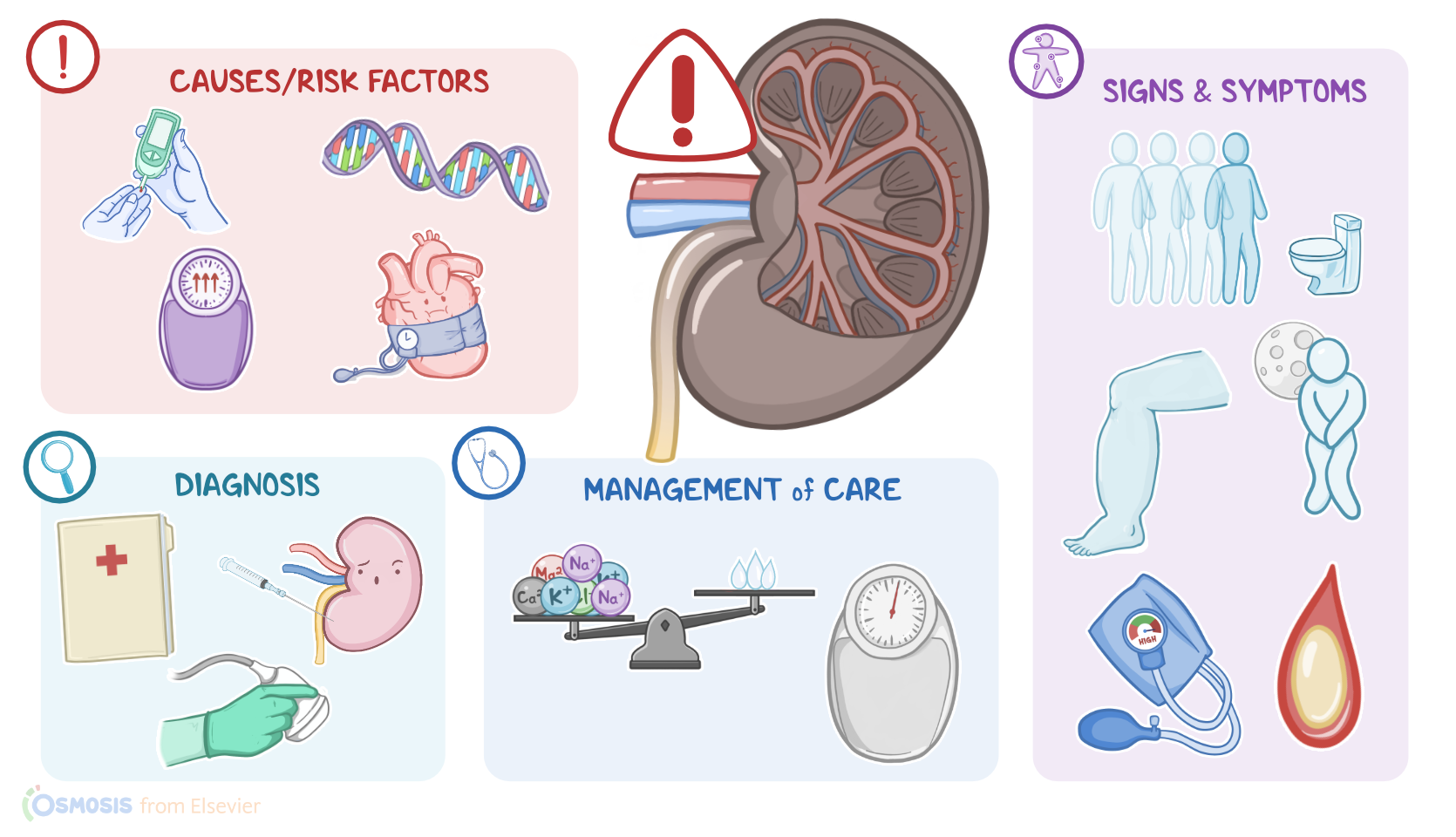
CAUSES AND RISK FACTORS
  • Causes
    • Diabetes mellitus
    • Hypertension
    • Rental artery stenosis
    • Glomerular disease
    • Polycystic renal disease
    • Tubulointerstitial diseases
    • Systemic disorders
    • Repeated episodes of pyelonephritis or obstructive uropathy
  • Modifiable risk factors
    • Obesity
    • Cardiovascular disease
    • Uncontrolled diabetes mellitus
    • Smoking
    • Nephrotoxic medications
  • Nonmodifiable risk factors
    • Age >60
    • Genetic predisposition

PATHOPHYSIOLOGY
  • Progressive and irreversible damage to kidneys leading to decline in kidney function
  • Kidney's lose ability to concentrate urine and excrete wastes and toxins
  • 5 stages
    • Stage 1: kidney function is normal
    • Stage 2: kidney function mildly decreased
    • Stage 3: kidney function moderately decreased
    • Stage 4: kidney function severely decreased
    • Stage 5: kidney function lost; ESRD

SIGNS AND SYMPTOMS
  • Asymptomatic
  • Symptomatic
    • Polyuria
    • Nocturia
    • Edema
    • Oliguria
    • Uremia
    • Uremic encephalopathy
    • Pericarditis
    • Uremic frost
    • Hypertension
    • Blood abnormalities
    • Renal osteodystrophy

DIAGNOSIS
  • History
  • Physical assessment
  • Laboratory tests
  • Abdominal ultrasound
  • Kidney biopsy

TREATMENT
  • Depends on stage
  • Stages 1 and 2
    • Adequate fluid balance
    • Lifestyle modifications
    • Treat hypertension
    • Treat hyperlipidemia
  • Stage 3
    • Correct blood abnormalities
    • Treat osteodystrophy
  • Stage 4
    • Renal replacement therapy
  • Stage 5
    • Renal replacement therapy
    • Kidney transplantation

MANAGEMENT OF CARE
  • Goals of care
    • Maintain fluid and electrolyte balance
    • Slow progression of disease
    • Manage disease complications
  • Assess
    • Kidney function
    • Fluid status
    • Daily weight
  • Report to HCP
    • Signs of fluid overload
    • Signs of pulmonary edema
    • Significant changes in weight
  • Fluid retention
    • Administer prescribed medications
    • Implement fluid restriction
  • Electrolyte imbalances
    • Administer prescribed medication
    • Monitor for signs of hyperkalemia
  • Renal replacement therapy
    • Assess insertion site
    • Assess circulation
    • Report to HCP
      • Inadequate circulation
      • Signs of clotting
      • Hematomas
      • Bleeding
      • Catheter dislodgement
      • Signs of infection
    • Avoid blood pressures and venipunctures in affected arm
  • Psychosocial needs
    • Assess
    • Assist with coping skills
    • Provide needed referrals

PATIENT AND FAMILY TEACHING
  • Explain condition, plan of care, and how to safely self-administer medications
  • Ways to reduce further kidney injury
    • Avoid nephrotoxic medication
    • Avoid exposure to radiographic contrast dye, pesticides, and heavy metals
  • Notify HCP
    • Starting medications
    • Weight gain > 2 kg (4.4 pounds)
    • Increased blood pressure
    • Shortness of breath
    • Swelling in extremities
    • Changes in urine appearance, frequency, or volume
  • Encourage regular follow-ups
  • Lifestyle modifications
    • Renal diet
    • Restrict fluids
    • Spread out fluid intake over 24-hours
    • Avoid certain foods
  • Refer to renal dietician

Transcript

Watch video only

Chronic kidney disease, or CKD for short, is a condition characterized by a slow and progressive decrease in kidney function, with a glomerular filtration rate, or GFR, of less than 60 mL/minute that develops over a minimum of three months.

Now, let’s take a closer look at how the kidneys work. We can think of the kidneys as the body’s natural blood filter. Their main function is to clear blood of metabolic wasteful substances and toxins by excreting them through urine. In addition, they secrete important hormones, and are essential in regulating the acid-base balance, pH, blood pressure, and electrolyte levels in the body.

Within each kidney, there are millions of tiny functional units called nephrons, which consist of a renal corpuscle and a set of renal tubules. The renal corpuscle is where blood filtration occurs, and it includes the glomerulus, a tiny bundle of capillaries, and the Bowman’s capsule, a cup-shaped structure that surrounds the glomerulus.

As blood flows through the glomerulus, water and small solutes dissolved in the blood are filtered into the Bowman’s capsule, creating an ultrafiltrate of blood. This filtrate then travels through the renal tubules, where urine is ultimately produced and modified according to the body’s necessities. The rate at which renal filtration takes place is called glomerular filtration rate, or GFR for short, and it is one of the main measures of kidney function. In a healthy adult, the estimated GFR is around 100-120 mL/min, and this value decreases slowly in all of us as we grow older.

All right, now, several conditions can speed up the rate at which GFR deteriorates, increasing the risk of developing CKD. In the United States, the leading causes of CKD are diabetes mellitus and hypertension, both of which are more common in elderly clients. Less common causes include renal artery stenosis, glomerular diseases, polycystic renal disease, tubulointerstitial diseases, and systemic disorders, like lupus or amyloidosis. Additionally, repeated episodes of pyelonephritis or obstructive uropathy, such as prostate disease, can also lead to CKD.

Regarding risk factors for CKD, modifiable ones include obesity, cardiovascular disease, uncontrolled diabetes mellitus, smoking, and exposure to nephrotoxic medications like NSAIDs or aminoglycosides; while nonmodifiable risk factors include being over 60 years of age and having a genetic predisposition to kidney disease.

Now, regardless of its cause, CKD results from progressive and irreversible damage to the kidneys, leading to a gradual decline in kidney function. As a result, the kidneys gradually lose their ability to concentrate the urine and excrete wasteful substances or toxins. So, CKD can be classified into five stages by determining the estimated GFR, which is measured in units of milliliters per minute per 1.73 square meters.

In clients at stage 1 CKD, there’s still a normal kidney function, since the remaining healthy nephrons are able to adapt, become larger, and work harder to maintain urine production. As the disease progresses into stage 2 CKD, kidney function is mildly decreased; while in stage 3 CKD, there’s a moderate decrease, and in stage 4 CKD, there’s a severe decrease in kidney function. Over time, as CKD progresses into stage 5, kidney function is completely lost, and clients develop renal failure, also known as end-stage kidney disease, or ESRD for short.

Now, the body is capable of coping with a significant reduction in kidney function without causing any symptoms, which makes CKD a largely silent disease. As the kidneys lose their ability to concentrate urine, clients may experience polyuria and nocturia. As damage progresses, though, fluid retention is more common and may result in edema and oliguria. In addition, as urine output decreases, wasteful substances or toxins like urea and creatinine begin to accumulate in the body; this can lead to uremia, which may cause general symptoms like fatigue, nausea, and loss of appetite.

As toxin levels build up, they can affect the functioning of the nervous system, resulting in uremic encephalopathy. This can cause asterixis, a tremor of the hand that appears when a client attempts to extend their wrists, along with ataxia and lethargy. The buildup of toxins can also affect the heart causing pericarditis. In some cases, clients can develop uremic frost, where urea crystals deposit in the skin giving the appearance of powdery snowflakes.

Additionally, clients with CKD often have hypertension due to sodium retention and activation of the renin-angiotensin system. Finally, clients may develop blood abnormalities, such as anemia, hyperkalemia, metabolic acidosis, and disorders of phosphate and calcium metabolism. Over time, resorption of calcium from the bones in an attempt to restore blood calcium levels can leave bones weak and brittle, which is known as renal osteodystrophy.

Ultimately, diagnosis of CKD is based on laboratory tests. These include blood tests showing an increase in blood creatinine, BUN, as well as a decrease in an estimated GFR. A urinalysis will show proteinuria, hematuria, white blood cells, glucose, and casts. If the exact cause of CKD is unknown, an abdominal ultrasound can be done to look for signs of scarring or polycystic kidneys, as well as obstructive uropathy. In long-term ESRD, X-rays may be done to check for renal osteodystrophy. Finally, a kidney biopsy can be done to look for inflammation, scarring or unusual deposits of a protein, and to determine how far CKD has advanced.

The goal of the treatment in CKD depends on the stage. In clients at stages 1 and 2, the main goal is making sure the client has an adequate fluid balance, as well as slowing down the decline in kidney function and preventing complications. This involves lifestyle modifications like smoking cessation, stopping any nephrotoxic medications, and maintaining a tight blood glucose control among clients with diabetes mellitus. Two of the main factors involved in the progression of CKD are hypertension and proteinuria, so clients often require treatment with an ACE inhibitor like enalapril or an ARB like losartan. Clients with hyperlipidemia should be started on lipid lowering agents to reduce cardiovascular risk.

On the other hand, in clients with stage 3 CKD, all blood abnormalities like anemia and electrolyte imbalances should be corrected as needed. If there’s evidence of renal osteodystrophy, additional measures, like calcium supplementation or treatment with phosphate binders, should be taken to prevent further loss of bone density. When clients reach stage 4, kidney function is severely reduced, so treatment involves getting them prepared for renal replacement therapy options, which include hemodialysis, peritoneal dialysis, and renal transplantation. Finally, in stage 5 CKD, the remaining kidney function is not enough to maintain life, so clients should be started on renal replacement therapy and consider kidney transplantation.

Now, let’s look at the nursing care of a client with chronic kidney disease. Keep in mind that nursing care may vary based on the progression and stage of kidney dysfunction that your client is experiencing. The overall goals are to maintain fluid and electrolyte balance, slow the progression of the disease, and manage any disease complications.