Epidural and subdural hematoma: Nursing
Notes
| EPIDURAL AND SUBDURAL HEMATOMA | ||
| KEY POINTS | NOTES | |
| DEFINITION |
| |
| PHYSIOLOGY |
| |
| CAUSES AND RISK FACTORS |
| |
| PATHOPHYSIOLOGY |
| |
| SIGNS AND SYMPTOMS |
| |
| DIAGNOSIS |
| |
| TREATMENT |
| |
| MANAGEMENT OF CARE |
| |
| PATIENT AND FAMILY TEACHING |
| |
Transcript
Intracranial hemorrhage or ICH for short, refers to bleeding inside the skull. Types of intracranial hemorrhage include epidural hematoma, subdural hematoma, subarachnoid hemorrhage, and intracerebral bleeding. Now, epidural hematoma is the collection of blood in the epidural space, which is the space between the dura mater and inner surface of the skull. In contrast, subdural hematoma is the collection of blood in the subdural space, meaning between the dura mater and the arachnoid mater.
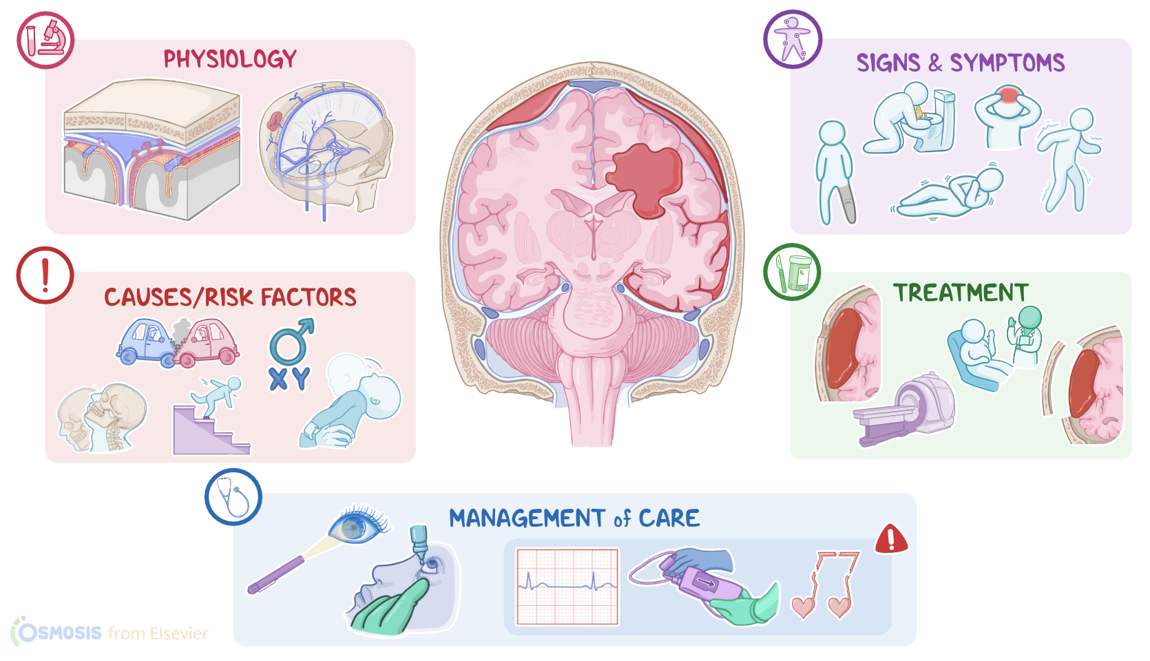
Let’s start by looking at the physiology of the meninges. This is a membrane that wraps around the brain and protects it from the outside environment. It’s made up of three meningeal layers. From outside to inside, these are the dura mater, arachnoid mater, and pia mater. Between the arachnoid and pia mater there’s the subarachnoid space, which houses the cerebrospinal fluid. The cerebrospinal fluid is a clear, watery liquid that cushions the brain from impact and bathes it in nutrients.
Now, the dura mater consists of an external and an internal layer. The external layer of the dura mater adheres tightly to the inner surface of the skull, so the epidural space is only a virtual space in the cranium, meaning the space isn’t appreciable unless pathology is present.
Between the external layer of the dura mater and the inner surface of the skull, there are meningeal arteries that supply meninges. The internal layer of the dura mater lies above the arachnoid mater, the two are separated by the subdural space. The subdural space plays a major role in venous blood drainage in the brain. The dura mater also forms dural venous sinuses which carry the venous blood from the brain. These sinuses are connected to the veins outside of the cranium by bridging veins.
Okay, let’s move onto epidural and subdural hematoma. The most common cause of epidural hematoma is rupture of the meningeal artery. This is often caused by blunt head trauma from falls or car crashes. The most common site for epidural hematoma is at the pterion which is the thinnest part of the skull, and it’s located right above the middle meningeal artery. Other causes of epidural hematoma may include hemorrhagic tumors, vascular malformations, infection or coagulation disorders. Risk factors for epidural hematoma include being assigned male at birth and age from 20 to 30 because the external layer of the dura mater hasn’t fully attached to the skull yet.
On the other hand, the main cause of a subdural hematoma is rupture of bridging veins located in the subdural space. Now, like with epidural hematoma, blunt head trauma is a major cause, but another important cause is acceleration-deceleration injury. This can occur when you step on the brakes while driving very fast, causing the head and brain to move forward and back violently, damaging the bridging veins. Another cause is shaken baby syndrome where a baby is violently shaken, making their head whip back and forth damaging the bridging veins.
Now, clients younger than 1 year of age and elderly clients are at higher risk because they often have relatively thin walled blood vessels that are susceptible to rupture. Old age, chronic alcohol use and neurodegenerative conditions like Alzheimer disease cause brain atrophy. When the brain shrinks, there’s more space around the bridging veins, leaving them exposed and unsupported.
Okay, moving on to pathology, with epidural hematoma once the meningeal artery is torn, blood will pool between the skull and the external layer of the dura mater. Now, what makes epidural hematomas so dangerous is that the dura mater is attached tightly to the sutures of the skull, so the blood can’t cross these sutures and becomes trapped. This can quickly increase the intracranial pressure or ICP and a large epidural hematoma on one side of the skull can cause a midline shift and push the brain towards the opposite side of the skull.
In the case of subdural hematoma, once a bridging vein is torn, the blood pools in the subdural space creating a hematoma. Because the source of the bleeding is venous, subdural hematomas usually grow slower than epidural hematomas which are caused by arterial bleeding. Also, since the blood isn’t restricted by sutures, it can be distributed over a larger area, so pressure doesn’t build up as quickly, unless the hematoma is very large.
Now, with both epidural and subdural hematoma, as blood accumulates over time the hematoma grows in size and increases the ICP. If not corrected, increased ICP can lead to complications, such as papilledema, which is the swelling of the optic disc where the optic nerve leaves the retina. The most severe complication, though, is brain herniation. Brain herniation occurs when a part of the brain is pushed into another space of the skull or even out of it through the foramen magnum, and this can affect other CNS structures, like the brainstem.
Clinical manifestations of an epidural hematoma most commonly include a loss of consciousness right after the head trauma. In a slow epidural bleed, this might be followed by a lucid interval of temporary improvement right after the initial trauma, but then hours later, they will deteriorate as the epidural hematoma grows and progresses to confusion and coma.
In the case of a subdural hematoma, there can also be a brief loss of consciousness followed by a lucid interval. However, depending on the size and speed of the bleed, this can last from days to weeks before symptoms develop. A subdural hematoma is considered acute if symptoms develop within 2 days of a head trauma, subacute if they develop between 2 days and 2 weeks of a head trauma, and chronic if they develop 2 weeks or more after a head trauma. Although symptom onset is slower than epidural hematoma, it’s actually more deadly since the blood can spread across a larger area around the brain, so deterioration can be quick once symptoms develop.
In both epidural and subdural hematomas, there can also be headaches, bouts of seizures and vomiting, as well as focal neurological symptoms like muscle weakness or sensory problems depending on the location of the hematoma. Additionally, there can be signs of increased ICP such as vomiting, a fixed dilated pupil on the same side as the hematoma, as well as the Cushing triad, which includes bradycardia, irregular respiratory pattern, and widening of pulse pressure where systolic pressure increase more than diastolic pressure.
Diagnosis of increased ICP starts with the client’s history and physical assessment, followed by a CT scan to confirm the diagnosis. In the case of an epidural hematoma, this typically shows a convex, “lens-shaped” collection of blood that does not cross the suture lines of the skull. In contrast, a subdural hematoma typically appears as a concave, “crescent-shaped” density that crosses the suture lines.
Once the diagnosis is confirmed, the client should be immediately sent to OR for surgery. Most epidural hematomas are treated by craniotomy, which is when part of the skull bone is removed in order to remove accumulated blood clot below. In some settings, a life-saving alternative is to drill a hole in the skull, which is called Burr hole trephination. Finally, non-surgical management with serial neurological assessments and CT scans might be considered for clients with small epidural hematomas and in good clinical condition.