Musculoskeletal injuries: Nursing process (ADPIE)

Last updated: January 26, 2022

Musculoskeletal injuries: Nursing process (ADPIE)

FINAL

FINAL

ACE inhibitors, ARBs and direct renin inhibitors
Thiazide and thiazide-like diuretics
Calcium channel blockers
Adrenergic antagonists: Beta blockers
Acyanotic congenital heart defects: Pathology review
Atherosclerosis and arteriosclerosis: Pathology review
Coronary artery disease: Pathology review
Peripheral artery disease: Pathology review
Valvular heart disease: Pathology review
Cardiomyopathies: Pathology review
Heart failure: Pathology review
Supraventricular arrhythmias: Pathology review
Ventricular arrhythmias: Pathology review
Heart blocks: Pathology review
Aortic dissections and aneurysms: Pathology review
Pericardial disease: Pathology review
Endocarditis: Pathology review
Hypertension: Pathology review
Shock: Pathology review
Vasculitis: Pathology review
Cardiac and vascular tumors: Pathology review
Dyslipidemias: Pathology review
Cardiac tamponade
Endocarditis
Myocarditis
Rheumatic heart disease
Heart failure
Cor pulmonale
Long QT syndrome and Torsade de pointes
Ventricular tachycardia
Premature ventricular contraction
Ventricular fibrillation
Atrial flutter
Premature atrial contraction
Atrial fibrillation
Atrioventricular nodal reentrant tachycardia (AVNRT)
Deep vein thrombosis
Hypotension
Orthostatic hypotension
Polycystic kidney disease
Pheochromocytoma
Cushing syndrome
Renal artery stenosis
Hypertension
Aneurysms
Aortic dissection
Peripheral artery disease
Angina pectoris
Unstable angina
Prinzmetal angina
Myocardial infarction
Stable angina
Arterial disease
ECG normal sinus rhythm
ECG cardiac hypertrophy and enlargement
ECG cardiac infarction and ischemia
ECG basics
ECG intervals
ECG axis
ECG QRS transition
ECG rate and rhythm
Cardiac conduction system
Cardiac conduction velocity
Normal heart sounds
Abnormal heart sounds
Cardiovascular changes during postural change
Cardiovascular changes during hemorrhage
Cardiac preload
Cardiac contractility
Cardiac afterload
Measuring cardiac output (Fick principle)
Thrombocytopenia: Clinical
Heparin-induced thrombocytopenia
Immune thrombocytopenia
Gout
Chronic kidney disease: Clinical
Traumatic brain injury: Pathology review
Traumatic brain injury: Clinical
Concussion and traumatic brain injury
Blood groups and transfusions
Blood products and transfusion: Clinical
HIV (AIDS)
Hodgkin lymphoma
Acromegaly
Musculoskeletal injuries: Nursing process (ADPIE)
Hemophilia: Nursing process (ADPIE)
Diabetes insipidus
Diabetes mellitus
Diabetes mellitus: Clinical
Diabetes mellitus: Pathology review
Diabetes mellitus (DM): Nursing process (ADPIE)
Diabetes insipidus: Nursing process (ADPIE)
Managing diabetes during the holidays: Information for patients and families
Hypoglycemics: Insulin secretagogues
Insulins
Epistaxis: Nursing process (ADPIE)
Appendicitis
Appendicitis: Clinical
Appendicitis: Pathology review
Appendicitis: Nursing process (ADPIE)
Hypothyroidism medications
Hyperosmolar hyperglycemic state (HHS): Nursing process (ADPIE)
Sympathomimetics: Direct agonists
Cushing syndrome and Cushing disease: Pathology review
Cushing syndrome: Clinical
Metabolic and respiratory alkalosis: Clinical
Metabolic and respiratory acidosis: Clinical
Conjunctivitis: Nursing process (ADPIE)
Stroke: Clinical
Stroke: Nursing process (ADPIE)
Peptic ulcer
Peptic ulcer disease (PUD): Nursing process (ADPIE)
Peptic ulcers and stomach cancer: Clinical
Gallbladder histology
Gallbladder disorders: Clinical
Acute cholecystitis
Oral cancer
Hepatitis A and Hepatitis E virus
Viral hepatitis: Clinical
Hepatitis medications
Seizures: Pathology review
Seizures: Clinical
Seizures and epilepsy
Febrile seizure
Seizure disorder: Nursing process (ADPIE)
Non-urothelial bladder cancers
Inflammatory bowel disease: Clinical
Inflammatory bowel disease: Pathology review
Anticoagulants: Heparin
Postoperative evaluation: Clinical
Trigeminal neuralgia
Trigeminal neuralgia: Nursing process (ADPIE)
Hypoparathyroidism
Pancreatitis: Pathology review
Pancreatitis: Clinical
Acute pancreatitis
Pancreatitis: Nursing process (ADPIE)
Chronic pancreatitis
Sickle cell disease (NORD)
Sickle cell disease: Clinical
Sickle cell disease: Nursing process (ADPIE)
Class IV antiarrhythmics: Calcium channel blockers and others
Hypertension: Clinical
Pulmonary hypertension
Hypertension: Nursing process (ADPIE)
Osteoarthritis
Joint pain: Clinical
Hyperthyroidism: Pathology review
Hyperthyroidism: Clinical
Deep vein thrombosis and pulmonary embolism: Pathology review
Hyperthyroidism
Hyperthyroidism medications
Hyperthyroidism: Nursing process (ADPIE)

Notes

MUSCULOSKELETAL INJURIES

KEY POINTS
NOTES
PATIENT REPORT
  • 24-year-old 
  • Urgent care
    • Reports of right ankle swelling and pain sustained while running

PATHOPHYSIOLOGY
  • Local musculoskeletal injuries 
  • Affect bones, joints, muscles, and ligaments 
  • Common types include 
    • Bone fractures 
    • Joint dislocations 
    • Muscle or tendon strains 
    • Ligament sprains 
  • Causes of sprains 
    • Force exceeds ligament elasticity 
    • Uneven surfaces 
    • Falls with twisting motion 
    • Awkward landings 
    • Sports-related contact 
  • Types of sprains 
    • Lateral sprains 
      • Most common type 
      • Caused by forced inversion 
      • Damages outer ligaments 
    • Medial sprains 
      • Caused by forced eversion 
      • Damages medial ligaments 
    • Risk factors for sprains 
      • Modifiable 
        • Inappropriate footwear 
        • Poor athletic condition 
        • No warm-up before activity 
        • Fatigue from overtraining 
      • Non-modifiable 
      • Biological males aged 15–24 
      • Biological females over age 30 
      • Previous ankle sprains 
      • Balance problems 
      • Foot misalignment  
    • Grades of sprains 
      • Grade one 
        • Mild ligament stretching 
        • Mild pain and swelling 
      • Grade two 
        • Partial ligament rupture 
        • Moderate pain, swelling, bruising 
        • Mild to moderate instability 
        • Painful walking and weight bearing 
      • Grade three 
        • Complete ligament tear 
        • Severe pain, swelling, bruising 
        • Significant instability and loss of function 
    • Complications of sprains
      • Acute 
        • Muscle or blood vessel injury 
        • Acute compartment syndrome 
      • Long-term 
        • Joint instability or stiffness 
        • Osteoarthritis 
        • Impaired range of motion

DIAGNOSIS AND TREATMENT
  • Diagnosis
    • History
    • Physical assessment
    • Diagnostic imaging
  • Treatment
    • PRICE method 
      • Protection and rest 
        • Use of brace or CAM boot 
      • Ice 
        • Reduces swelling and pain 
      • Compression 
        • Helps control inflammation 
      • Elevation 
        • Promotes fluid drainage and reduces swelling 
    • Additional interventions 
      • Below-knee casts may shorten recovery time 
      • Pain management
      • Physical therapy 
      • Gradual exercises
      • Surgical intervention 

ASSESSMENT
  • Vital signs 
    • Temperature 98.2°F (36.8°C) 
    • Heart rate 62 beats per minute 
    • Respiratory rate 16 breaths per minute 
    • Blood pressure 115/70 mmHg 
    • SpO2 100% on room air 
    • Pain 6/10 
  • Right foot and ankle assessment 
    • Edematous with ecchymosis on lateral side 
    • Warm and tender around lateral malleolus 
    • Capillary refill < 3 seconds 
    • Able to wiggle toes 
    • Strong posterior tibial and dorsalis pedis pulses 
    • Able to bear some weight  

NURSING DIAGNOSES
  • Risk for peripheral neurovascular dysfunction related to edema
  • Impaired physical mobility related to musculoskeletal injury
  • Acute pain related to tissue injury

PLANNING
  • Before leaving clinic, patient will verbalize the signs of neurovascular complications and demonstrate safe mobilization
  • Over 4 weeks
    • Patient will return to baseline activity level
    • Pain will be managed at 3/10 

IMPLEMENTATION
  • Administered ordered NSAID  
  • Neurovascular education 
    • Taught signs of neurovascular compromise 
      • Numbness or tingling below ankle 
      • Severe pain or swelling 
      • Inability to move foot or wiggle toes 
      • Toes becoming pale, blue, or cold 
  • PRICE method 
    • Wrapped foot and ankle with elastic bandage 
    • Applied and adjusted ankle brace for support 
    • Demonstrated proper brace fit and security 
    • Instructed to wear brace when moving 
  • Activity and mobility 
    • Advised to avoid discomfort-causing activities 
    • Encouraged range-of-motion exercises daily 
    • Instructed to elevate ankle while resting 
    • Recommended icing ankle 15–20 minutes several times daily 
    • Continue medication 
    • Crutch training 
  • Discharge and follow-up 
    • Provided printed instructions summarizing care plan 
    • Encouraged attending physical therapy 
    • Recommended follow-up with HCP
      • Pain or swelling does not improve in 72 hours 
      • Neurovascular symptoms develop 
      • Weight bearing remains difficult after 4 days

EVALUATION
  • Pain level decreased to 3/10 
  • Foot has normal warmth and circulation 
  • Able to wiggle toes 
  • Patient correctly summarized teaching provided 
  • Verbalized signs of neurovascular complications 
  • Agreed to seek immediate care if symptoms occur 
  • Will call HCP for follow-up 
  • Plans to attend physical therapy sessions 
  • Correctly demonstrated crutch use 

Transcript

Watch video only

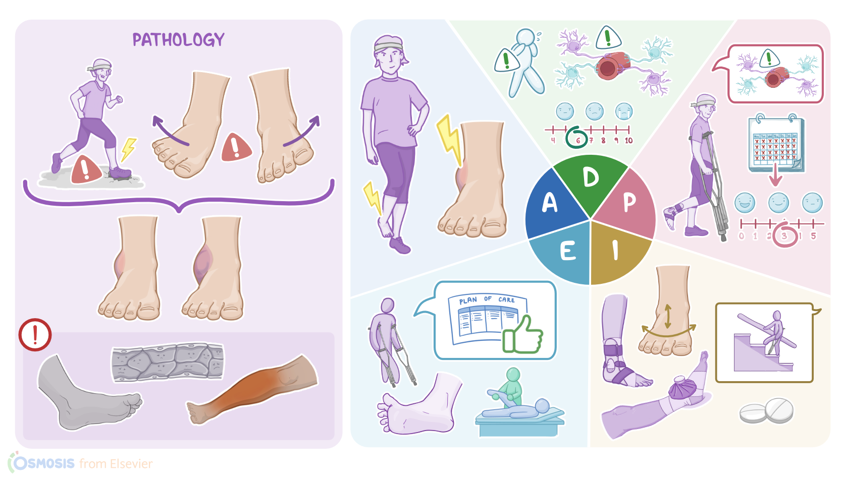
Liam Warner is a 24-year-old male who is dropped off at the urgent care clinic with right ankle pain and swelling.  This morning Liam was running in a 10K run when  his foot landed on an uneven surface, injuring his ankle. He says “My foot didn’t land right and I could feel it rolling  inward. I wasn’t able to finish because I could barely walk.”  While in the clinic, an ankle X-ray series rules out a bone fracture, and a grade 2 sprain is suspected.  

Local musculoskeletal injuries mainly include conditions that affect bones, joints, muscles, and ligaments. The most common ones include bone fractures; joint dislocations; muscle or tendon strains; and finally, ligament sprains. Now, sprains typically occur when the physical force applied to the joint exceeds the elasticity of the surrounding ligaments. And this is particularly common in ankle injuries, when there’s incorrect positioning of the foot at landing, like when a person is walking or running on an uneven surface; or they fall and twist their ankle; another frequent scenario involves jumping and awkwardly landing on their foot; or being stepped on during a sports game.

Now, there are two main types of ankle sprains. Lateral sprains are the most common ones and they are typically caused by forced inversion of the foot. In other words, a person’s sole rotates inwards or medially, but too much, eventually damaging the ligaments located on the outer side of the joint. On the other hand, medial sprains are caused by forced eversion of the foot, meaning a sole rotates outwards or laterally, eventually damaging the medial ligaments. 

Alright, there are some factors that can put a person at risk for ankle sprains. Modifiable risk factors include wearing inappropriate footwear, having poor athletic condition, not warming up before training, and fatigue from overtraining or intense physical activity. On the other hand, non-modifiable risk factors include being male between the ages of 15 to 24, or being female over the age of 30, as well as previous history of ankle sprains; balance problems; and conditions associated with foot misalignment, such as pes cavovarus, often referred to as overarched foot.

Depending on how severe the injury is, ankle sprains can be classified into one of three grades. Grade one ankle sprains refers to mild stretching of the ligament, and it’s typically associated with mild pain and swelling. Grade two ankle sprains involve a partial rupture of the ligaments and present with moderate pain and swelling, as well as bruising. There is mild to moderate joint instability, with some restriction in ankle motion, and both walking and weight bearing are painful. Finally, grade three ankle sprains involve complete ligament tears, and present with severe pain, swelling, and bruising; as well as significant joint instability with loss of function and weight bearing. 

Now, most ankle sprains heal without causing any complications. Rarely though, some clients may develop acute complications from associated muscle or blood vessel injuries, such as acute compartment syndrome. This is a serious condition in which there’s a rapid increase in the pressure within an enclosed compartment, leading to tissue damage due to hypoxia and ischemia. On the other hand, long-term complications may occur years after the injury. The most common ones include joint instability or stiffness, as well as osteoarthritis, all of which can ultimately result in impaired range of motion.

In most cases, diagnosis is based on history and physical examination. If a fracture is suspected, it can be confirmed by performing an X-ray. An MRI can be done to evaluate the condition of the surrounding soft tissue or detect a hidden fracture. 

Treatment of ankle sprains is usually symptomatic and includes the PRICE method, which stands for protection and rest, by immobilizing the ankle with a brace or controlled ankle motion boot or CAM boot; as well as ice, compression, and elevation of the affected ankle to reduce the swelling. Additionally, below-knee casts can be used to reduce the length of the recovery period; while pain and inflammation can be managed with acetaminophen or NSAIDs like ibuprofen. This is followed by mobilization of the ankle with physical therapy and gradual exercises to recover the full mobility and strength of the ankle. Fortunately, surgery is rarely needed for ankle sprains.

After introducing yourself as his nurse you begin your assessment.  Liam’s vital signs are oral temperature 98.2° F or 36.8° C; heart rate 62 beats per minute, respiratory rate 16 breaths per minute, blood pressure 115/70 mmHg, SpO2 100% on room air and pain 6/10.  Liam’s right foot and ankle are edematous with ecchymosis on the lateral side of his foot. The ankle is warm and tender to palpation around the lateral malleolus. Capillary refill in less than 3 seconds, he can wiggle his toes, and you palpate strong posterior tibial and dorsalis pedis pulses

He is able to bear toe-touch weight and take 5 limping steps. Next, you elevate Liam’s ankle above the level of his heart and apply an ice pack. After documenting your assessment findings, you let Liam know the physician assistant, or PA, will be in to examine him shortly.