Nephrotic syndrome: Nursing
Introduction0:00–0:17
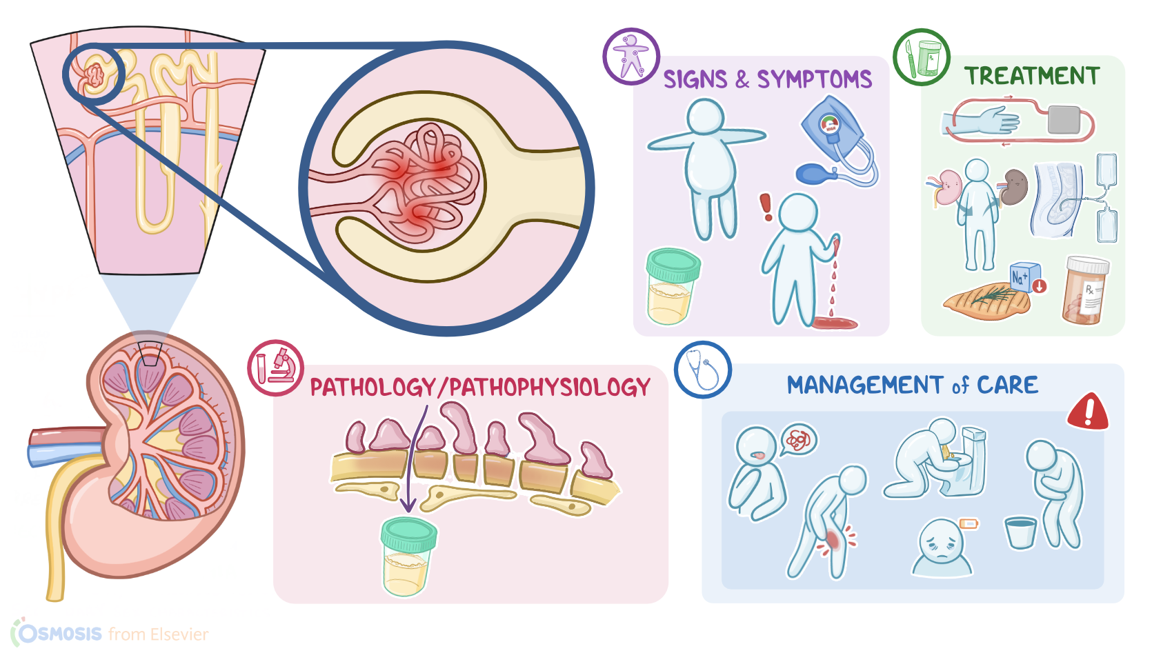
Nephrotic syndrome is a disorder characterized by a constellation of signs and symptoms caused by conditions that affect the tiny glomeruli in the kidneys, resulting in excessive loss of protein through urine, called proteinuria.
First, let’s quickly review some kidney physiology! Remember that the nephron is the functional unit of the kidney, and its role is to filter waste products and water from the blood.
Physiology0:17–2:11
Each nephron is made up of a renal corpuscle and a set of renal tubules, which are, in order: the proximal convoluted tubule, the loop of Henle, the distal convoluted tubule and finally, the collecting ducts which drain urine into the renal papillae and eventually empty into the renal pelvis.
The renal corpuscle is made up of the glomerulus, which is a bundle of capillary loops, and Bowman’s capsule, which surrounds the glomerulus.
Now, blood enters the glomerulus through the afferent arteriole, then inside the glomerulus, glomerular filtration occurs.
Then, the remaining blood exits the glomerulus through the efferent arteriole. The glomerular capillary wall is a semi-permeable membrane with three layers: the endothelium, which is the inner layer; the basement membrane; and the epithelium, which is the outermost layer.
For filtration to occur, the endothelial and epithelial cells that line the capillary wall are separated by small pores that filter water and small particles from the blood and into the Bowman capsule.
Only small particles are filtered from the blood and never proteins, since they are larger molecules. Also, normally, there aren’t any red blood cells in the urine.
For glomerular filtration to occur, the hydrostatic pressure created by the blood pressure needs to be greater than opposing forces, such as the tubular filtrate and oncotic pressure, which is the pressure of proteins.
The final product of filtration is called filtrate or primary urine, which will flow through the Bowman capsule into the proximal convoluted tubule.
Ok, now, some of the most common causes of nephrotic syndrome includea cute glomerulonephritis, focal segmental glomerulosclerosis, and minimal change disease, as well as diabetes and systemic lupus erythematosus.
Causes & risk factors2:11–2:44
Other causes include some cancers such as Hodgkin lymphoma; certain infections like those caused by Streptococcus species or HIV; as well as some drugs, such as NSAIDs and penicillamine.
Lastly, nephrotic syndrome that occurs during the first year of life has a genetic basis. And now, let’s look at the pathology of nephrotic syndrome.
Pathology2:44–4:20
Regardless of cause, the hallmark feature of nephrotic syndrome is damage to the glomerular basement membrane, which increases its permeability.
This allows proteins to make their way past the membrane and into the urine. Proteinuria, hypoalbuminemia, edema, hypertension, and hyperlipidemia develop as a consequence.As total serum protein levels, specifically the protein albumin, decrease, there’s a lower oncotic pressure in blood vessels, which causes edema that can eventually progress to generalized swelling in the entire body, called anasarca.
Additionally, the diminished oncotic pressure stimulates lipoprotein synthesis in the liver, causing hyperlipidemia. Other proteins, like antibodies, are also lost in the urine This means the immune response is also altered in nephrotic syndrome, so the client is at risk for infections.Additionally, important anticoagulant proteins are lost through urine, so another result of nephrotic syndrome is a state of hypercoagulability, which can increase the risk for arterial and venous thromboembolism, like deep vein thrombosis, renal vein thrombosis or even pulmonary embolism.
Paradoxically, there’s also an increased risk of bleeding with nephrotic syndrome, because proteins such as coagulation factors are also lost through the urine.
Finally, calcium also tends to be lost through urine, causing hypocalcemia. Now let’s translate that into a clinical picture.
Clinical manifestations4:20–5:02
Edema initially manifests as periorbital or facial edema. Fluid retention can become generalized or build up in the abdominal cavity, which causes ascites.
And all that gradual accumulation of fluid in the body causes weight gain. Additionally, because of hyperlipidemia, fat bodies or fatty casts often appear in the urine, causing foamy urine, and urine output may decrease.
Finally, clients can develop hypertension, though blood pressure may be decreased if the client is hypovolemic, and they may bleed for longer than normal following an injury.
Diagnosis5:02–5:46
The diagnosis of nephrotic syndrome starts with the client’s history and physical assessment. A 24 hour urine collection shows severe proteinuria, with more than 3.5 g/dL of protein in a 24 hour urine sample.
Clients also have serum albumin levels below 3 g/dL as well as hyperlipidemia. There are also abnormalities in the coagulation panel, such as an increased PT, aPTT, and INR.
Creatinine and blood urea nitrogen are also usually increased, and GFR is decreased. Last but not least, a kidney biopsy can be done to establish the cause of nephrotic syndrome.
Treatment of nephrotic syndrome depends on the cause. General treatment includes nutrition therapy, immunosuppressants, and symptomatic treatment.
Treatment5:46–7:08
Nutrition therapy includes a low sodium diet, moderate protein intake and fluid restriction in the case of significant edema.
Corticosteroids such as prednisone, as well as other immunosuppressants like cyclophosphamide can be used to help induce remission.ACE inhibitors or ARBs, which help reduce proteinuria.
Treatment of hyperlipidemia includes lipid-lowering agents, like statins. Anticoagulant therapy could be needed if there’s thrombosis.
Diuretics, especially loop diuretics like furosemide can improve edema. If a client also has diabetes, then managing diabetes is an important consideration for nephrotic syndrome.
Plasma expanders like salt-poor human albumin might be needed for clients with severe massive edema. Now, if all those treatment options are ineffective, nephrotic syndrome can progress to end-stage renal disease.
In this case, renal replacement therapy is needed, which can be achieved through hemodialysis, peritoneal dialysis, or kidney transplantation.
Alright, let’s look at the nursing care you’ll provide for a client with nephrotic syndrome. Overall, your priority nursing goals are to promote normal kidney function, decrease peripheral edema, minimize complications, and provide psychosocial support to promote quality of life.First, assess your client’s kidney function by monitoring their laboratory test results, including urine specific gravity, urine protein levels, and serum albumin, calcium, BUN, and GFR.
Management and care7:08–10:05
Administer corticosteroids, ACE inhibitors or ARBs as prescribed. Report to the healthcare provider signs that indicate worsening nephrotic syndrome, including increased proteinuria or uremia, which includes symptoms like, slurred speech, lethargy, nausea, vomiting, or muscle cramps.Also keep a close eye on your client’s fluid balance by assessing their blood pressure, heart rate, weight, fluid intake and urine output, abdominal girth, and the presence of edema.
Also, monitor their hydration status, including skin turgor, status of mucous membranes, and capillary refill. Administer diuretics and plasma expanders as prescribed.
Protect edematous skin from injury by cleaning edematous areas carefully, avoiding exposure to temperature extremes, and repositioning every 2 hours to avoid prolonged pressure.
Also, elevate edematous extremities to promote venous return of excess fluid. If your client is on fluid restriction, provide frequent oral care.
Be sure to consult the registered dietician to develop a diet with low sodium and moderate protein for your client. Report to the healthcare provider signs of dehydration or worsening edema.Take steps to minimize complications by monitoring your client for hyperlipidemia and administering prescribed lipid-lowering agents.
Routinely assess for signs of infection, such as fever, sore throat, cough, or diarrhea. Also, assess for unusual bleeding as well as signs of thromboembolism, including weakness on one side of the body, speech or vision changes, facial asymmetry, headache, chest pain, dyspnea, hemoptysis, or pain or swelling in the lower extremities.
Monitor laboratory test results including aPTT, PT, and INR. Report to the healthcare provider signs of unusual bleeding or thromboembolism, and prepare to administer anticoagulant therapy if needed.
Finally, offer supportive listening and encourage participation in care to help your client cope with body image changes caused by edema or ascites.Okay, let’s switch gears to client and family teaching.
Instruct them to follow a low sodium, moderate protein diet, and remind them to include more protein intake during times of high proteinuria.
General client and family teaching10:05–11:38
If loss of appetite occurs, encourage them to eat small, frequent meals to ensure nutritional adequate intake. Explain the need to restrict fluids during times of severe edema; and advise clients to monitor fluid balance by weighing themselves daily, preferably at the same time, on the same scale, wearing the same clothes, as well as measuring their abdominal girth, checking for edema, and noting decreased urine output or changes in their urine’s appearance, such as turning a dark color or becoming frothy.
Stress the importance of minimizing their risk of infection by avoiding persons with known infections and performing diligent handwashing.
Remind them to be gentle with their edematous skin by bathing in water that is not too warm or cold, using mild soap, patting skin gently afterwards, and using a moisturizer.
Also advise them not to scratch or rub their skin vigorously. Finally, instruct them to contact their healthcare provider if they have worsening edema, if they have chest pain, trouble breathing, symptoms of infection, or unusual bleeding.
Alright, as a quick recap… Nephrotic syndrome is a kidney disorder that increases glomerular permeability. Some of the most common causes of nephrotic syndrome include acute glomerulonephritis, focal segmental glomerulosclerosis, and minimal change disease, as well as diabetes and systemic lupus erythematosus; as well as infections with Streptococcus species and HIV; and certain medications, like penicillamine or NSAIDs.
Review11:38–13:30
The hallmark pathological feature of nephrotic syndrome is damage to the glomerular basement membrane and proteinuria. Proteinuria, in turn, leads to hypoalbuminemia, edema, hyperlipidemia, increased risk of infections and bleeding, hypercoagulability, and hypocalcemia.
Clinical manifestations include edema, changes in urine appearance, hypertension, and unusual bleeding. Diagnosis begins with the client’s history and physical assessment, and also includes a 24 hour urine collection, serum albumin, lipid levels, a coagulation panel, kidney function tests, and, to determine the cause, a kidney biopsy.
Treatment, depending on the cause, can include nutritional therapy, psychological support, non-immune mediated therapy, immune-mediated therapy, and, if it progresses to end-stage renal disease, renal replacement therapy.
Nursing management focuses on promoting normal kidney function, decreasing peripheral edema, minimizing complications, and providing psychosocial support.
Client and family teaching involves explaining treatment and side effects, dietary modifications, self-monitoring, and when to seek medical attention.
determine the cause a kidney biopsy. Treatment depending on the cause can include nutritional therapy, psychological support non immune-mediated therapy, immune-mediated therapy and as it progresses to end-stage.
Renal disease, renal replacement therapy, nursing management focuses on promoting normal kidney function, decreasing peripheral edema, minimizing complications and providing psychosocial support client, a family teaching, involves explaining treatment and side effects, dietary modifications self-monitoring, and when to seek medical attention,
| NEPHROTIC SYNDROME | ||
| KEY POINTS | NOTES | |
| DEFINITION |
| |
| PHYSIOLOGY |
| |
| CAUSES AND RISK FACTORS |
| |
| PATHOPHYSIOLOGY |
| |
| SIGNS AND SYMPTOMS |
| |
| DIAGNOSIS |
| |
| TREATENT |
| |
| MANAGEMENT OF CARE |
| |
| PATIENT AND FAMILY TEACHING |
| |
No notes for this video yet
Try adding a note below