Omphalocele and gastroschisis: Nursing

Omphalocele and gastroschisis: Nursing

Watch later

Watch later

Infection prevention and control - Precautions and personal protective equipment (PPE): Nursing skills
Urinary tract infections (UTIs): Nursing process (ADPIE)
Modes of infectious disease transmission
Epstein-Barr virus (Infectious mononucleosis)
Pneumonia
Wound healing
Palliative and hospice care: Nursing
Postoperative care: Nursing
Biology of cancer: Nursing
Biliary atresia: Nursing
Cholecystitis: Nursing
Cholelithiasis: Nursing
Colorectal cancer: Nursing
Diarrhea: Nursing
Diverticular disease: Nursing
Esophageal cancer: Nursing
Gastric cancer: Nursing
Hepatitis: Nursing
Inflammatory bowel disease - Crohn disease and ulcerative colitis: Nursing
Intestinal obstruction: Nursing
Irritable bowel syndrome (IBS): Nursing
Jaundice: Nursing
Laryngeal cancer: Nursing
Liver cancer: Nursing
Pancreatic cancer: Nursing
Esophageal atresia and tracheoesophageal fistula: Nursing
Geriatric considerations - Gastrointestinal: Nursing
Hirschsprung disease: Nursing
Hyperemesis gravidarum: Nursing
Necrotizing enterocolitis: Nursing
Omphalocele and gastroschisis: Nursing
Appendicitis: Nursing process (ADPIE)
Celiac disease: Nursing process (ADPIE)
Cirrhosis: Nursing process (ADPIE)
Gastroesophageal reflux disease (GERD): Nursing process (ADPIE)
Hiatal hernia: Nursing process (ADPIE)
Pancreatitis: Nursing process (ADPIE)
Peptic ulcer disease (PUD): Nursing process (ADPIE)
Antacids: Nursing pharmacology
Antidiarrheals: Nursing pharmacology
Antiemetics: Nursing pharmacology
Antispasmodics (GI): Nursing pharmacology
Gallstone-dissolving agents: Nursing pharmacology
Gastric mucosal protective agents: Nursing pharmacology
Histamine H2 antagonists: Nursing pharmacology
Laxatives: Nursing pharmacology
Medications for hepatic encephalopathy: Nursing pharmacology
Pancreatic enzyme replacements: Nursing pharmacology
Proton pump inhibitors (PPIs): Nursing pharmacology
Treatment for Helicobacter pylori: Nursing pharmacology
Weight loss medications: Nursing pharmacology
Bladder tumors: Nursing
Chronic kidney disease (CKD): Nursing
Dialysis care: Nursing
Epididymitis: Nursing
Glomerulonephritis: Nursing
Nephrotic syndrome: Nursing
Polycystic kidney disease (PKD): Nursing
Prostate cancer: Nursing
Pyelonephritis: Nursing
Renal and urinary calculi: Nursing
Renal cancer: Nursing
Testicular cancer: Nursing
Urinary retention: Nursing
Bladder exstrophy: Nursing
Circumcision: Nursing
Cryptorchidism: Nursing
Enuresis: Nursing
Geriatric considerations - Urinary: Nursing
Hypospadias and epispadias: Nursing
Acute kidney injury (AKI): Nursing process (ADPIE)
Benign prostatic hyperplasia (BPH): Nursing process (ADPIE)
Urinary incontinence - Stress: Nursing process (ADPIE)
Antispasmodics (GU): Nursing pharmacology
Cholinergic therapy (GU): Nursing pharmacology
Diuretics - Osmotic and carbonic anhydrase inhibitors: Nursing pharmacology
Diuretics - Thiazide, thiazide-like, loop, and potassium-sparing diuretics: Nursing pharmacology
Anemia - Aplastic: Nursing
Anemia - Iron-deficiency: Nursing
Anemia - Macrocytic: Nursing
Arterial blood gas (ABG) - Overview: Nursing
Arterial blood gas (ABG) - Metabolic acidosis: Nursing
Arterial blood gas (ABG) - Metabolic alkalosis: Nursing
Arterial blood gas (ABG) - Respiratory acidosis: Nursing
Arterial blood gas (ABG) - Respiratory alkalosis: Nursing
Blood cultures: Nursing
Cardiac biomarkers - Creatine kinase (CK): Nursing
Cardiac biomarkers - Troponin: Nursing
Coagulation studies - Partial thromboplastin time (PTT): Nursing
Complete blood count (CBC) - Hemoglobin and hematocrit: Nursing
Complete blood count (CBC) - Platelets: Nursing
Complete blood count (CBC) - Red blood cells (RBC): Nursing
Complete blood count (CBC) - White blood cells (WBC) and differential: Nursing
Complete metabolic panel (CMP) - Blood urea nitrogen (BUN) and creatinine (Cr): Nursing
Complete metabolic panel (CMP) - Chloride: Nursing
Complete metabolic panel (CMP) - Estimated glomerular filtration rate (eGFR): Nursing
Complete metabolic panel (CMP) - Glucose: Nursing
Complete metabolic panel (CMP) - Liver function tests (LFT): Nursing
Complete metabolic panel (CMP) - Potassium: Nursing
Complete metabolic panel (CMP) - Sodium: Nursing
Complete metabolic panel (CMP) - Total protein: Nursing
Disseminated intravascular coagulation (DIC): Nursing
Multiple myeloma: Nursing
Neutropenia: Nursing
Polycythemia: Nursing
Thalassemia: Nursing
Thrombocytopenia: Nursing
Leukemia: Nursing process (ADPIE)
Anticoagulants - Direct thrombin and factor Xa inhibitors: Nursing pharmacology
Anticoagulants - Heparin: Nursing pharmacology
Anticoagulants - Warfarin: Nursing pharmacology
Antihyperlipidemics - Bile acid sequestrants and cholesterol absorption inhibitors: Nursing pharmacology
Antihyperlipidemics - Fibrates: Nursing pharmacology
Antihyperlipidemics - Miscellaneous: Nursing pharmacology
Antihyperlipidemics - Statins: Nursing pharmacology
Antiplatelet agents: Nursing pharmacology
Blood products: Nursing pharmacology
Hematopoietic growth factors: Nursing pharmacology
Hemostatics: Nursing pharmacology
Iron preparations: Nursing pharmacology
Thrombolytics: Nursing pharmacology
Autoimmunity: Nursing
Human immunodeficiency virus (HIV) and acquired immunodeficiency syndrome (AIDS): Nursing
Hypersensitivity reactions - Type I: Nursing
Hypersensitivity reactions - Type II: Nursing
Hypersensitivity reactions - Type III: Nursing
Hypersensitivity reactions - Type IV: Nursing
Immune response - Adaptive: Nursing
Immune response - Innate: Nursing
Immunodeficiency disorders - Primary: Nursing
Immunodeficiency disorders - Secondary: Nursing
Inflammatory process: Nursing
Scleroderma: Nursing
Shock - Anaphylactic: Nursing
Sjögren syndrome: Nursing
Systemic lupus erythematosus (SLE): Nursing
Toxic shock syndrome (TSS): Nursing
Erythema infectiosum (Fifth disease): Nursing
Fever: Nursing
Infectious mononucleosis: Nursing
Mumps (Parotitis): Nursing
Neonatal sepsis: Nursing
Pertussis: Nursing
Poliomyelitis: Nursing
Postpartum infections: Nursing
Roseola (Exanthem subitum): Nursing
Rubella (German measles): Nursing
Rubeola (Measles): Nursing
Smallpox: Nursing
Zika virus: Nursing
Anaphylaxis: Nursing process (ADPIE)
Lyme disease: Nursing process (ADPIE)
Rheumatoid arthritis (RA): Nursing process (ADPIE)
Antirejection immunosuppressants: Nursing pharmacology
Biologic agents: Nursing pharmacology
Disease-modifying therapy for multiple sclerosis: Nursing pharmacology
Immunoglobulins: Nursing pharmacology
Immunomodulators: Nursing pharmacology
Immunosuppressants for autoimmune diseases: Nursing pharmacology
Non-biologic disease-modifying antirheumatic drug (DMARD) therapy: Nursing pharmacology
Vaccines: Nursing pharmacology
Acne: Nursing
Animal and snake bites: Nursing
Burn injury: Nursing
Cutaneous fungal infections: Nursing
Erysipelas and cellulitis: Nursing
Folliculitis, carbuncles, and furuncles: Nursing
Herpes simplex virus (HSV): Nursing
Herpes zoster: Nursing
Impetigo: Nursing
Insect stings and bites: Nursing
Pediculosis and scabies: Nursing
Psoriasis: Nursing
Rocky Mountain spotted fever (RMSF): Nursing
Skin cancer - Basal cell carcinoma, squamous cell carcinoma, and melanoma: Nursing
Urticaria: Nursing
Geriatric considerations - Integumentary: Nursing
Atopic dermatitis: Nursing process (ADPIE)
Frostbite: Nursing process (ADPIE)
Methicillin-resistant Staphylococcus aureus (MRSA): Nursing process (ADPIE)
Pressure injury: Nursing process (ADPIE)
Antibiotics - Topical: Nursing pharmacology
Antifungals - Topical: Nursing pharmacology
Corticosteroids - Topical: Nursing pharmacology
Debridement agents: Nursing pharmacology
Keratolytics: Nursing pharmacology
Medications for acne vulgaris: Nursing pharmacology
Osteoarthritis: Nursing
Osteomyelitis: Nursing
Osteoporosis: Nursing
Hip fractures: Nursing
Developmental dysplasia of the hip: Nursing
Acute compartment syndrome: Nursing process (ADPIE)
Fractures: Nursing process (ADPIE)
Gout: Nursing process (ADPIE)
Musculoskeletal injuries: Nursing process (ADPIE)
Buerger disease: Nursing
Raynaud phenomenon: Nursing
Peripheral arterial disease (PAD): Nursing process (ADPIE)
Peripheral venous disease (PVD): Nursing process (ADPIE)
Atelectasis: Nursing
Geriatric considerations - Respiratory: Nursing
Venous thromboembolism (VTE): Nursing process (ADPIE)
Asthma: Nursing process (ADPIE)
Bacterial pneumonia: Nursing process (ADPIE)
Bronchiolitis and respiratory syncytial virus (RSV): Nursing process (ADPIE)
Carbon monoxide poisoning: Nursing process (ADPIE)
Chronic obstructive pulmonary disease (COPD): Nursing process (ADPIE)
Epiglottitis: Nursing process (ADPIE)
Foreign body aspiration and upper airway obstruction: Nursing process (ADPIE)
Laryngotracheobronchitis (LTB) and croup: Nursing process (ADPIE)
Smoke inhalation injury: Nursing process (ADPIE)
Papulosquamous and inflammatory skin disorders: Pathology review

Notes

OMPHALOCELE AND GASTROSCHISIS

KEY POINTS
NOTES
DEFINITION
  • Congenital abdominal wall defects
  • Omphalocele
    • Abdominal contents protrude into external peritoneal sac
  • Gastroschisis
    • Abdominal contents slip outside the abdomen without a sac

PHYSIOLOGY
  • Abdominal wall development
    • Formed by skin, superficial fascia, muscles, deep fascia, and parietal peritoneum
    • 3rd-4th week
      • Embryonic folding occurs allowing the gut to form within the abdominal cavity
    • 6th week
      • Midgut herniates through umbilical ring into umbilical cord
    • 10th week
      • Midgut comes back from umbilical cord

CAUSES AND RISK FACTORS
  • Causes
    • Unknown
  • Risk factors
    • Genetic and chromosomal abnormalities
    • Fetal exposure to nitrosamines or cyclooxyrgenase inhibitors
    • Maternal age less than 20 or more than 40
    • Multiple gestation
    • Family history
    • Assigned male at birth
    • Young mother with first child
    • Prematurity

PATHOPHYSIOLOGY
  • Omphalocele
    • Abdominal wall fails to close during development
    • Midgut fails to return to cavity
    • Complications
      • Intestinal atresia or stenosis
      • Herniated organs
      • Bowel perforation 
      • Necrosis
      • Malrotation
      • Pulmonary hypoplasia
  • Gastroschisis
    • Abdominal wall fails to form
    • Hole remains to right of umbilicus
    • Abdominal contents protrude through wall
    • Complications
      • Malabsorption
      • Infarction of intestinal wall 

SIGNS AND SYMPTOMS
  • Mass protruding from abdomen
  • Difficulty feeding or passing stool
  • Respiratory distress

DIAGNOSIS
  • History
  • Physical assessment
  • Intrauterine ultrasound or MRI
  • Amniocentesis
  • Maternal laboratory tests

TREATMENT
  • Supportive care
  • Silastic silo
  • Surgery

MANAGEMENT OF CARE
  • Goals of care
    • Minimize heat and fluid loss
    • Prevent complications
    • Promote comfort
  • Place newborn in sterile bowel bag
  • Place in isolette
  • Monitor temperature
  • Administer medications as prescribed
  • Place catheter
  • Monitor fluid output
  • Maintain NPO status
  • Place nasogastric tube
  • Administer prescribed parenteral nutrition
  • Position infant on side
  • Assist with placement of silo
  • Monitor perfusion of bowel
    • Notify HCP
      • Dusky, dry, or cool bowel
  • Decrease environmental stimuli
  • Assess pain

PATIENT AND FAMILY TEACHING
  • Explain condition, plan of care, and how to safely administer medications
  • Teach caregivers about medications and feedings at home
  • Notify HCP
    • Isn't eating well
    • Not gaining weight
    • Difficulty with bowel movements
  • Seek emergency care
    • Signs of bowel obstruction

Transcript

Watch video only

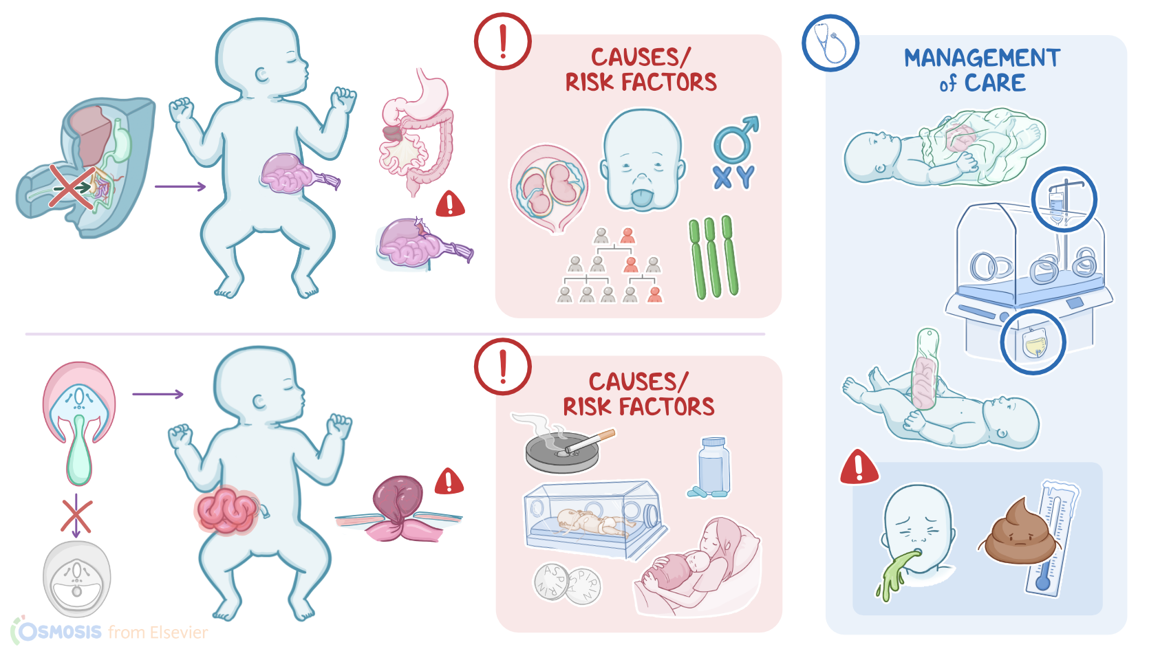
Omphalocele and gastroschisis are both congenital abdominal wall defects. An omphalocele is an umbilical ring defect where the abdominal contents, such as the gastrointestinal tract or liver, can protrude into an external peritoneal sac. Alternatively, in gastroschisis, the abdominal contents slip outside the abdomen without a sac.

Alright, let’s quickly review the physiology of the development of the abdominal wall. From the outside in, the abdominal wall is formed by the skin, superficial fascia, muscles, the deep fascia, and the parietal peritoneum which lines the abdominal cavity. During the third to fourth week of fetal development, the embryo starts to change shape from a flat, three-layer disc to something more shaped like a cylinder, a process called embryonic folding. In the horizontal plane, the two lateral folds eventually come together and close off at the midline, except for at the umbilicus, where the umbilical cord connects the fetus to the placenta. This folding allows for the formation of the gut within the abdominal cavity.

During around the sixth week of development, the liver and intestines grow really quickly, and because the abdominal cavity is still pretty small, there’s limited space, which causes the midgut to herniate through the umbilical ring into the umbilical cord, and this happens normally. At about week 10, though, the abdominal cavity typically has grown enough to allow the midgut to come back from the umbilical cord.

Now, the causes of omphalocele and gastroschisis are mostly unknown, but they are associated with certain conditions or environmental exposures. Omphaloceles are associated with genetic and chromosomal abnormalities such as Trisomy 13, 18, or 21, or Beckwith-Wiedemann syndrome which is a genetic condition where the infant grows larger than normal. Meanwhile, gastroschisis is rarely associated with genetic conditions but instead is related to fetal exposure to nitrosamines in tobacco and cyclooxygenase inhibitors like aspirin and ibuprofen.

Risk factors for omphalocele include maternal age less than 20 or more than 40, multiple gestation, family history, and clients assigned male at birth. In contrast, the risk of gastroschisis is increased for young mothers with their first child. Finally, additional risk factors for gastroschisis include prematurity.

Moving on to pathology, omphalocele develops when the abdominal wall fails to close properly during development and then this large abdominal wall defect results in the midgut failing to rotate and return into the cavity. Other contents, like the liver, might then also herniate into the umbilical cord. Over time, this can lead to several complications including injury to herniated organs such as liver injury, intestinal atresia or stenosis, bowel perforation, and necrosis.

Another complication of omphalocele is malrotation with volvulus, which occurs when the midgut doesn’t rotate in the way it should and twists around itself, leading to intestinal obstruction, infarction, and necrosis. The abdominal cavity may also become malformed or the sac around the herniated organs may rupture. A large omphalocele can even affect the respiratory system, such as inducing pulmonary hypoplasia where lungs do not grow to full capacity.

In contrast, gastroschisis occurs when the abdominal wall fails to form normally during the embryonic period, leaving a hole typically to the right of the umbilicus. This makes it easy for the abdominal contents, like the intestines, stomach, and liver, to protrude through the abdominal wall. In contrast to omphaloceles, these are not covered by a peritoneal layer and are exposed to amniotic fluid. As a result, the intestines may get irritated and inflamed, which can lead to malabsorption issues. Finally, sometimes, when the bowels herniate, it can compromise their blood supply, which can lead to infarction of the intestinal wall.

Now, the clinical manifestations for both omphalocele and gastroschisis include a mass protruding from the abdomen. In omphalocele, the abdominal mass is typically in the anterior midline at the site of the umbilical cord. It is often covered by a membrane of amnion and peritoneum and can contain intestinal loops or parts of other abdominal organs like the liver.

In contrast, gastroschisis is a full-thickness para-umbilical abdominal wall defect, typically to the right of the umbilicus. The bowel that is protruding out often is thick, matted, and doesn’t have any membrane covering it. In addition, some infants may experience difficulty feeding or passing stool, as well as respiratory distress if pulmonary hypoplasia is present.

The diagnosis of omphalocele and gastroschisis starts with the client’s history and physical assessment. When still in utero, additional tests involved include both imaging, such as intrauterine ultrasonography or MRI, and fluid testing, including amniocentesis and a blood test for maternal serum alpha-fetoprotein or MSAFP which is typically increased.