Pulmonary edema: Nursing
Introduction0:00–0:18
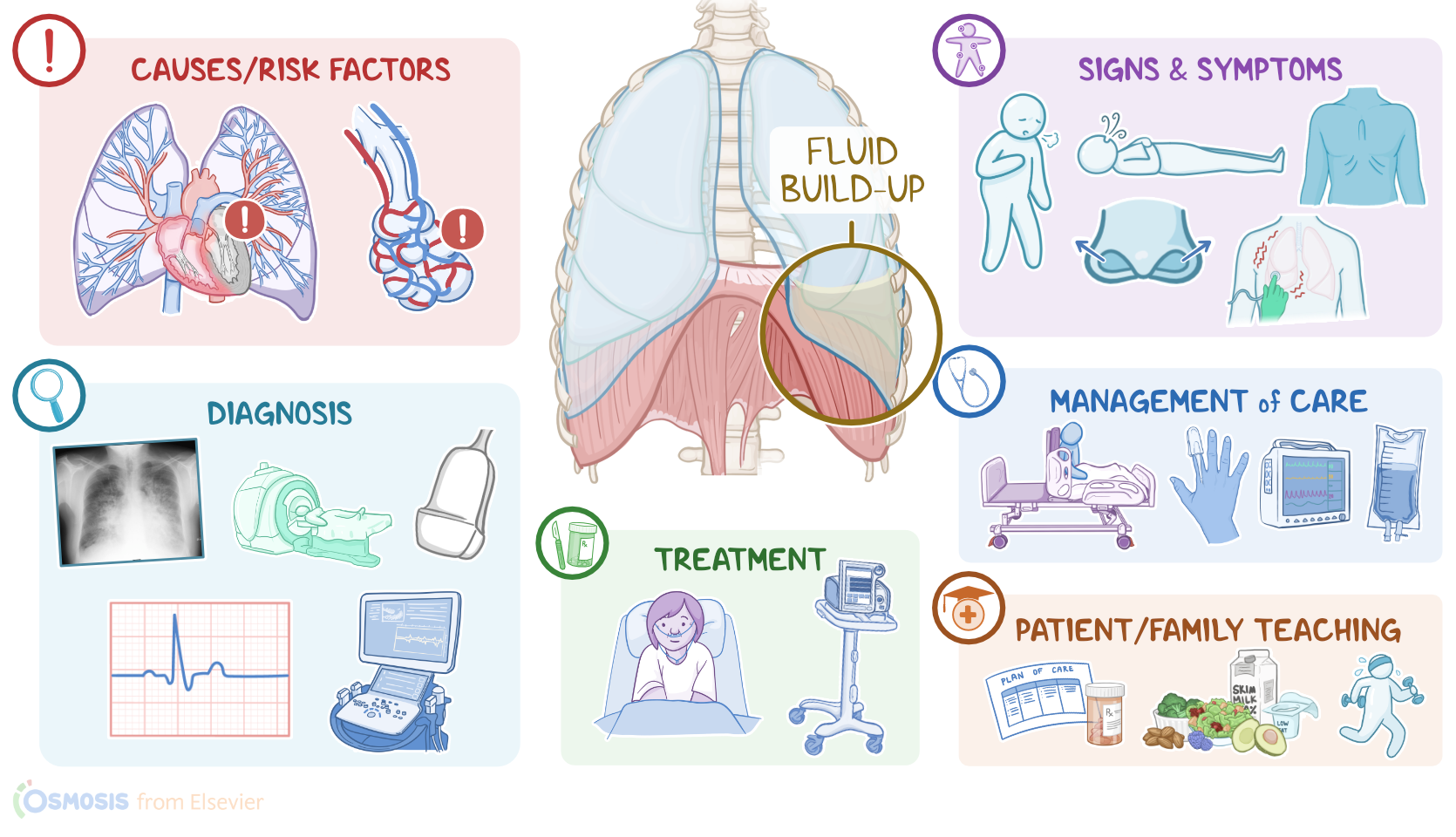
Pulmonary edema refers to the buildup of fluid in the lungs; and this can be further classified as cardiogenic pulmonary edema, which occurs as a result of heart disease; and noncardiogenic pulmonary edema, which usually occurs in the presence of damaged pulmonary capillaries.To understand pulmonary edema, first, let’s quickly review the lower respiratory tract, which includes the lower part of the trachea, and the lungs containing the bronchi, bronchioles, alveolar ducts, and finally the alveoli.
Physiology0:18–1:23
Alveoli are tiny air-filled sacs that are surrounded by tiny blood vessels, called capillaries. Zooming in even more, we can see a tiny space between the alveolar wall and the capillary wall, called the interstitial space, which normally contains a small amount of interstitial fluid.
And that’s where most gas exchange occurs. So, each time we breathe in, the inhaled oxygen moves through the alveolar membrane, the interstitial space, and then through the capillary membrane to reach the blood, which is brought back through the pulmonary veins into the left heart and further into the systemic circulation.
On the other hand, carbon dioxide is carried from the right heart, through pulmonary arteries, into the lungs, where it diffuses from the capillaries through the interstitial space and into the alveoli, and then gets breathed out.Now, cardiogenic pulmonary edema is caused by inadequate pumping of the heart, which usually occurs because of left-sided heart failure, ventricular arrhythmias, myocarditis, myocardial infarction, or valvular disorders, as well as severe hypertension and cardiomyopathy.
Causes & risk factors1:23–2:17
On the flip side, noncardiogenic pulmonary edema can be caused by damage to the lungs, which can occur because of pulmonary infections; acute respiratory distress syndrome, or ARDS for short; sepsis; pneumonia; as well as inhalation of toxic substances or aspiration of gastric contents.
Other important conditions associated with noncardiogenic pulmonary edema include conditions that cause decreased circulating proteins, such as malnutrition, liver failure, and nephrotic syndrome.
Now, let’s switch our focus to pathology. With cardiogenic causes, such as left-sided heart failure, the left ventricle can’t pump blood effectively into the systemic circulation, meaning that blood starts to back up in the left atrium, and subsequently in the pulmonary veins and capillaries.
Pathology2:17–3:51
The extra blood in the pulmonary capillaries results in increased pulmonary hydrostatic pressure.. Now, increased pressure within pulmonary capillaries pushes fluid into the interstitial space of the lungs, eventually causing pulmonary edema.
On the flip side, noncardiogenic causes like pneumonia are associated with an inflammatory process that makes nearby capillaries more permeable.
As a result, proteins and fluid from the blood enter the interstitial space, leading to pulmonary edema. Other noncardiogenic causes, such as nephrotic syndrome can cause pulmonary edema by decreasing protein production or increasing protein loss.
Fewer blood proteins lower the oncotic pressure, and subsequently cause the shift of the fluid from the blood vessels into the interstitial space.
Now, once pulmonary edema occurs, that makes the gas exchange difficult because oxygen and carbon dioxide now have to diffuse through a wide layer of interstitial fluid to get from the alveoli to the capillaries.
Less oxygen in the blood keeps tissues deprived of oxygen, which is known as hypoxia. Eventually, pulmonary edema can cause life-threatening complications, including respiratory failure.Clients with pulmonary edema typically present with anxiety, decreased level of consciousness, short, shallow, and rapid breathing, as well as nasal flaring and intercostal or substernal retractions during inspiration.
Clinical manifestations3:51–4:55
In severe cases, they may also experience shortness of breath, and cyanosis. If there’s left-sided heart failure, clients can also experience orthopnea, which refers to the shortness of breath while lying flat.
This happens because lying flat pulls back more blood from the lower extremities back into the right side of the heart and eventually to the lungs.
Finally, in severe cases, some individuals can have a cough associated with blood-tinged or frothy sputum.Auscultation typically reveals crackles, which is the sound of collapsed alveoli popping open with inspiration; while on percussion, there’s dullness over the affected area; and on palpation, there might be decreased fremitus.Now, diagnosis of pulmonary edema is based on the client’s history and physical assessment, followed by a chest X-ray, which can be used to reveal the presence of fluid in the interstitial space.
Diagnosis4:55–5:20
Additional imaging studies can be used to assess the heart function and look for cardiogenic causes of pulmonary edema, such as an ultrasound, CT scan, ECG, or echocardiogram .
Treatment5:20–6:00
Alright, now the treatment for pulmonary edema depends on the underlying condition, so clients with cardiogenic causes require inotropic medications that increase the strength of cardiac contractions, such as digoxin or dobutamine; or decrease the blood pressure, such as diuretics.
Because gas exchange is affected, supportive care for breathing is really important, usually in the form of supplemental oxygen and noninvasive respiratory support with either bilevel positive airway pressure, called BiPAP for short; or continuous positive airway pressure, called CPAP for short.
In severe cases, clients might require intubation and mechanical ventilationAlright, let's move onto the nursing care you will provide for a client with pulmonary edema.
Management and care6:00–7:32
Your priority nursing goals are to promote adequate oxygenation and provide supportive care to treat the underlying cause.
Begin by positioning your client in a high Fowler position to facilitate breathing, and institute pulse oximetry. Assess your client’s vital signs, and perform a focused respiratory assessment, noting their respiratory rate, work of breathing, breath sounds, color of skin and mucous membranes, oxygen saturation, and level of consciousness.
Now, if your client is diagnosed with cardiogenic pulmonary edema due to left ventricular failure, insert an indwelling urinary catheter and keep a close eye on their intake and output.
Then, initiate ECG monitoring, establish IV access, and provide a minimal infusion of fluids, as ordered. Administer the prescribed medications, including a diuretic to decrease cardiac preload and a positive inotropic medication such as dobutamine.
Continue to monitor your client closely, and report to the healthcare provider if your client becomes confused, has nasal flaring or respiratory retractions, or an oxygen saturation is less than 90%.
Administer oxygen via nasal cannula and encourage them to take slow deep breaths through their nose, and prepare to provide BiPAP, CPAP or assist with mechanical ventilation, as indicated.
Okay, moving on to client and family teaching for a client with pulmonary edema related to heart failure. Begin by explaining that pulmonary edema is a condition where fluid builds up in the lungs, which interferes with their body getting the oxygen it needs.
General client and family teaching7:32–8:39
Review their plan of care, and let them know how their prescribed medications can help rid their body of extra fluid and help their heart pump more efficiently.
Be sure to reinforce teaching regarding managing their condition, including maintaining a healthy weight, physical activity as tolerated, and dietary modifications to reduce sodium and increase fiber, as well as fruits and vegetables.Then, instruct them to contact their healthcare provider if they notice they have gained weight quickly, or if they notice any swelling in their legs, ankles, or feet.
Also stress the importance of getting immediate emergency medical help if they develop shortness of breath; if they trouble sleeping due to difficulty breathing; if they cough up pink, frothy sputum; or if they feel they can’t get enough air.
All right, as a quick recap.... Pulmonary edema refers to the buildup of fluid in the lungs which interferes with gas exchange.
Review8:39–9:53
It can be classified as cardiogenic pulmonary edema, which occurs as a result of problems like inadequate pumping of the heart; and noncardiogenic pulmonary edema, which is usually caused by damaged pulmonary capillaries.
Clinical manifestations of pulmonary edema include dyspnea, orthopnea, nasal flaring, and respiratory retractions. During auscultation, crackles are often heard.
Diagnosis is based on the client’s history and physical assessment, followed by a chest X-ray, which can be used to reveal the presence of fluid in the interstitial space, as well as other tests to identify the underlying cause.
Priority nursing goals are to promote adequate oxygenation and provide supportive care. Client and family education is focused on learning about their condition, home management, and when to seek medical care.
| PULMONARY EDEMA | ||
| KEY POINTS | NOTES | |
| DEFINITION |
| |
| PHYSIOLOGY |
| |
| CAUSES AND RISK FACTORS |
| |
| PATHOPHYSIOLOGY |
| |
| SIGNS AND SYMPTOMS |
| |
| DIAGNOSIS |
| |
| TREATMENT |
| |
| MANAGEMENT OF CARE |
| |
| PATIENT AND FAMILY TEACHING |
| |
No notes for this video yet
Try adding a note below