Shock - Cardiogenic: Nursing
Introduction0:00–0:22
Cardiogenic shock is a lifethreatening condition where the heart is unable to pump enough blood to the rest of the body.
Due to cardiac dysfunction, this leads to acute hypoperfusion and hypoxia of the tissues and organs despite the presence of an adequate intravascular volume in the cardiovascular system.
Physiology0:22–1:47
All right, let's go over the physiology of the cardiovascular system, which consists of the heart and blood vessels. The heart pumps out blood to the body's organs and tissues with each heartbeat, which consists of two phases, systole, which is when the heart contracts and pumps the blood out and diastole, which is when the heart relaxes and fills with blood.
The stroke volume, meaning the amount of blood pumped out by the heart in a single heartbeat is influenced by the cardiac contractility.
Preload and afterload. Contractility is how strongly the heart is contracting during systole preload is how much the heart's smooth muscle is stretched at the end of diastole.
And this is mainly dependent upon how much blood is filling the heart. The more they're stretched, the more force they can generate during contraction.
Kind of like a rubber band. Afterload refers to the resistance that the heart must overcome to pump out blood during systole.
And this is affected mainly by peripheral vascular resistance under nonpathological conditions. This is mainly determined by the vasodilation and vasoconstriction of the blood vessels.
If we multiply the stroke volume by the heart rate, we'll get the cardiac output, which is the amount of blood pumped out by the heart in one minute.
And it's the main measure of the heart's function. Ok.
Cardiogenic shock can be caused by any condition that prevents the heart from pumping enough oxygenated blood, thus affecting the cardiac output.
Causes & risk factors1:47–3:23
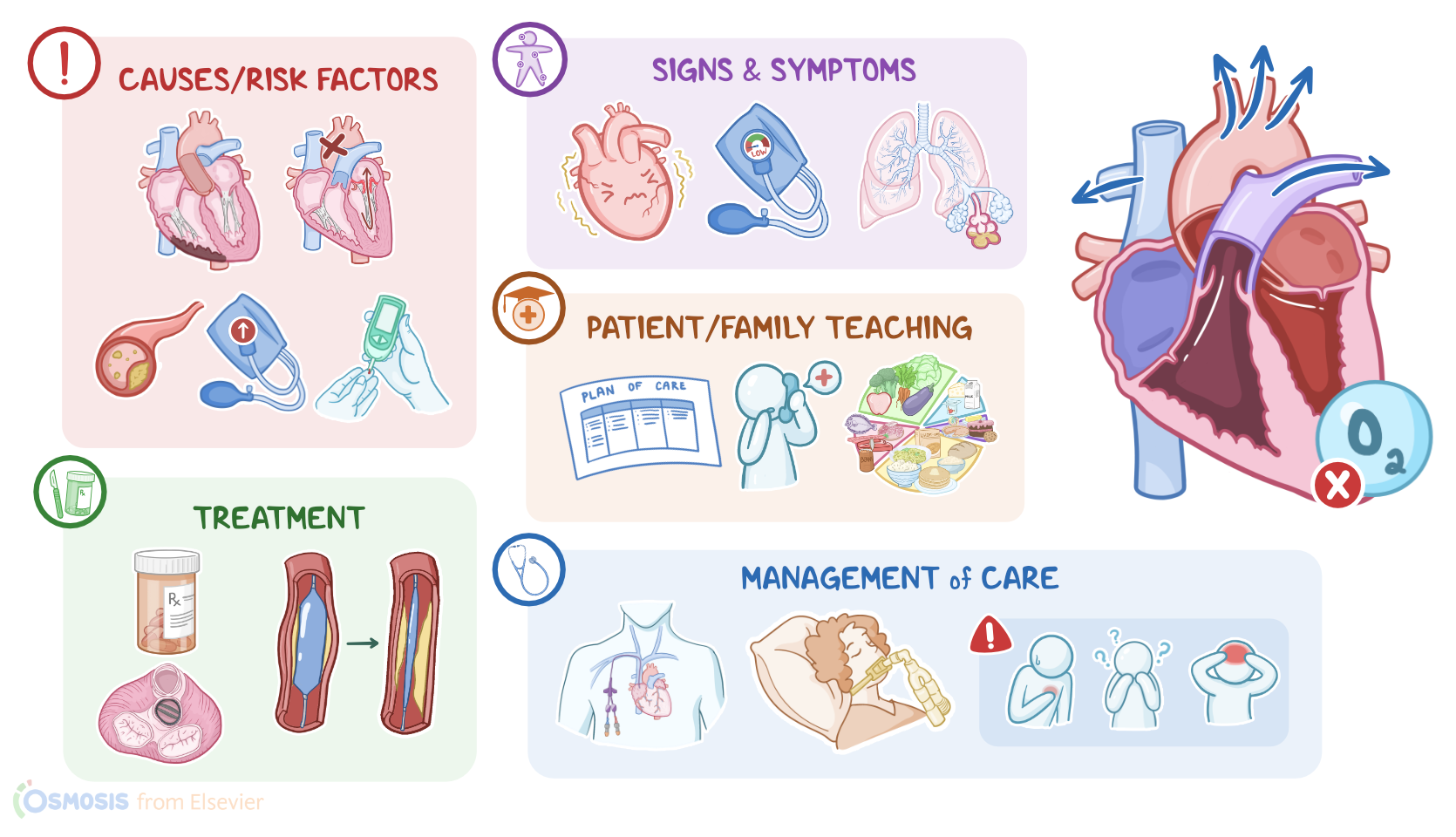
The most common causes involve damage to the heart tissue. The most important is acute myocardial infarction where ischemia and necrosis of cardiac tissue impairs its ability to contract other disorders that damage the heart can also lead to cardiogenic shock.
Common ones include myocarditis, acute heart failure, and myocardial contusion from trauma. Next, arrhythmias including tachycardia, bradycardia and heart block can all decrease cardiac output.
Finally, there are mechanical causes like aortic and mitral valve insufficiency. For example, with mitral valve insufficiency, the valve doesn't close properly.
So, during systole, some of the blood goes from the left ventricle back into the left atrium. Instead of getting pumped out the aorta.
Risk factors for cardiogenic shock include advancing age as well as having existing heart problems such as chronic heart failure.
A prior myocardial infarction or multiple cardiovascular risk factors such as longstanding high BP, coronary heart disease or diabetes.
Mellitus also taking medications that decrease cardiac contractility like beta blockers or having electrolyte imbalances that can affect heart rhythm, like hyper or hypokalemia can be a risk factor for cardiogenic shock.
Now, the pathology of cardiogenic shock develops when the heart becomes unable to pump blood effectively. As a result, there's a decrease in stroke volume which in turn decreases cardiac output, causing BP to drop and ultimately causing hypoperfusion of organs and tissues.
Pathology3:23–4:50
So, in order to increase cardiac output, the body releases vasoconstrictor molecules into the bloodstream. These include catecholamines like epinephrine and norepinephrine, which cause vasoconstriction, increased heart rate and contractility to maintain BP and blood flow as well as A DH, which acts on the kidneys to increase fluid retention and angiotensin two, which also constricts blood vessels and causes sodium retention in the kidneys.
In turn raising BP. If the cause of shock is not managed in time, these compensatory mechanisms may begin to fail, leading to severe tissue hypoxia, vital organs like the heart brain and kidneys may begin to shut down leading to multiple organ failure at the same time.
Since blood isn't being pumped forward into the systemic circulation, it starts backing up into the pulmonary and systemic blood vessels.
Eventually fluid can be pushed out of the circulation and into the lungs and tissues leading to complications like pulmonary and peripheral edema.
Usually the initial clinical manifestations of cardiogenic shock are associated with tissue hypoperfusion and include compensatory tachycardia, tachypnea and hypotension.
Clinical manifestations4:50–7:48
During the initial stage, compensatory mechanisms such as increased heart rate and vascular constriction are sufficient to maintain cardiac output within the normal range.
Cardiac output can best be seen by obtaining the mean arterial pressure or the average pressure in one cardiac cycle because it is a more accurate indicator of perfusion than a normal BP.
In this stage, the map is decreased by less than 10 millimeters of mercury. From baseline, clients may experience tachycardia but compensatory mechanisms keep BP at around the normal range.
So it may be difficult to detect shock at this stage. During the compensation stage.
The compensatory mechanisms are fully active but the map is decreased and is about 10 to 15 millimeters of mercury below baseline, the client's skin can become cold and clammy indicating that blood flow is being redirected to vital organs like the brain and heart.
Other symptoms include pallor severe hypotension and tachycardia decreased peripheral pulses and oliguria as blood starts backing up in the body.
Clients may develop jugular venous distension or the jugular vein that's close to the heart becomes enlarged and distended as well as pulmonary edema, which typically presents with dyspnea and crackles and peripheral edema causing the limbs to swell in the progressive stage.
Clients may develop organ failure because the compensatory mechanisms can no longer guarantee adequate blood flow to vital organs.
The map is sustained at more than 20 millimeters of mercury below baseline. The client may experience anxiety, altered level of consciousness from decreased perfusion to the brain cyanosis, increased respirations, decreased oxygen, saturation from lung failure, profound hypotension, bradycardia and irregular heart rhythm from heart failure as well as Anura which occurs due to kidney failure.
In the refractory stage. Cell death occurs in the vital organs due to the lack of oxygen reaching the tissues.
The map is still sustained at more than 20 millimeters of mercury from baseline At this point, the damaged organs cannot respond to treatment leading to multiple organ dysfunction.
The client may experience a sudden loss of consciousness, shallow respirations, unmeasurable oxygen saturation, nonpalpable pulses or death.
Now, the diagnosis of cardiogenic shock starts with the client's history and physical assessment. The client should be monitored closely to detect signs of hypoperfusion such as a drop in oxygen saturation, low or declining BP or a decreased urinary output.
Diagnosis7:48–8:28
This is usually followed by additional tests including an ecg an echocardiogram and a chest X ray. To identify the precipitating cause of cardiogenic shock.
Laboratory tests can reveal decreased oxygen levels, electrolyte abnormalities and signs of end organ damage like elevated lactate levels.
Definitive treatment of cardiogenic shock involves addressing the underlying cause when possible. This can include revascularization of the heart muscle in cases of acute myocardial infarction, surgical repair or replacement of a valve in valvular heart disease or treatment of an arrhythmia.
Treatment8:28–9:26
Before definitive treatment can be administered, the client's heart may need some temporary support to function. Inotropic medications like dobutamine or Milone can be administered at low doses to improve cardiac contractility.
But this can also increase map and cause a decrease to cardiac output. In addition, a mechanical support device like an intra aortic balloon pump can be placed into the aorta.
This balloon pump inflates during early diastole, increasing blood flow to the heart and then deflates during systole, reducing the afterload and dramatically improving cardiac output.
All right, let's take a look at the nursing care you'll be providing for a client with cardiogenic shock. Your priority, nursing goals are to maintain cardiac output.
Monitor for complications of cardiogenic shock and monitor for improved hemodynamic stability. First, assist in maintaining adequate cardiac output, assess your client's vital signs.
Management and care9:26–12:51
L oc peripheral pulses and auscultate. Their heart and lung sounds, also assess their pain level, noting the severity and location, then initiate ecg monitoring to assess for arrhythmias.
Administer the prescribed IV fluids to help maintain adequate perfusion. Immediately report to the healthcare provider.
If your client experiences chest pain, low BP, S3 heart sounds on auscultation or pulsus paradoxus. Prepare to assist with treating the underlying cause and administer medications as prescribed, which could include inotropes, vasopressors or antidysrhythmic.
Also prepare your client for insertion of a central venous catheter to accurately monitor your client's cardiac output. And for cardiac catheterization to increase myocardial perfusion as indicated, then assess your client for complications of shock.
Review your client's ABG results to check for acidosis and check your client's L OC heart rate and respirations, notify the healthcare provider immediately.
If your client experiences tachycardia, tachypnea, confusion or headache or if the arterial ph is less than 7.35 or bicarbon, it is less than 21 million equivalents per liter.
Prepare your client for endotracheal intubation and mechanical ventilation. Continue to watch your client closely noting signs of decreased perfusion.
Insert an indwelling urinary catheter and monitor your client's intake and output to assess renal perfusion, then check for decreased cerebral perfusion by assessing their L oc speech patterns and pupillary responses.
Notify the healthcare provider immediately. If your client experiences minimal urine output, altered L oc cold or clammy skin or decreased peripheral pulses, standby to assist with pulmonary catheter insertion for advanced hemodynamic monitoring.
Finally continue to assess your client for indications of improved hemodynamic status and improved perfusion including capillary refill, increased BP, improving skin color and temperature, regular heart rate and rhythm and increased urine output.
Also monitor your client's CVP level to determine if it is in the normal range of 2 to 5 millimeters of mercury. Be sure to assess your client's map as well, which should be within 10 millimeters of mercury of their baseline reading.
If your client responds to the therapeutic interventions, prepare to reduce the rate of fluids and begin weaning medications as indicated.
Ok, moving on to client and family teaching first explain that cardiogenic shock is a condition that is caused by heart damage, preventing their heart from pumping out enough blood to meet the body's needs.
General client and family teaching12:51–13:57
Then review the plan of care. Teach them about the treatments they received and allow time for them to ask questions.
Finally educate your client on health promotion strategies that promote heart health such as smoking cessation, increasing daily activity and eating a healthy diet.
Be sure to provide resources to assist your client in transitioning to home safely including smoking, cessation, counseling and resources.
Referrals to a registered dietitian and cardiac rehabilitation. Teach your client how to recognize early signs of cardiac shock and to immediately notify their healthcare provider if they experience problems such as tachycardia, tachypnea, chest pain, shortness of breath confusion or dizziness.
All right, as a quick recap, cardiogenic shock is a condition in which the heart is unable to pump adequate blood to the rest of the body due to cardiac dysfunction.
Clients with M I are at risk for developing cardiogenic shock as well as clients with acute episodes of congestive heart failure, cardiac dysrhythmias, myocarditis, valvular diseases or trauma to the myocardium.
Review13:57–14:58
Risk factors associated with cardiogenic shock are previous cardiovascular conditions and chronic conditions such as diabetes, hypertension and high cholesterol.
Early signs and symptoms of cardiogenic shock are tachycardia hypotension and pulmonary congestion. Treatment of cardiogenic shock involves correcting the underlying cause while temporarily supporting the LV function and promoting myocardial perfusion.
Nursing goals of care include maintaining adequate cardiac output monitoring for complications of cardiogenic shock. And assessing for hemodynamic stability, client and family education is focused on promoting a healthy lifestyle and when to seek medical care.
| SHOCK - CARDIOGENIC | ||
| KEY POINTS | NOTES | |
| DEFINITION |
| |
| PHYSIOLOGY |
| |
| CAUSES AND RISK FACTORS |
| |
| PATHOPHYSIOLOGY |
| |
| SIGNS AND SYMPTOMS |
| |
| DIAGNOSIS |
| |
| TREATMENT |
| |
| MANAGEMENT OF CARE |
| |
| PATIENT AND FAMILY TEACHING |
| |
- "Shock: causes, assessment and investigation. " Anaesthesia & Intensive Care Medicine. (2023;24(2):99-107. )
- "Not all Shock States Are Created Equal. " Anesthesiology Clinics. (2023;41(1):1-25. )
- "Mosby's diagnostic and laboratory test Reference (15th ed). ISBN: 978-0-323-67519-2 " Elsevier (2021)
- "Damage control approach to refractory neurogenic shock: a new proposal to a well-established algorithm. " Colomb Med (Cali) (2021;52(2):e4164800. Published 2021 Jun 30. )
- "Fundamentals of nursing (10th ed.). ISBN 978-0323810340 " Elsevier (2021)
- "Critical care nursing: Diagnosis and management (9th ed). ISBN 978-0323642958 " Elsevier (2022)
- "Hemodynamic monitoring in cardiogenic shock. " Curr Opin Crit Care. (2021;27(4):454-459. )
No notes for this video yet
Try adding a note below