Menkes Disease

What It Is, Causes, Signs, Symptoms, and More

Author: Georgina Tiarks
Editor: Alyssa Haag
Editor: Ian Mannarino, MD, MBA
Editor: Kelsey LaFayette, DNP
Illustrator: Jessica Reynolds, MS
Modified: Jan 06, 2025

What is Menkes disease?

Menkes disease is a severe neurodegenerative hereditary connective tissue disorder that affects copper transport and metabolism. It typically presents at birth and worsens throughout one’s lifespan. A range of pathologic variants may present, however, the most severe variants cause death within the first few years of life. On the other hand, a less severe form known as occipital horn syndrome,  may reduce one’s lifespan to the fifth decade. Menkes disease is typically inherited in an X-linked recessive manner, mainly impacting genetic males, though de novo mutations do occur. Both Menkes disease and Wilson disease impact copper homeostasis, however, Menkes disease causes a deficiency, whereas Wilson disease results in excessive accumulation.
n infographic detailing the background, risk factors, symptoms, diagnosis, and treatment of Menkes disease; including DNA strand with mutation.

What causes Menkes disease?

Menkes disease is caused by mutations in the ATP7A gene, which is typically responsible for developing a transmembrane copper-transporting P-type ATPase (ATP7A) protein that delivers and exports copper within cells. ATP7A protein is found everywhere except the liver, which primarily uses ATP7B protein instead. The liver synthesizes ceruloplasmin using the ATP7B protein, which then binds to copper in the blood. Copper is essential for many enzymatic processes, such as neurotransmitter synthesis, collagen and keratin production, elastin cross-linking, free radical scavenging, iron transport, and melanin production. Without the functioning copper transport proteins, these processes cease to occur. The phenotypic outcome and severity of Menkes disease are dependent on what enzymatic processes are disturbed.

What are the signs and symptoms of Menkes disease?

The signs and symptoms of Menkes disease can vary. At birth, cephalohematomas (i.e., blood accumulation underneath the scalp) and spontaneous fractures may occur. Infants may present with jaundice, temperature instability, hypoglycemia, hernias (e.g., umbilical, inguinal), loose and pale skin, and failure to thrive. Connective tissue defects and bone abnormalities (e.g., pectus excavatum, pectus carinatum, frontal bossing, micrognathia, long arched palate) are common, leading to multiple fractures and tortuous blood vessels that cause fluctuating blood supply. Hair is normal at birth but is replaced by fine, sparse, kinky, wiry, or steel wool-like hair (i.e., pili torti) over time. This gives Menkes disease its colloquial name, “kinky hair disease.” 

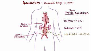
The less severe form of Menkes disease, occipital horn syndrome, primarily affects connective tissue. In this form, skeletal abnormalities such as kyphosis, scoliosis, pectus excavatum, and pectus carinatum are common. Individuals may also have hypermobility of their joints, vascular abnormalities (e.g., aneurysms, varicose veins, orthostatic hypotension), urogenital abnormalities (e.g., bladder diverticula, urinary tract infections), and low intellectual aptitude. OHS has characteristic facial features including a long, thin face; down-slanting eyes; long philtrum; large ears; and loose skin. Their life expectancy varies from early childhood to 50 years of age.

In the more severe type of Menkes disease, infants may progress through normal psychomotor development for the first 2 to 4 months before neurologic regression begins. They may then experience unrelenting seizures despite treatment, drowsiness, lethargy, spasticity, weakness, and joint hypermobility.  At later stages, vascular abnormalities may occur (e.g., aneurysms, subdural hematoma), as well as anemia, blindness, and respiratory failure. Death typically occurs within the first three years of life due to cerebral hemorrhage or neurodegeneration.

How is Menkes disease diagnosed?

Menkes disease may be suspected after a thorough history and physical examination by a healthcare professional. A physical examination may reveal the characteristic clinical features. Once a clinical assessment has been made, a healthcare professional may order serum copper levels and ceruloplasmin levels, which are typically low in Menkes disease. However, these blood tests may not be accurate in neonates, therefore, plasma catecholamine concentrations may be assessed instead, as copper is essential to their enzymatic processes. High ratios of the initial substrate compared to the end product may indicate reduced enzyme activity. For example, high DOPA (i.e., the initial substrate) to dihydroxyphenylglycol (i.e., the end product) displays low dopamine β-hydroxalase enzyme activity indicative of a disruption in the enzymatic process due to Menkes disease. Genetic testing may also be completed, indicating a mutation to the ATP7A gene.

In addition to diagnosing Menkes disease, it is important to assess for concomitant pathologies. A cystourethrogram may be useful in identifying urogenital abnormalities. Vascular studies can be used to diagnose aneurysms. X-ray or CT imaging studies may show osteoporosis, fractures, or cranial Wormian bones. In OHS, radiographic studies might show occipital bone protrusions that mimic horns. A light microscopy hair analysis can also show characteristics of Menkes disease.

How is Menkes disease treated?

Menkes disease has no definitive, curative treatment, therefore, treatment focuses on symptomatic care and treating coexistent disease processes to improve survival and quality of life. Vascular complications such as aneurysms may be treated with conservative management, and if unstable, clipping or endovascular coiling may be necessary. Physical therapy and pain medication (e.g., acetaminophen, ibuprofen) may be used for skeletal abnormalities. 

Although seizures in Menkes disease are often treatment-resistant, some studies have shown that copper supplementation may decrease seizure activity by treating the underlying cause. In recent years, supplemental copper administration has been utilized in the form of copper-histidine. Copper cannot be ingested due to its poor intestinal absorption, however, administering copper histidine, or copper histidinate, subcutaneously or parenterally has been shown to lessen the disease burden and neurodegeneration. Copper-histidine supplementation works best when a partially functional transporter is present and if initiated within days of birth.

What are the most important facts to know about Menkes disease?

Menkes disease is a hereditary copper transport disease that causes a copper deficiency. Copper is an essential component of enzymatic processes throughout the body, therefore, deficiency can cause nervous system, vascular, skeletal, and connective tissue abnormalities. Menkes disease is caused by an X-linked recessive mutation of the ATP7A gene responsible for developing a copper transporter protein. The symptoms of Menkes disease can vary depending on the mutations present. A mild form of Menkes disease exists, is called occipital horn syndrome, and individuals can live up to 50 years of age. However, more severe forms result in detrimental neurodegeneration, which can cause a severe reduction in lifespan. Menkes disease can be diagnosed through a clinical examination and blood testing. Most often, a low serum copper or ceruloplasmin level is diagnostic. Treatment of Menkes disease is primarily supportive, however, supplementing with copper-histidine has been found to improve survival in some people. Neonatal diagnosis and treatment allow for the best clinical outcomes. 

References


Bankier A. Menkes disease. J Med Genet. 1995;32(3):213-215.     


Olivieri JF, Jeyakumar A, Shivaram GM, Koo KSH, Monroe EJ. Emergent embolization of a ruptured splenic artery aneurysm complicating Menkes disease. Radiol Case Rep. 2018;13(6):1267-1270. doi:10.1016/j.radcr.2018.08.032


Rizk T, Mahmoud A, Jamali T, Al-Mubarak S. Menkes disease presenting with epilepsia partialis continua. Case Rep Neurol Med. 2014;2014:525784. doi:10.1155/2014/525784


Tümer Z, Møller LB. Menkes disease. European Journal of Human Genetics. 2010;18(5):511. doi:10.1038/ejhg.2009.187


Vairo FP e, Chwal BC, Perini S, Ferreira MAP, de Freitas Lopes AC, Saute JAM. A systematic review and evidence-based guideline for diagnosis and treatment of Menkes disease. Molecular Genetics and Metabolism. 2019;126(1):6-13. doi:10.1016/j.ymgme.2018.12.005