A
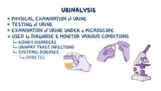
urinalysis tests for the chemical and microscopic properties of the urine. The exact tests included vary depending on the dipstick test, but common ones include urine pH, specific gravity, blood, proteins, glucose, ketones, white blood cells (leukocyte esterase), nitrites,
bilirubin, and urobilinogen. Microscopic examination can determine the presence of solid elements in the urine, such as red or white blood cells, epithelial cells, urinary casts, microorganisms, and crystals.
pH
Urine pH is usually slightly acidic, though it can vary from as low as 4.5 to as high as 8.0 depending on the body’s acid-base balance. The formation of kidney stones is highly influenced by urinary pH. Acidic urine favors the crystallization of uric acid or cystine stones, whereas alkaline urine promotes calcium and phosphate-containing stones. Alkaline urine can result from infection with urea-splitting bacteria—such as Proteus mirabilis, Klebsiella, or Pseudomonas—which break urea molecules into ammonium and carbon dioxide, thereby increasing urine pH to neutral or alkaline values. Urinary pH level is also influenced by dietary factors. A high protein diet can give rise to acidic urine, whereas eating a vegetarian diet can give rise to alkaline urine.
Specific gravity (SG)
Specific gravity measures the kidney’s ability to concentrate or dilute urine by comparing its density to that of distilled water (1.000). Because urine contains various solutes, it has a higher specific gravity than water—usually ranging from 1.005 to 1.025. A high specific gravity represents concentrated urine and may indicate dehydration or increased levels of solutes in the urine. Low specific gravity, on the other hand, represents diluted urine and may occur with a high fluid intake, diabetes insipidus, or due to kidney disease when the kidney’s ability to concentrate urine is impaired.
Blood
The presence of blood in the urine is known as hematuria. It can be classified as gross hematuria when it is visible by the naked eye or microhematuria when it can only be detected using a microscope. Hematuria can occur due to a variety of causes, including damage to the kidney’s glomerular filtration barrier, which typically prevents blood from passing into the urine; lesions to the urinary tract, such as urinary tract infections, kidney stones, or tumors; as well as systemic disorders, like vasculitis, lupus, and tuberculosis among others. In menstruating individuals, blood in the urine can occur due to contamination of the urine specimen.
Protein
In healthy individuals, urine only contains a small amount of proteins because most protein molecules, such as albumin, are too large to pass through the glomerular filtration barrier. When a significant amount of protein appears in the urine, it is known as proteinuria, and it is generally a sign of glomerular damage. Benign causes of proteinuria include fever, strenuous exercise, dehydration, emotional stress, and acute illness. More serious causes include glomerulonephritis and multiple myeloma.
Glucose
Glycosuria, or glucose in urine, is a common finding in individuals with uncontrolled diabetes mellitus though it may also occur when glucose levels are normal, such as during pregnancy, in cases of physiological stress, and when taking corticosteroids. There is a group of hypoglycemic medications called sodium-glucose co-transporter-2 (SGLT-2) inhibitors (e.g. canagliflozin, empagliflozin, dapagliflozin) that act on the kidneys to promote the excretion of glucose in the urine, which also leads to glycosuria.
Ketones
Ketones are metabolic products of fatty acid metabolism. Breakdown of fat may occur in states of prolonged starvation, persistent vomiting or diarrhea, chronic alcohol use, and with a low-carbohydrate diet (e.g. “keto” or ketogenic diets). More importantly, ketones may build up in the blood of individuals with poorly controlled diabetes mellitus (especially diabetes mellitus type 1) and cause acidosis. This is known as diabetic ketoacidosis (DKA) and constitutes a potentially life-threatening emergency. DKA can be suspected when high levels of ketones are detected in the blood or urine in individuals with diabetes.
White Blood Cells, Leukocyte Esterase, and Nitrites
The presence of white blood cells in the urine, or pyuria, is a common sign of urinary tract infection (UTI). Because white blood cells can only be assessed by microscopic examination, dipstick tests use leukocyte esterase (i.e. an enzyme inside white blood cells) to estimate the concentration of white blood cells in urine. In cases where a UTI is suspected, a urine culture is obtained to confirm the diagnosis and identify the underlying organism. If no bacterial cells are detected, the individual is said to have sterile pyuria—a typical finding in urinary tuberculosis.
Most species of Gram-negative bacteria that colonize the urinary tract produce nitrate reductase, an enzyme that converts dietary nitrates into nitrites. Both urinary nitrites and leukocyte esterase provide a good screening test for urinary tract infections; however, since not all bacteria produce nitrites, a negative nitrite test alone does not rule out a urinary tract infection.
Bilirubin and Urobilinogen
Bilirubin is a breakdown product of red blood cell metabolism that is usually not found in the urine. If bilirubin is found in the urine, liver disease or
biliary obstruction (where there is a problem with drainage of bile into the gut) may be suspected. On the other hand, there may be small amounts of urobilinogen in the urine of healthy individuals. Urobilinogen is the end-product of
conjugated bilirubin after it passes through the bile duct and has been
metabolized by bacteria in the intestines. Part of this urobilinogen is reabsorbed into the
portal circulation and eventually filtered by the kidneys. Individuals with increased red blood cell destruction (i.e.
hemolytic anemia) or liver disease may have elevated urobilinogen levels in the urine. On the other hand, if the bile duct is obstructed, less bilirubin enters the intestine, and ultimately, less urobilinogen is found in the urine.