Flail chest: Nursing

1,457views

Flail chest: Nursing

Acute Final

Acute Final

Endocrine system anatomy and physiology
Antepartum assessment - Fetus: Nursing
Assessment of gestational age: Nursing
Fetal circulation: Nursing
Fetal development: Nursing
Group B streptococcus (GBS) infection in pregnancy: Nursing
Hepatitis B virus (HBV) infection in pregnancy: Nursing
Hyperemesis gravidarum: Nursing
Large for gestational age (LGA) infant: Nursing
Preeclampsia and eclampsia: Nursing
Prenatal screening: Nursing
Placenta previa: Nursing process (ADPIE)
Placental abruption: Nursing process (ADPIE)
Birth-related procedures: Nursing
Cesarean birth: Nursing
Intrapartum assessment - Fetal heart rate patterns: Nursing
Intrapartum assessment - Uterine activity: Nursing
Premature rupture of membranes (PROM): Nursing
Shoulder dystocia: Nursing
Prolapsed umbilical cord: Nursing process (ADPIE)
Stages of labor: Nursing
Assessment - Postpartum: Nursing
Perinatal depression: Nursing
Physiology of lactation: Nursing
Postpartum infections: Nursing
Postpartum hemorrhage: Nursing
Biliary atresia: Nursing
Cleft lip and palate: Nursing
Congenital diaphragmatic hernia: Nursing
Congenital heart defects - Acyanotic: Nursing
Congenital heart defects - Cyanotic: Nursing
Esophageal atresia and tracheoesophageal fistula: Nursing
Craniosynostosis: Nursing
Hemolytic disease of the fetus and newborn: Nursing
Hyperbilirubinemia: Nursing process (ADPIE)
Infant of a diabetic mother (IDM): Nursing
Meconium aspiration syndrome: Nursing
Neonatal respiratory distress syndrome (NRDS): Nursing
Neonatal sepsis: Nursing
Neural tube defects: Nursing
Newborn adaptation to extrauterine life: Nursing
Persistent pulmonary hypertension of the newborn (PPHN): Nursing
Physical assessment - Neonate: Nursing
Small for gestational age (SGA) infant: Nursing
Postterm infant: Nursing
Thermoregulation - Neonate: Nursing
Arterial blood gas (ABG) - Overview: Nursing
Arterial blood gas (ABG) - Metabolic acidosis: Nursing
Arterial blood gas (ABG) - Metabolic alkalosis: Nursing
Arterial blood gas (ABG) - Respiratory acidosis: Nursing
Arterial blood gas (ABG) - Respiratory alkalosis: Nursing
Adrenal insufficiency (Addison disease): Nursing
Anemia - Iron-deficiency: Nursing
Anemia - Aplastic: Nursing
Anemia - Macrocytic: Nursing
Case study - Hypothyroidism: Nursing
Case study - Iron-deficiency anemia: Nursing
Case study - Sickle cell anemia: Nursing
Complete blood count (CBC) - Hemoglobin and hematocrit: Nursing
Complete blood count (CBC) - Red blood cells (RBC): Nursing
Complete blood count (CBC) - Platelets: Nursing
Complete metabolic panel (CMP) - Blood urea nitrogen (BUN) and creatinine (Cr): Nursing
Complete metabolic panel (CMP) - Estimated glomerular filtration rate (eGFR): Nursing
Complete metabolic panel (CMP) - Liver function tests (LFT): Nursing
Cushing syndrome and Cushing disease: Nursing
Hematopoietic growth factors: Nursing pharmacology
Hyperparathyroidism: Nursing
Hyperthyroidism: Nursing process (ADPIE)
Hypoparathyroidism: Nursing
Hyperpituitarism: Nursing
Hypopituitarism: Nursing
Hypothyroidism: Nursing process (ADPIE)
Medications affecting the parathyroid glands: Nursing pharmacology
Medications for growth hormone disorders: Nursing pharmacology
Medications for thyroid disorders: Nursing pharmacology
Neutropenia: Nursing
Polycythemia: Nursing
Thrombocytopenia: Nursing
Acute kidney injury (AKI): Nursing process (ADPIE)
Benign prostatic hyperplasia (BPH): Nursing process (ADPIE)
Case study - Cholecystitis: Nursing
Case study - Cirrhosis: Nursing
Case study - Chronic kidney disease (CKD): Nursing
Case study - Benign prostatic hyperplasia (BPH): Nursing
Case study - Gastroesophageal reflux disease (GERD): Nursing
Case study - Pediatric appendicitis: Nursing
Case study - Pyelonephritis: Nursing
Cholecystitis: Nursing
Cholelithiasis: Nursing
Chronic kidney disease (CKD): Nursing
Cirrhosis: Nursing process (ADPIE)
Diverticular disease: Nursing
Gastroesophageal reflux disease (GERD): Nursing process (ADPIE)
Hemolytic uremic syndrome: Nursing
Hirschsprung disease: Nursing
Intestinal obstruction: Nursing
Irritable bowel syndrome (IBS): Nursing
Nephrotic syndrome: Nursing
Pyloric stenosis: Nursing process (ADPIE)
Renal and urinary calculi: Nursing
Urinary incontinence - Stress: Nursing process (ADPIE)
Diabetes insipidus: Nursing process (ADPIE)
Dialysis care: Nursing
Case study - Diabetic ketoacidosis (DKA): Nursing
Case study - Pediatric diabetes mellitus type 1: Nursing
Diabetes mellitus (DM): Nursing process (ADPIE)
Hyperosmolar hyperglycemic state (HHS): Nursing process (ADPIE)
Diabetic ketoacidosis (DKA): Nursing process (ADPIE)
Case study - Epilepsy: Nursing
Case study - Head injury: Nursing
Epidural and subdural hematoma: Nursing
Case study - Stroke: Nursing
Hemorrhagic stroke - Intracranial hemorrhage (ICH) and subarachnoid hemorrhage (SAH): Nursing
Increased intracranial pressure (ICP): Nursing
Hydrocephalus: Nursing process (ADPIE)
Intracranial aneurysm: Nursing
Seizure disorder: Nursing process (ADPIE)
Stroke: Nursing process (ADPIE)
Jaundice: Nursing
Nutrition - Enteral: Nursing skills
Nutrition - Newborn: Nursing
Nutrition - Parenteral: Nursing skills
Phenylketonuria (PKU): Nursing
Arterial embolism: Nursing
Disseminated intravascular coagulation (DIC): Nursing
Hemophilia: Nursing process (ADPIE)
Acute respiratory distress syndrome (ARDS): Nursing
Asthma: Nursing process (ADPIE)
Atelectasis: Nursing
Bacterial pneumonia: Nursing process (ADPIE)
Bronchiolitis and respiratory syncytial virus (RSV): Nursing process (ADPIE)
Case study - Acute respiratory distress syndrome (ARDS): Nursing
Care of an intubated client: Nursing skills
Case study - Chronic obstructive pulmonary disease (COPD): Nursing
Case study - Impaired gas exchange: Nursing
Case study - Pediatric asthma: Nursing
Chest tube care: Nursing
Chronic obstructive pulmonary disease (COPD): Nursing process (ADPIE)
Cystic fibrosis: Nursing
Epiglottitis: Nursing process (ADPIE)
Flail chest: Nursing
Intraoperative care: Nursing
Pleural effusion: Nursing
Pneumothorax and hemothorax: Nursing
Pulmonary edema: Nursing
Smoke inhalation injury: Nursing process (ADPIE)
Tracheostomy: Nursing
Venous thromboembolism (VTE): Nursing process (ADPIE)
Arrhythmias - Asystole: Nursing
Arrhythmias - Atrial flutter (Aflutter): Nursing
Arrhythmias - Premature atrial contractions (PACs): Nursing
Arrhythmias - Heart blocks: Nursing
Arrhythmias - Atrial fibrillation (Afib): Nursing
Arrhythmias - Premature ventricular contractions (PVCs): Nursing
Arrhythmias - Sinus tachycardia and sinus bradycardia: Nursing
Arrhythmias - Supraventricular tachycardia (SVT): Nursing
Arrhythmias - Ventricular fibrillation (Vfib): Nursing
Arrhythmias - Ventricular tachycardia (Vtach): Nursing
Cardiac biomarkers - Troponin: Nursing
Case study - Acute coronary syndrome (ACS): Nursing
Case study - Atrial fibrillation (Afib): Nursing
Case study - Heart failure with reduced ejection fraction (HFrEF): Nursing
Case study - Deep vein thrombosis (DVT): Nursing
Case study - Hypertension: Nursing
Case study - Hypovolemic shock: Nursing
Coronary artery disease (CAD) and angina pectoris: Nursing process (ADPIE)
Electrocardiogram (ECG) - Normal sinus rhythm (NSR): Nursing
Heart defects that decrease pulmonary blood flow - Nursing considerations & client education: Nursing
Hypertension: Nursing process (ADPIE)
Left-sided heart failure: Nursing process (ADPIE)
Myocardial infarction (MI): Nursing process (ADPIE)
Pericardial effusion and cardiac tamponade: Nursing process (ADPIE)
Peripheral arterial disease (PAD): Nursing process (ADPIE)
Rheumatic heart disease: Nursing process (ADPIE)
Shock - Cardiogenic: Nursing
Shock - Neurogenic: Nursing
Shock - Obstructive: Nursing
Shock - Septic: Nursing
Sickle cell disease: Nursing process (ADPIE)
Valvular heart disease: Nursing

Notes

FLAIL CHEST

KEY POINTS
NOTES
DEFINITION
  • Multiple consecutive rib fractures creating a free floating section of ribs and connective tissue that moves out of sync with rest of chest

PHYSIOLOGY
  • Thoracic cage
    • 12 pairs of ribs
    • Sternum
    • Protect heart, lungs, and blood vessels
  • Intercostal muscles
    • Between each set of ribs 
  • Diaphragm
    • Covers bottom of thoracic cage

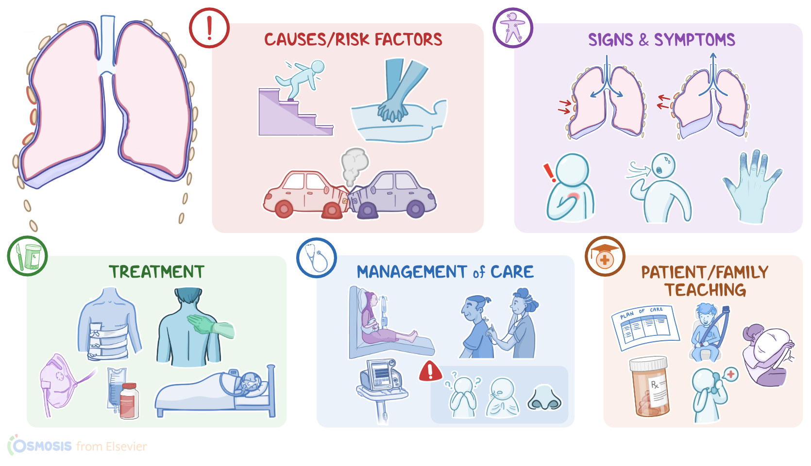
CAUSES AND RISK FACTORS
  • Causes
    • Blunt chest trauma
  • Risk factors
    • Intoxication
    • Contact sports
    • Advanced age
    • Assigned male at birth

PATHOPHYSIOLOGY
  • Three or more rib fractures in two or more places
  • Bilateral detachment of ribs from sternum 
  • Ribs not mechanically attached to rest of chest wall 
  • Chest moves paradoxically and uncoordinated
  • Lungs unable to fully expand
  • Severe pain further impairs breathing
  • Complications
    • Pulmonary contusion
    • Atelectasis
    • Acute respiratory distress syndrome
    • Pneumothorax
    • Blood vessel or organ injury

SIGNS AND SYMPTOMS
  • Paradoxical breathing
  • Pain
  • Splinting
  • Shortness of breath
  • Cyanosis 
  • Refractory hypoxemia
  • Hemodynamic instability
  • Diminished breath sounds
  • Crepitus

DIAGNOSIS
  • History
  • Physical assessment
  • CT
  • X-ray

TREATMENT
  • Splinting
  • Oxygen
  • IV fluids
  • Pain management
  • Chest physiotherapy
  • Positive airway pressure
  • Intubation and mechanical ventilation
  • Surgery

MANAGEMENT OF CARE
  • Goals of care 
    • Maintain oxygenation and ventilation
    • Manage pain
    • Monitor for complications
  • Assist to high Fowler position
  • Splint flail segment
  • Apply pulse oximeter
  • Administer oxygen and/or positive pressure 
  • Perform pulmonary assessment
  • Assess chest
  • Examine rest of body for other injuries
  • Encourage deep breaths
  • Assist with incentive spirometer
  • Provide pain management
  • Maintain bed rest and limit oxygen demands
  • Monitor arterial blood gas results and vital signs
    • Notify HCP
      • Respirations below 12 or above 20
      • Decrease oxygen saturation
      • Increased work of breathing

PATIENT AND FAMILY TEACHING
  • Explain condition, plan of care, and how to safely self-administer medications
  • Full recovery timeline
  • Safety precautions

Transcript

Watch video only

Flail chest is a serious condition, often caused by severe blunt chest trauma. It occurs when multiple consecutive ribs are fractured, creating a separate, free floating section of ribs and connecting tissue that moves out of sync with the rest of the chest during breathing.

First, let’s quickly look at the anatomy and physiology of the thoracic cage. The thoracic cage is made up of 12 pairs of ribs and the sternum, which together create the shape of the chest wall and protect thoracic structures, such as the heart, lungs, and blood vessels.

Now, the key muscles involved in breathing are the intercostal muscles, which are located between each set of ribs, and the diaphragm, which covers the whole bottom of the thoracic cage. When a person inhales the diaphragm contracts, moving downward and the intercostal muscles contract to move the rib cage out. This increases the volume while decreasing pressure in the thoracic cavity. The change in pressure pulls air down into the lungs. In contrast, when a person exhales the diaphragm and intercostal muscles relax which decreases the volume, while increasing pressure in the thoracic cavity. This makes air move out of the lungs.

Now, the main cause of a flail chest is blunt trauma to the chest, which can be caused by falls, abuse, car crashes, crush injuries, or even cardiopulmonary resuscitation.

Risk factors for a flail chest include modifiable ones, such as intoxication, and practicing contact sports; as well as non-modifiable ones, such as advanced age, and being assigned male sex at birth.

Okay, now, the pathology of a flail chest begins when a blunt trauma causes three or more ribs to fracture in two or more places, or when there’s bilateral detachment of ribs from the sternum. This creates a segment of the ribs that is not mechanically attached to the rest of the chest wall, As a result of these two pathologies, the chest moves in a paradoxical or uncoordinated fashion. This means that when the person breathes in, the chest wall expands, pulling outwards, but intrathoracic pressure becomes more negative, pulling the unattached segment inwards. On the other hand, when the person breathes out, the chest wall contracts, pulling inwards, but intrathoracic pressure becomes more positive, which pushes the separated section outwards.

Damage to the thoracic wall also prevents the lungs from expanding fully which impairs respiration. In addition,the intercostal muscles pull on the broken rib segment during breathing which causes severe pain and further impairs breathing.

Now, flail chest is also associated with several complications, the most common of which is pulmonary contusion, or injury of the lung parenchyma. This can cause atelectasis or collapse of the alveoli in the lung. These collapsed alveoli can’t participate in gas exchange, causing shunting, so blood doesn't become oxygenated as it moves through the lungs.

This can result in acute, refractory hypoxemia, meaning that the client’s oxygenation does not improve even when they receive higher concentrations of oxygen; and ultimately, clients develop acute respiratory distress syndrome, or ARDS. Since clients with flail chest have trouble breathing or coughing up airway secretions, they are also at an increased risk of developing pneumonia. Another complication is pneumothorax, which is when there’s air in the pleural cavity, and that doesn’t allow the lung on that side to expand properly. Finally, in severe cases, the broken ribs can tear the aorta, damage the diaphragm, or injure internal organs like the spleen, liver, or kidneys.

Typically, the clinical manifestations of flail chest include paradoxical breathing, along with pain at the sites of the fractures, which intensifies with breathing, moving, coughing, or even sneezing. Some clients also splint, meaning that they take short, shallow, rapid breaths in an effort to limit chest movement and reduce pain. In severe cases, they may also experience shortness of breath, cyanosis, and refractory hypoxemia. Additionally, clients can develop hemodynamic instability, which presents as hypotension, tachycardia, or arrhythmias.

During auscultation of the chest, there are diminished breath sounds, and palpation of the chest typically reveals crepitus, which refers to crunching or grinding sounds, when the site of the fracture is moved.