Hiatal hernia: Nursing process (ADPIE)
Client Report0:00–0:50
Mary Fowler, aged 68, presents to the emergency department, or ED with vomiting and abdominal pain and distention. Mary has a history of paraesophageal hiatal hernia with symptoms of gastroesophageal reflux disease, or GEERD, which she normally manages with lifestyle modifications and acid-reducing medications.
Her symptoms worsened over the last two days, and she's been unable to keep food or liquid down over the last 24 hours. Mary is diagnosed with a large incarcerated paraesophageal hiatal hernia confirmed with an abdominal X-ray and computerized tomography or CT scan.
Pathology0:50–4:46
Underneath the diaphragm, the esophagus then connects with the stomach to form the gastroesophageal junction and the lower esophageal sphincter, or LES, a ring of muscles that opens to allow food and liquids into the stomach and closes to keep gastric contents from coming back up through the esophagus.
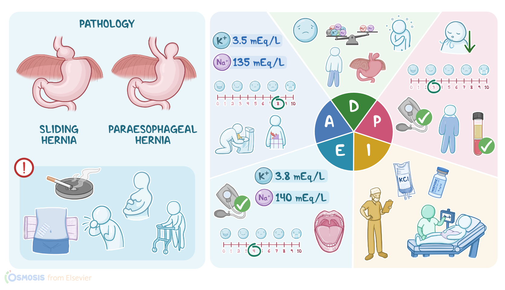
There are two main types of hiatal hernias. The most common type is a sliding hernia, where the gastroesophageal junction slides up and down through the hiatus.
Less common are paraesophageal hernias, where parts of the stomach roll up through the hiatus, forming a pocket next to the esophagus.
The risk of developing a hiatal hernia increases if the diaphragm is weakened from trauma, abdominal surgery, or smoking.
Likewise, anything that increases pressure inside the abdomen like coughing, obesity, pregnancy, straining, or heavy lifting increases the risk of hernia development.
The risk also increases with age due to age-related widening of the hiatus, or in those who are born with an unusually large hiatal opening.
Because hiatal hernias can interfere with the function of the LES, signs and symptoms are often similar to GERD. Acidic gastric contents can reflux into the esophagus and cause heartburn and pain in the upper abdomen or chest.
Stomach acid can also reflux up into the larynx, irritating the vocal folds and causing hoarseness. If gastric contents are aspirated into the airway, it can cause respiratory complications, such as pneumonia or asthma-like symptoms.
Persistent reflux can lead to esophageal inflammation or esophagitis, and it can even erode the lining of the esophagus, creating an ulcer, which can lead to bleeding or perforation.
Fibrosis or scarring can occur, resulting in oesophageal narrowing or strictures, which make swallowing difficult. Eventually, the cells lining the esophagus change, a condition known as Barrett's esophagus, which is associated with a higher risk of esophageal adenocarcinoma.
Now, a hiatal hernia can become incarcerated, meaning the herniated part of the stomach becomes trapped between structures in the chest cavity, like between the diaphragm, esophagus, and lungs.
If the blood flow to the hernia is cut off, the hernia becomes strangulated, which then leads to ischemia and necrosis and clinical manifestations like pain, difficulty swallowing, and vomiting.
Strangulation of any hernia is a surgical emergency that requires immediate treatment. A rare and life-threatening complication of a hiatal hernia is the formation of gastric volvulus, seen more often with paraesophageal hernias.
This involves the stomach twisting on itself more than 180 degrees with the hiatus acting as a pivot point. This results in complete obstruction and strangulation.
Clinical manifestations include abdominal pain and distention, along with wretching without vomiting. A gastric volvulus is a surgical emergency.
Diagnosis of a hiatal hernia is based on the client's medical history and physical examination. CT scanning can visualize part of the stomach and the chest.
A barium X-ray, also known as an upper GI series or direct visualization with an upper endoscopy, are also used. Supportive care and symptom management are often the treatments for GERD, including lifestyle changes and acid-reducing medications.
Anti-reflux surgery, known as Nissen fundoplication, is often required for symptomatic cases. After introducing yourself to Mary as her nurse, confirming her identity, and performing hand hygiene, you begin your assessment.
Assessment4:46–6:14
Her vital signs are a tympanic temperature of 97.9 °F or 36.6 °C, heart rate of 102 BPM and regular, respirations, 24 per minute and shallow, BP of 98/60 millimeters of mercury, and oxygen saturation 96% on room air.
When you ask Mary about her pain, she places her hand over her epigastric region a few inches above her belly button. She describes the pain as tightness and rates it as an 8 out of 10.
Her oral mucosa is pink and dry. You note that her skin is tenting after gently pinching a fold of skin on her upper chest.
Her abdomen is soft, distended, and tender to palpation. You review Mary's recent lab values and note that her potassium is 3.5 milliequivalents per liter, and her sodium is 135 milliequivalents per liter.
After documenting your assessment findings, you let Mary know that you will return shortly, after updating the surgical team.
Using the assessment data you've gathered so far, you form these nursing diagnoses to guide your nursing care for Mary. Acute pain related to gastric obstruction, ineffective breathing pattern related to abdominal pain, fluid volume deficit related to vomiting, risk for electrolyte imbalance related to GI losses, risk for ineffective GI tissue perfusion related to incarcerated hiatal hernia.
Diagnosis6:14–6:37
Now, it's time to develop a plan of care. Your goals for Mary are that her pain will be managed pre and post-operatively at a level of 3 out of 10, which Mary identifies as a tolerable level.
Planning6:37–7:28
Her respirations will be in the normal range of 12 to 20 per minute with adequate depth. Her fluid balance will be normalized with evidence of moist mucous membranes and improved BP.
And her pre-operative labs will show sodium and potassium levels well within normal range. After she returns from surgery, Mary will be free of signs and symptoms of GI ischemia, including pain, nausea, and vomiting.
As you continue to monitor Mary, you will alert the healthcare team for changes such as a drop in BP, difficulty breathing, new onset of pain, or wretching without vomiting.
After collaborating with Mary's surgical team, it's time to implement the plan of care. As ordered by Mary's surgeon, you initiate an IV fluid bolus of 0.9% normal saline with added potassium chloride.
Implementation7:28–8:15
You explain these interventions will help to hydrate her, improve her BP, and replenish her electrolytes. Next, you administer morphine by slow IV push.
And you coach Mary in taking slow, deep breaths, explaining how using a pillow to splint her belly will improve her comfort while deep breathing after surgery.
After you've carried out these initial orders, Mary's surgical team arrives to review the operative plan and obtain informed consent for a surgical repair of her incarcerated hiatal hernia.
It's time to evaluate Mary's response to your intervention so far. Your assessments before she leaves for surgery include a tympanic temperature of 98.4 °F or 36.9 °C, heart rate of 80 BPM and regular, respiratory rate of 20 with adequate depth, BP, 110/68 millimeters of mercury, pain, 4 out of 10, oxygen saturation, 97% on room air.
Evaluation8:15–9:13
Mary's oral mucous membranes still appear pink and moist, and her skin continues to tint when gently pinched. After documenting these findings, you review Mary's repeat lab work.
Her potassium is now 3.8 milliequivalents per liter, and her sodium is 140 milliequivalents per liter. And when it's time for Mary's surgical procedure, you transfer her care to the perioperative team, and they will continue applying the nursing process to evaluate her care.
All right. There's a quick recap.
Summary9:13–10:08
Your client is 68 year old Mary, who's been diagnosed with an incarcerated paraesophageal hiatal hernia, in which part of the stomach bulges up through the hiatus and becomes trapped within the chest.
Your assessment identified pain and signs of dehydration. The nursing diagnoses you identified were acute pain, ineffective breathing pattern, fluid volume deficit, risk for electrolyte imbalance, and risk for ineffective GI tissue perfusion.
The goals you identified when planning care for Mary included management of her pain, restoration of fluid and electrolyte balance, stabilization of her vital signs, and a resolution of her incarcerated hernia.
You collaborated with Mary and the surgical team to implement interventions to reach these goals. Afterwards, you evaluated if Mary's goals were met, and Mary's healthcare team will continue to reassess and revise the plan of care as needed to achieve the goals of care.
| HIATAL HERNIA | ||
| KEY POINTS | NOTES | |
| PATIENT REPORT |
| |
| PATHOPHYSIOLOGY |
| |
| DIAGNOSIS AND TREATMENT |
| |
| ASSESSMENT |
| |
| NURSING DIAGNOSES |
| |
| PLANNING |
| |
| IMPLEMENTATION |
| |
| EVALUATION |
| |
- "Ackley and Ladwig’s Nursing Diagnosis Handbook: An Evidence-Based Guide to Planning Care, 13th edition" Mosby (2022)
- "Hydrocephalus: historical analysis and considerations for treatment" Eur J Med Res (2022)
- "Medical-Surgical Nursing: Concepts for Interprofessional Collaborative Care, 10th Edition" Elsevier (2020)
- "Harrison’s Principles of Internal Medicine, 21st edition" McGraw Hill / Medical (2022)
- "Gastric volvulus in a patient with hiatal hernia" Visual Journal of Emergency Medicine (2022)
- "All That Wheezes Is Not Asthma: Giant Hiatal Hernia" Am J Med (2022)
- "Critical Care Nursing: Diagnosis and Management, 9th edition" Elsevier (2021)
- "Syndromic Hydrocephalus" Neurosurg Clin N Am (2022)
No notes for this video yet
Try adding a note below