Inflammatory bowel disease - Crohn disease and ulcerative colitis: Nursing
Introduction0:00–0:25
Inflammatory bowel disease or IBD is a condition characterized by chronic gastrointestinal tract inflammation due to autoimmune reactions, as well as systemic symptoms like fatigue, fever, and unintentional weight loss.
There are two types of IBD, Crohn disease and ulcerative colitis.The gastrointestinal tract consists of a long tube, where food travels through, which runs from the mouth to the anus.
Physiology0:25–2:18
Now if we zoom into a cross-section of the gastrointestinal tract, the walls are typically lined by the same four layers of tissue.
From the inside out, there’s the mucosa, which absorbs nutrients and secretes mucus and digestive enzymes; the submucosa, a dense layer of tissue that contains blood vessels, lymphatics, and nerves; the muscularis externa, which contracts to move food through the bowel; and the outermost layer is either the adventitia or the serosa, and faces the abdominal or peritoneal cavity.So when we eat a big meal, the food travels from the mouth, down the pharynx and esophagus, to reach the stomach, which has a handful of glands that secrete hydrochloric acid and pepsin to help digest the food, now called a bolus.
Then the bolus passes towards the small intestine, or small bowel, made of the duodenum, jejunum, and ileum, where nutrients are absorbed.
Examples are - monosaccharides, amino acids, fatty acids, electrolytes, minerals, vitamins, and water. To increase the surface for absorption, the mucosa in the small intestine has lots of tiny ridges and grooves, each of which has little finger-like projections called villi.
And in turn, each villus is covered in tiny little microvilli. Then, whatever isn’t absorbed travels to the large intestine, made of the cecum, colon, rectum, and anal canal.
Here, there are trillions of bacteria, collectively called the gut microbiome, which help produce essential B and K vitamins.
Now, in the large intestine, a little more water and electrolytes are absorbed to finally form the feces that are eliminated through the anus.Now, the exact cause of IBD is unknown, but there’s a genetic component because it runs in families.
Causes & risk factors2:18–2:44
So, risk factors include family history, northern European descent, and the presence of genetic and inflammatory disorders like cystic fibrosis, psoriasis, or multiple sclerosis.
In addition, smoking and Ashkenazi Jewish descent are associated with an increased risk for Crohn disease.Now, the pathology of inflammatory bowel disease starts in the gastrointestinal tract of a genetically susceptible client when the immune system is triggered by an antigen.
Pathology2:44–6:02
initiating an excessive inflammatory response. This ends up damaging the mucosal epithelium, which provides a perfect environment for bacteria to invade and grow.
More immune cells come to the scene, and release cytokines, that cause further inflammation and damage. In Crohn disease, the inflammation can occur anywhere from the mouth to the anus.
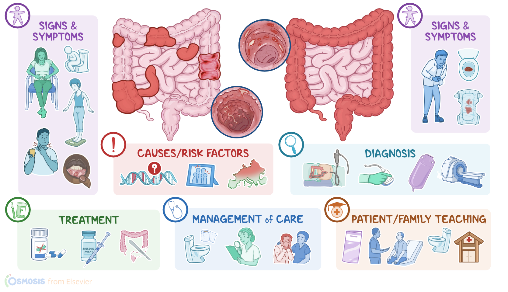
They appear most commonly in the terminal ileum and colon, but usually spares the rectum. There’s typically large areas of healthy tissue in between damaged areas, so they’re called “skip lesions.” The lesions caused by the inflammation involve the entire depth of the intestinal wall, which is referred to as being transmural.
These deep lesions and ulcers often run longitudinally along the tissue with sections of normal mucosa in between, thus creating a “cobblestone” appearance.
On the flip side, with ulcerative colitis, the inflammation most often starts at the rectum, and spreads continuously along the large intestine.
The damage is limited to the mucosa and submucosa of the intestinal wall. Over time, both Crohn disease and ulcerative colitis may lead to several complications.
With both Crohn disease and ulcerative colitis, there might be acute or chronic bleeding and anemia due to iron deficiency.
Sometimes, a phlegmon can form, which is where there’s a localized area of inflammation in the intestinal wall that can get infected and become an abscess.
Now Crohn disease can result in strictures, which may eventually cause bowel obstruction. Other complications include micro-perforations and fistulas, which are communications between two epithelial organs.
If the small intestine is affected, there’s reduced absorption of bile salts which increases the risk of gallstones. Decreased fat absorption will cause the excess fat to bind to calcium in the small intestine.
This prevents oxalate from binding to the calcium so they’re absorbed into the body and increase the risk of developing calcium-oxalate kidney stones.Now, ulcerative colitis is more likely to cause toxic megacolon as a complication but this could also occur in Crohn disease.
In severe cases, it can lead to perforation with peritonitis. Other conditions that often occur with ulcerative colitis include inflammatory arthritis, such as ankylosing spondylitis, skin lesions like pyoderma gangrenosum and erythema nodosum, as well as primary sclerosing cholangitis, which is when there’s fibrosis and inflammation of both the intra- and extrahepatic bile ducts.
Finally, clients with inflammatory bowel disease are at increased risk for developing colorectal cancer.In Crohn disease, gastrointestinal symptoms include crampy abdominal pain, watery diarrhea that may or may not be bloody, and sometimes malabsorption symptoms like steatorrhea, or B12 deficiency, causing malnutrition as well as weight loss.
Clinical manifestations6:02–6:02
Clinical manifestations6:02–6:49
A very frequent finding are aphthous ulcers in the mouth. Some clients may also present esophageal involvement, with odynophagia or painful swallowing, and dysphagia.Clients with ulcerative colitis usually present with gastrointestinal symptoms that include colicky abdominal pain, which is often relieved after defecation; bloody diarrhea; rectal bleeding; and tenesmus, which is pain when defecating.
There may also be malaise, anorexia, and fever.All right, diagnosis of IBD starts with the client’s history, and physical assessment, followed by endoscopy or colonoscopy with biopsy to confirm the diagnosis.
Diagnosis6:49–7:23
Laboratory tests may also show elevated ESR or PCR and fecal calprotectin, which indicate an inflammatory process in the gastrointestinal tract.
Finally, imaging studies like barium enema, ultrasound, CT, and MRI can be performed to assess the extent of inflammation in and beyond the bowel wall.Now, the choice of treatment depends on the type and severity of IBD.
Treatment7:23–8:22
Usually, treatment involves the use of corticosteroids, methotrexate or 5-aminosalicylic acid, or 5-ASA for short. An alternative is antibiotics like ciprofloxacin and metronidazole, which are thought to help control symptoms by reducing bacteria levels in the intestine and by curbing the immune response.
Finally, more severe cases might need a biologic agent like infliximab to suppress the immune system, which can be combined with an immunomodulator like azathioprine.
Beyond that, another treatment option involves surgery and removal of the affected tissue. In Crohn disease, surgery is reserved for clients with complications, since it doesn’t cure the disease, because inflammation can occur anywhere along the GI tract.
On the other hand, since ulcerative colitis only affects the large intestine, a colectomy that removes the entire colon can treat the disorder.
Alright, let’s look at the nursing care you’ll provide for a client with inflammatory bowel disease. Your priority nursing goals are to promote normal bowel elimination, monitor for complications, and provide psychosocial support.
Management and care8:22–11:01
Begin by performing an abdominal assessment, noting bowel sounds, and any distension or bloating. Then, ask about current bowel elimination patterns, including the color and consistency of the stool, as well as for the presence of blood.
Then, administer the prescribed medications. Okay, moving on to complications.
First, assess their current pain level, as well as it’s onset, and determine if it occurs before or after meals. Assist them into a position of comfort and administer prescribed analgesics.
Be sure to promote a restful environment and place your client on bedrest to help decrease their intestinal activity. Report to the healthcare provider any indications of a bowel perforation or peritonitis, this can include abdominal distension, pain, and guarding; nausea and vomiting, or increased temperature.
Prepare your client for further intervention, as indicated. Next, to monitor your client for a fluid imbalance, assess their vital signs and mental status; monitor their intake and output, and check their most recent laboratory test results.
Report any indications of dehydration, which can include hypotension, tachycardia, confusion or other alteration in mental status as well as; oliguria and increased urine specific gravity; and increased BUN and creatinine.
Also be sure to report hypokalemia immediately, since electrolyte imbalance can accompany fluid loss. Then, administer ordered IV fluids, and continue monitoring.Also perform a nutritional assessment, making note of your client’s current weight.
Maintain their NPO status and provide nutrition by administering the prescribed total enteral nutrition, as tolerated. Consult with a registered dietician for dietary recommendations to meet your client's caloric and nutritional needs.
Be prepared to progress your client’s diet as their symptoms decrease and they can tolerate oral nutrition. Lastly, assess your client’s psychosocial needs, such as anxiety and depression that surround their diagnosis.
Encourage them to express their feelings, and provide them with information about community and online support groups for IBD that can offer them additional support in coping with the psychological impact of the condition.
Okay, let's move on to client and family education. First, explain to your client that Inflammatory bowel disease is a chronic condition that is caused by their own immune system attacking their gastrointestinal tract, causing inflammation.
General client and family teaching11:01–12:42
Review the plan of care, teach them about the purpose of their prescribed medications and supplements, and instruct them to take them exactly as directed.
Lastly, emphasize the importance of keeping follow-up appointments after discharge for continued monitoring.Then, let them know about lifestyle modifications they can do that can help to decrease the occurrance exacerbations.
Teach them about stress reduction techniques, and encourage them to find the techniques that work best for them. Talk about the benefits of engaging in regular physical activity as tolerated, and remind them of the importance of staying well hydrated.
For dietary modifications, suggest they keep a food diary to help identify and eliminate foods that tend to exacerbate their symptoms.
Generally, advise them to avoid sugary foods and high fat foods, and eat smaller, frequent meals throughout the day instead of three big meals.
Also advise them to limit caffeine and carbonated beverages. Lastly, if your client has a history of smoking, stress the importance of avoiding all products containing nicotine, and provide them with smoking cessation resources and counseling.Finally, advise your client to notify their healthcare provider immediately if their symptoms start to worsen, if they continue to lose weight, or if they develop any severe abdominal pain, fever, vomiting, or notice blood in their stool.
Alright, as a quick recap…. IBD is a condition characterized by chronic gastrointestinal tract inflammation due to autoimmune reactions, as well as systemic symptoms like fatigue, fever, and unintentional weight loss.
Review12:42–14:51
The two types are Crohn disease and ulcerative colitis. The cause of IBD is unknown but there is a genetic component.
Risk factors include family history, northern European descent, and inflammatory disorders. Both disorders are caused by chronic inflammation of the gastrointestinal tract but Crohn disease can occur anywhere in the GI tract, usually spares the rectum, and forms skip lesions, while Ulcerative colitis develops in the rectum and large intestine, then spreads continuously.
The lesion in Crohn disease is transmural and can have a cobblestone appearance, while the lesion in ulcerative colitis only affects the mucosa and submucosa.
Crohn disease may present clinically with symptoms like crampy abdominal pain, watery diarrhea that is sometimes bloody, malnutrition, weight loss, aphthous mouth ulcers, odynophagia, and dysphagia.
Clients with ulcerative colitis usually present with colicky abdominal pain that is often relieved by defecation, bloody diarrhea, rectal bleeding, and tenesmus.
Diagnosis typically includes endoscopy or colonoscopy with biopsy to confirm the diagnosis. Imaging studies like barium enema, ultrasound, CT and MRI can help assess the extent of disease.
Now, treatment usually involves corticosteroids and antibiotics, and for more severe cases biologic agents to suppress the immune system.
In addition, affected tissue can be surgically removed. Priority nursing goals include promoting normal bowel elimination, monitoring for complications, and providing psychosocial support.
Client and family teaching focuses on learning about the disorder, lifestyle modifications, and when to seek medical attention.
| INFLAMMATORY BOWEL DISEASE | ||
| KEY POINTS | NOTES | |
| DEFINITION |
| |
| PHYSIOLOGY |
| |
| CAUSES AND RISK FACTORS |
| |
| PATHOPHYSIOLOGY |
| |
| SIGNS AND SYMPTOMS |
| |
| DIAGNOSIS |
| |
| TREATMENT |
| |
| MANAGEMENT OF CARE |
| |
| PATIENT AND FAMILY TEACHING |
| |
No notes for this video yet
Try adding a note below