Peptic ulcer disease (PUD): Nursing process (ADPIE)
Client Report0:00–0:42
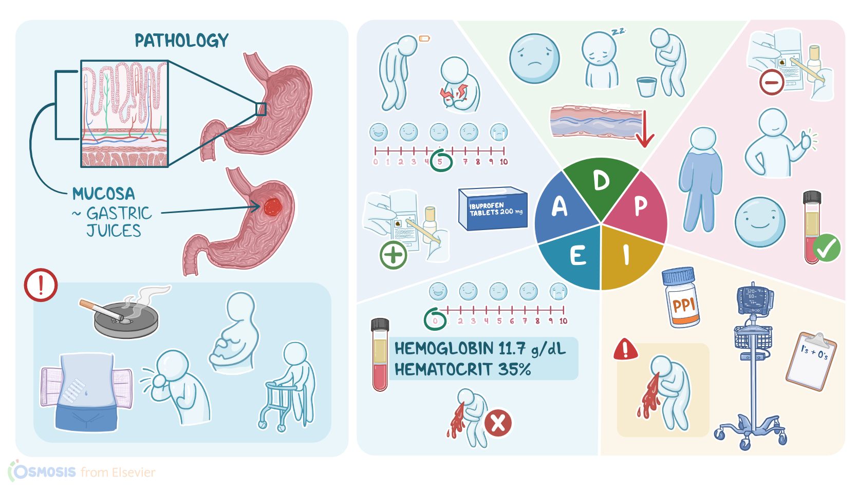
Paula Salazar is a 56-year-old Hispanic female client admitted to your unit after an esophagogastroduodenoscopy or EGD revealed a bleeding gastric ulcer which was controlled by thermal coagulation.
Her recent history includes intermittent upper abdominal pain and a recent weight loss of 5 lbs because of nausea and decreased appetite.
After experiencing an episode of hematemesis, her primary care physician or PCP ordered an EGD. A biopsy taken during the procedure is negative for Helicobacter pylori, or H.
pylori for short. Her other health problems include fatigue and chronic back pain.
Pathology0:42–4:55
Peptic ulcer disease or PUD is an erosion of the lining of the gastrointestinal or GI tract, most commonly in the proximal duodenum and stomach.
The lining of the GI tract is made of four layers, the mucosa, which is the innermost layer, followed by the submucosa, muscularis, and serosa.
Cells in the mucosa secrete harsh gastric juices like hydrochloric acid and pepsin, which help digest food. Normally, the mucosa is protected from self-digestion due to tightly joined epithelial cells that resist penetration and by prostaglandins that stimulate secretion of a protective layer of mucus and bicarbonate.
When the gastric juices overcome these protective mechanisms and damage the gastric lining, a peptic ulcer develops. Sometimes PUD is asymptomatic, but often the exposure to irritating gastric juices can cause burning epigastric pain.
Typically, pain from gastric ulcers increases 15 to 30 minutes after a meal, while pain from duodenal ulcers increases 2 to 3 hours after a meal and at night.
Other common symptoms include bloating, abdominal fullness, and nausea. If the erosion extends down to the muscularis level, it can damage blood vessels, resulting in an upper GI bleed, hematemesis, or melina, when blood passes through the lower GI tract, producing a dark, tarry stool.
Anemia from extensive blood loss causes symptoms like fatigue, pallor, and shortness of breath. Sometimes, an ulcer can perforate through all four layers of the gastric lining and create an opening into the peritoneal cavity.
Free air and gastric contents that enter the abdomen cause peritonitis and findings like severe abdominal pain, abdominal rigidity, and tachycardia.
It is important to keep an eye out for these findings because a perforation requires emergent surgical management. A major risk factor for developing PUD is infection with H.
pylori, which causes an inflammatory response that disrupts protective mechanisms in the gastric mucosa. Another common cause of PUD is NSAID use, because these drugs work by inhibiting prostaglandin synthesis, which is essential for gastric protection.
Reducing the level of prostaglandins over a prolonged period of time leaves the gastric mucosa susceptible to damage, and over time, ulcers can begin to develop.
There are also certain lifestyle choices that can contribute to PUD. Smoking reduces blood flow to the gastric mucosa and is associated with cell death and poor healing.
While alcohol consumption increases cell permeability and weakens the protective mucus barrier, other risk factors are African-American or Hispanic ethnicity, and having a first-degree relative with PUD.
Finally, a rare cause of PUD is Zollinger-Ellison syndrome, which is a gastrin-secreting tumor that increases gastric acid production.
Diagnosis of PUD is based on history, physical examination, and by EGD which visualizes the lining of the stomach and duodenum.
During the procedure, a biopsy is obtained and analyzed for the presence of H. pylori.
H. pylori may also be detected by a urea breath test.
Finally, labs like a CBC or testing for fecal occult blood are used to identify common complications of PUD like bleeding or anemia.
Treatment of PUD depends on the underlying cause and severity of the disease. NSAIDs, if used, are discontinued.
Medications like proton pump inhibitors or PPIs and H2 receptor antagonists or H2RAs are used to decrease production of stomach acid.
If H. pylori is detected, a triple or quadruple regimen is prescribed that may include a combination of PPI, certain antibiotics, and a bismuth subsalicylate.
For some clients, a prostaglandin analog like misoprostol is prescribed, while sucralfate is administered in some cases because it forms a coating over the ulcer to protect it while it heals.
OK, it's time to start your assessment of Mrs. Salazar, who has just returned from the EGD where hemostasis of her bleeding ulcer was achieved.
Assessment4:55–6:25
As you enter the room, you introduce yourself, perform hand hygiene, and confirm her identity. You note that Mrs.
Salazar does not appear to be in acute distress, but seems tired. Her vital signs are temperature 98.9 °F or 37.2 °C, heart rate 98 BPM, respirations, 18 per minute, BP 98/62 millimeters of mercury, and oxygen saturation 95% with oxygen per nasal cannula at 2 L per minute.
She rates her abdominal pain as 5 out of 10. Her skin is cool and dry.
An IV in her left arm is intact and running 0.9% normal saline at 150 mL per hour. You note her most recent hemoglobin is 11.5 g per deciliter, and her hematocrit is 35%.
Fecal occult blood is positive. As you review her current medication, you note she takes ibuprofen for back pain.
She needs to take it daily to get through her day due to chronic back pain, and she adds, I make sure I follow the directions on the bottle before exiting the exam room.
You document your findings and let her know the physician is making rounds and will be in shortly to continue her examination.
All right, after assessing Mr. Salazar, you are ready to make your nursing diagnoses, which include fluid volume deficit related to GI bleeding, fatigue related to blood loss.
Diagnosis6:25–6:47
Acute pain related to mucosal injury, nausea related to mucosal injury, and ineffective health management related to continued use of NSAIDs for musculoskeletal pain.
All right, after you collaborate with Mrs. Salazar and the interdisciplinary team, you plan goals for her care.
Planning6:47–7:16
Goals to be met before discharge include that Mrs. Salazar will be free from signs and symptoms of GI bleeding, including restoration of a normal fluid volume, hemoglobin, and hematocrit levels returning to a normal range, and decreased fatigue.
Her GI pain will be reduced, and finally, she will participate in an appropriate pain management plan to address her chronic back pain.
Implementation7:16–8:40
Next, it's time to implement actions to help Mrs. Salazar reach her goals.
You ask the certified nursing assistant or CNA to record strict INO, vital signs every 4 hours, and to let you know immediately if Mrs.
Salazar experiences hematemesis. Next, you administer the prescribed proton pump inhibitor, pantoprazole, explaining how it will help decrease the acid in her stomach, promote healing, and reduce pain and nausea.
A few hours later, you check in on Mrs. Salazar.
She is awake and resting comfortably. So you take the opportunity to provide some health promotion teaching.
Her physician has prescribed the proton pump inhibitor, omeprazole after discharge. So, you review how the medication works and potential adverse effects like changes in elimination, such as constipation or diarrhea.
You advise her to avoid foods that may trigger abdominal pain, like caffeine, alcohol, and highly processsed fatty foods.
And you explain the importance of including foods that contain iron, such as dark green leafy vegetables, meat, enriched breads, legumes, and cereals.
For pain management, she should avoid NSAIDs like ibuprofen. Instead, her physician is recommending acetaminophen, and she has a referral to meet with a physical therapist for additional help with her back pain.
Evaluation8:40–9:35
Now it's near the end of your shift, and before you sign out, you review Mrs. Salazar's care plan and response to treatment so far.
Mrs. Salazar's latest vital signs are temperature 98.6 °F or 37 °C, heart rate 80 BPM, respiratory rate 16 per minute, BP, 110/68 millimeters of mercury, and pain is 0 out of 10.
She continues to be on oxygen per nasal cannula, 2 L per minute, and her oxygen saturation is now 97%. The most recent labs show a hemoglobin of 11.7 g per deciliter and a hematocrit of 35%.
She has not had another episode of hematemesis during your shift. Mrs.
Salazar is starting to make some improvement, and her plan of care will continue to be evaluated and adjusted as needed in preparation for her discharge home.
Summary9:35–10:34
All right, as a quick recap, you have been caring for Mrs. Salazar, who was diagnosed with a bleeding peptic ulcer after an episode of hematemesis.
Peptic ulcer disease, or PUD is an erosion in the gastric lining that happens when gastric juices overcome the mechanisms that protect the gastric mucosa.
H. pylori and chronic NSAID use are the two main causes of PUD.
Your nursing assessment reveals Mrs. Salazar is showing signs and symptoms of blood loss from her bleeding ulcer.
The nursing diagnoses you develop address her bleeding, fluid volume, pain, nausea, and fatigue. Together with the interdisciplinary team, you create a plan of care and implement actions aimed at healing her ulcer and preventing further blood loss.
Restoring her fluid volume, managing her pain and fatigue, and adopting lifestyle modifications to prevent further ulcer development.
Throughout her illness, you continually evaluate her responses to your interventions and adapt the care plan as needed to achieve optimal outcomes.
| PEPTIC ULCER DISEASE (PUD) | ||
| KEY POINTS | NOTES | |
| PATIENT REPORT |
| |
| PATHOPHYSIOLOGY |
| |
| DIAGNOSIS AND TREATMENT |
| |
| ASSESSMENT |
| |
| NURSING DIAGNOSES |
| |
| PLANNING |
| |
| IMPLEMENTATION |
| |
| EVALUATION |
| |
- "Ackley and Ladwig’s Nursing Diagnosis Handbook: An Evidence-Based Guide to Planning Care, 13th edition" Mosby (2022)
- "Harrison’s Principles of Internal Medicine, 21st edition" McGraw Hill / Medical (2022)
- "Evidence-based clinical practice guidelines for peptic ulcer disease 2020" J Gastroenterol (2021)
- "Clinical Guidelines for Drug-Related Peptic Ulcer, 2020 Revised Edition" Gut Liver (2020)
- "Perforated and bleeding peptic ulcer: WSES guidelines" World J Emerg Surg (2020)
- "Health Assessment for Nursing Practice, 7th edition" Elsevier (2021)
- "Upper Gastrointestinal Bleeding in Adults: Evaluation and Management" Am Fam Physician (2021)
No notes for this video yet
Try adding a note below