Polycystic kidney disease (PKD): Nursing

Polycystic kidney disease (PKD): Nursing

Watch later

Watch later

Hypokalemia: Clinical
Movement of water between body compartments
The role of the kidney in acid-base balance
Blood histology
Blood components
Respiratory acidosis
Metabolic acidosis
Respiratory alkalosis
Metabolic alkalosis
Metabolic and respiratory alkalosis: Clinical
Metabolic and respiratory acidosis: Clinical
Bronchodilators: Beta 2-agonists and muscarinic antagonists
Chronic obstructive pulmonary disease (COPD): Clinical
Chronic obstructive pulmonary disease (COPD): Nursing process (ADPIE)
Adrenergic antagonists: Alpha blockers
Sympatholytics: Alpha-2 agonists
Alpha-2 adrenergic agonists: Nursing pharmacology
Adrenergic receptors
Adrenergic antagonists: Beta blockers
Diabetes mellitus: Clinical
Diabetes mellitus (DM): Nursing process (ADPIE)
Diabetes mellitus
Diabetes mellitus: Pathology review
Stroke: Nursing process (ADPIE)
Stroke: Clinical
Ischemic stroke
Intracerebral hemorrhage
Peptic ulcers and stomach cancer: Clinical
GERD, peptic ulcers, gastritis, and stomach cancer: Pathology review
Sickle cell disease (NORD)
Sickle cell disease: Nursing process (ADPIE)
Sickle cell disease: Clinical
Blood groups and transfusions
Erythropoietin
Blood products: Nursing pharmacology
Oxygen binding capacity and oxygen content
Blood products and transfusion: Clinical
Hemophilia
Hemophilia: Nursing process (ADPIE)
Leukemia: Nursing process (ADPIE)
Chronic leukemia
Leukemias: Pathology review
Acute leukemia
Leukemia: Clinical
Lymphomas: Pathology review
Lymphoma: Clinical
Non-Hodgkin lymphoma
Hodgkin lymphoma
Lymphatic system anatomy and physiology
Anatomy of the abdominal viscera: Liver, biliary ducts and gallbladder
Liver anatomy and physiology
Cirrhosis
Cirrhosis: Pathology review
Cirrhosis: Clinical
Cirrhosis: Nursing process (ADPIE)
Hepatitis C virus
Hepatitis A and Hepatitis E virus
Liver cancer: Nursing
Cholestatic liver disease
Non-alcoholic fatty liver disease
HIV (AIDS)
HIV and AIDS: Pathology review
Antiretrovirals for HIV/AIDS - Protease inhibitors: Nursing pharmacology
Antiretrovirals for HIV/AIDS - NRTIs and NNRTIs: Nursing pharmacology
Antiretrovirals for HIV/AIDS - Integrase strand transfer inhibitors: Nursing pharmacology
Anatomy of the abdominal viscera: Pancreas and spleen
Pancreatitis: Nursing process (ADPIE)
Glaucoma: Nursing process (ADPIE)
Glaucoma
Eye conditions: Refractive errors, lens disorders and glaucoma: Pathology review
Acute respiratory distress syndrome (ARDS): Nursing
Anatomy of the coronary circulation
Coronary artery disease: Pathology review
Coronary artery disease: Clinical
ECG basics
ECG normal sinus rhythm
ECG rate and rhythm
ECG axis
ECG intervals
ECG QRS transition
ECG cardiac infarction and ischemia
Cardiac cycle
Arterial disease
Delirium
Dementia and delirium: Clinical
Vascular dementia
Frontotemporal dementia
Dementia with Lewy bodies
Dementia: Pathology review
Tumor lysis syndrome (TLS): Nursing Process (ADPIE)
Cholelithiasis: Nursing
Coronary artery disease (CAD) and angina pectoris: Nursing process (ADPIE)
Breast cancer: Nursing process (ADPIE)
Ovarian cancer: Nursing
Cervical cancer: Nursing
Hormones and hormone modulators for cancer: Nursing pharmacology
Endometriosis: Nursing
Heart failure
Left-sided heart failure: Nursing process (ADPIE)
Heart failure: Pathology review
Pneumonia: Pathology review
Pneumonia
Bacterial pneumonia: Nursing process (ADPIE)
Respiratory stimulants: Nursing pharmacology
Corticosteroids - Inhaled: Nursing pharmacology
Pneumothorax and hemothorax: Nursing
Chest trauma: Clinical
Pleural effusion: Nursing
Tuberculosis (TB): Nursing
Parkinson disease: Nursing process (ADPIE)
Huntington disease
Multiple sclerosis (MS): Nursing
Multiple sclerosis
Guillain-Barré syndrome: Nursing
Guillain-Barre syndrome
Myasthenia gravis: Nursing
Acute kidney injury (AKI): Nursing process (ADPIE)
Acute kidney injury: Clinical
Chronic kidney disease (CKD): Nursing
Chronic kidney disease
Chronic kidney disease: Clinical
Polycystic kidney disease (PKD): Nursing
Renin-angiotensin-aldosterone system
Osteoarthritis: Nursing
Osteoarthritis
Rheumatoid arthritis (RA): Nursing process (ADPIE)
Rheumatoid arthritis: Clinical
Rheumatoid arthritis
Rheumatoid arthritis and osteoarthritis: Pathology review
Systemic lupus erythematosus
Systemic lupus erythematosus (SLE): Pathology review
Systemic lupus erythematosus (SLE): Clinical
Systemic lupus erythematosus (SLE): Nursing
Mycobacterium tuberculosis (Tuberculosis)
Peripheral venous disease (PVD): Nursing process (ADPIE)
Peripheral arterial disease (PAD): Nursing process (ADPIE)
Buerger disease: Nursing
Raynaud phenomenon: Nursing
Aortic dissections and aneurysms: Pathology review
Aortic aneurysms and dissections: Clinical
Aortic dissection
Aortic aneurysm: Nursing process (ADPIE)
Venous thromboembolism (VTE): Nursing process (ADPIE)
Benign prostatic hyperplasia (BPH): Nursing process (ADPIE)
Benign prostatic hyperplasia
Prostate cancer: Nursing
Prostate cancer
Testicular cancer: Nursing
Testicular cancer
Diabetic ketoacidosis (DKA): Nursing process (ADPIE)
Endocrine system anatomy and physiology
Vitamin B12 deficiency
Thalassemia: Nursing
Anemia - Iron-deficiency: Nursing
Anemia - Macrocytic: Nursing
Anemia - Aplastic: Nursing
Thyroid hormones
Medications for thyroid disorders: Nursing pharmacology
Hyperthyroidism: Nursing process (ADPIE)
Hypothyroidism: Nursing process (ADPIE)
Hypoparathyroidism: Nursing
Hyperparathyroidism: Nursing
Anxiety disorders: Nursing process (ADPIE)
Spinal cord injury (SCI): Nursing
Cluster A personality disorders
Smoke inhalation injury: Nursing process (ADPIE)
Shock - Anaphylactic: Nursing
Shock - Obstructive: Nursing
Shock - Neurogenic: Nursing
Shock - Hypovolemic: Nursing
Shock - Cardiogenic: Nursing
Shock - Septic: Nursing
Pulmonary edema: Nursing
Burn injury: Nursing

Notes

POLYCYSTIC KIDNEY DISEASE (PKD)

KEY POINTS
NOTES
DEFINITION
  • Fluid-filled sacs form in the kidney
    • Infantile 
    • Adult 
    • Acquired 

PHYSIOLOGY
  • Kidneys
    • Outer cortex
    • Inner medulla
    • Nephrons
      • Functional unit of the kidney
  • Minor calyces
  • Major calyces
  • Renal pelvis
  • Ureters
  • Hormone production
    • Renin
    • Erythropoietin

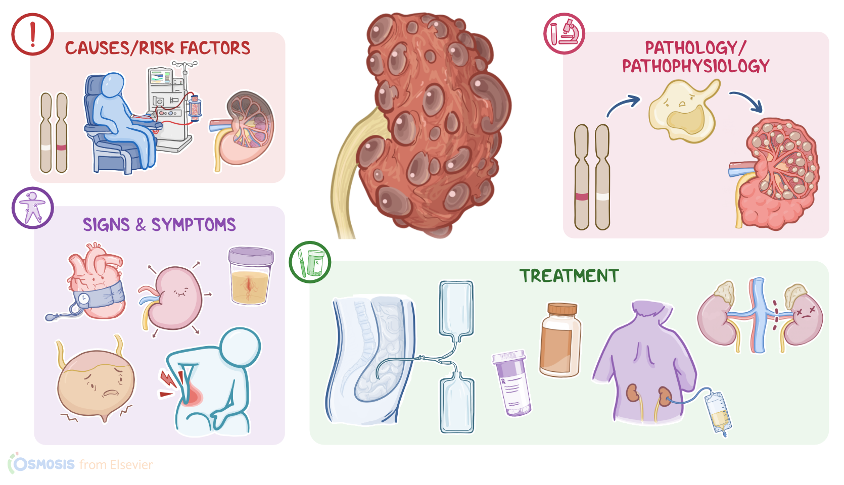
CAUSES AND RISK FACTORS
  • Causes
    • Depends on type of disease
      • Infantile 
        • Genetic mutation
      • Adult 
        • Genetic mutation
      • Acquired 
        • Chronic kidney disease
        • Long-term dialysis
        • Kidney failure

PATHOPHYSIOLOGY
  • Infantile and adult 
    • Genetic mutation
    • Loss of cellular regulation and uncontrolled cell division
    • Formation of cysts in the nephrons
    • Cysts enlarge and colect fluid
    • Damage kidney
    • Damage blood vessels
    • Release of renin
    • Enlargement of the kidney
    • Impaired renal function
  • Acquired
    • Mechanism unknown
    • Chronic uremia from chronic kidney disease
    • New cysts damage kidneys
    • Loss of renal function
  • Complications of infantile and adult 
    • Renal failure
    • Hypertension
    • Preeclampsia
    • Hematuria
    • Infections
    • Kidney stones
    • Cyst development in other organs
  • Complications of acquired 
    • Cyst infection
    • Cyst rupture

SIGNS AND SYMPTOMS
  • Differ for each type
  • Infantile
    • Oligohydramnios
    • Small for gestational age
    • Enlarged kidneys
    • Portal hypertension
    • Breathing difficulties
  • Adult
    • Flank pain
    • Hypertension
    • Kidney stones
    • Urinary tract infections
    • Hematuria
    • Nocturia
    • Dilute urine
  • Acquired 
    • Asymptomatic
    • Symptomatic
      • Fever
      • Back pain
      • Hematuria
  • All types
    • Enlarged kidneys

DIAGNOSIS
  • History
  • Physical assessment
  • Abdominal ultrasound
  • CT
  • MRI
  • Laboratory tests
  • Urinalysis

TREATMENT
  • Medications
    • Tolvaptan
    • Antihypertensives
    • Calcium channel blockers
    • Beta blockers
    • Opioids and acetaminophen 
    • Potassium citrate
    • Antibiotics
  • Avoid NSAIDs and aspirin
  • Percutaneous drainage
  • Nephrectomy
  • Dialysis
  • Kidney transplantation

MANAGEMENT OF CARE
  • Goals of care
    • Slow disease progression
    • Monitor for complications
  • Slow disease progression
    • Manage blood pressure
    • Review laboratory results
    • Administer prescribed antihypertensives
  • Monitor for complications
    • Intake and output
    • Signs of hyponatremia
    • Signs of hyperkalemia
    • Urine color
    • Assess pain
    • Liver function tests
    • Renal function
    • Notify HCP
      • Nausea or vomiting
      • Headaches
      • Confusion
      • Muscle weakness or twitching
      • Seizures
      • Palpitations
      • Low serum sodium
      • Elevated potassium
      • Signs of bleeding
      • Severe pain
      • Pallor
      • Unstable vital signs
      • Abdominal distention
      • Anorexia
      • Dyspnea
      • Decreasing eGFR
      • Increasing serum creatinine
      • Increasing urine albumin

PATIENT AND FAMILY TEACHING
  • Explain condition, plan of care, and how to safely self-administer medications
  • How to control blood pressure
  • Lifestyle modifications
  • Monitor blood pressure
  • Avoid dehydration
  • Avoid NSAIDs and other medications that damage kidneys
  • Dietary modifications
  • Avoid tobacco and provide smoking cessation
  • Avoid factors that increase risk of urinary tract infections
  • Notify HCP
    • Changes in blood pressure
    • Pain or burning during urination
    • Foul smelling urine
    • Urinary frequency or urgency
    • Fever
    • Blood in urine
    • Severe abdominal pain
    • Severe headache
    • Changes in mental status

Transcript

Watch video only

With polycystic kidney disease, poly- means multiple and cystic refers to fluid-filled sacs. So, polycystic kidney disease describes a condition in which fluid-filled sacs form in the kidney. There are three types of polycystic kidney disease: infantile polycystic disease, which appears in infancy or childhood; adult polycystic disease, which appears in adulthood; these first two conditions are inherited. The third type is not inherited, and is therefore called acquired polycystic disease.

First, let’s quickly review the anatomy and physiology of the kidneys. These organs are made up of an outer cortex and an inner medulla. The cortical tissue extends towards the medulla, forming renal columns that divide the medulla into pyramidal-shaped structures called the renal pyramids. Now, the cortex and the medulla house the functional units of the kidney, called the nephrons, which filter the blood and create urine.

This urine drains from the tips of renal pyramids to the minor calyces, which then drain into the major calyces. The major calyces then merge to form the renal pelvis, which drains urine into the ureters. The two ureters carry urine to the urinary bladder, which is a pelvic organ that stores urine.

During urination, urine passes from the bladder to the urethra and to the outside of the body. The kidneys can also produce hormones, such as renin and erythropoietin. Renin raises blood pressure when it falls below normal, while erythropoietin stimulates red blood cell production from the bone marrow.

Now, the causes of polycystic kidney disease depends on the type of disease. Infantile polycystic disease is caused by a mutation of the polycystic kidney and hepatic disease gene, or PKHD1 gene for short. This type is inherited in an autosomal recessive pattern, meaning that the individual needs to receive one gene mutation from each parent to have the disease, so it’s also known as autosomal recessive polycystic kidney disease, or ARPKD.

On the other hand, adult polycystic kidney disease is caused by a mutation in the polycystic kidney disease 1 gene or the polycystic kidney disease 2 gene, called PKD1 and PKD2 genes. In contrast to the infantile type, adult polycystic disease is inherited in an autosomal dominant pattern, meaning that the individual needs to receive only one gene mutation from one of the parents to have the disease, so it’s also called autosomal dominant polycystic kidney disease or ADPKD. Finally, acquired polycystic disease is typically caused by chronic kidney disease and long-term dialysis related to kidney failure.

Alright, now the pathology of infantile and adult polycystic kidney diseases starts with PKHD gene mutation, which causes loss of cellular regulation and uncontrolled cell division. This leads to the formation of cysts in the nephrons, and these cysts enlarge over time by collecting fluid.

As cysts grow, they cause kidney damage by putting pressure on the nearby healthy kidney tissue. These cysts can also put pressure on the nearby blood vessels, lowering their blood pressure. This drop in blood pressure triggers the release of renin, which increases the systemic blood pressure as an attempt to increase the blood flow to the kidneys. In addition, growing cysts can cause enlargement of the kidney with less healthy tissue.

The result is chronic kidney disease and impairs renal functions like fluid and electrolyte balance. One of these is salt wasting where the kidneys can not retain sodium, leading to hyponatremia. The client will also have impaired urine production and waste elimination. When it comes to acquired polycystic disease, the exact mechanism is unknown, but it seems to be associated with the chronic uremia caused by chronic kidney disease. These new cysts can damage the kidneys even further, leading to additional loss of renal function.

Now, the main complications of infantile and adult polycystic kidney diseases are renal failure and hypertension, which can also manifest as preeclampsia during pregnancy. Other complications include hematuria, infections, and kidney stones that could cause urinary tract obstruction.

In some clients, cysts can also develop in the liver, blood vessels, or other organs causing cystic liver disease; cerebral aneurysms that can rupture; heart valve disorders; in addition to diverticulosis in the colon. These cysts can also enlarge, putting pressure on the lungs, which can impair breathing. Finally, for acquired cystic disease, the complications typically include cyst infection and rupture.

The clinical manifestations of polycystic kidney disease differ for each type. Signs and symptoms of infantile polycystic kidney disease can start during intrauterine life, and these include low levels of amniotic fluid, called oligohydramnios; infants that are small for gestational age; enlarged kidneys; portal hypertension, and breathing difficulties. On the other hand, clients with adult polycystic disease typically present with flank pain, hypertension, kidney stones, and urinary tract infections, in addition to hematuria, nocturia, and inability to concentrate urine. Clients with acquired polycystic disease are usually asymptomatic, but some present with fever, back pain, or hematuria. With all types, the kidneys are typically enlarged on physical assessment.

The diagnosis of polycystic kidney disease starts with the client’s history and physical assessment. Next, an abdominal ultrasound is the main method used for diagnosis. This can be followed by CT or MRI for confirmation. Imaging tests will show enlarged kidneys with multiple cysts. Lab work includes a CBC, blood urea nitrogen, or BUN for short, and creatinine levels, and the latter can be used to calculate the estimated glomerular filtration rate, or eGFR for short. Increased BUN and creatinine levels and a decrease in eGFR typically signify disease progression. Finally, urinalysis can show hematuria; proteinuria; as well as pyuria, or increased WBCs in the urine, when there’s a urinary tract infection. Serum inflammatory markers, like CRP, along with serum WBCs can also be increased with a systemic infection.

Treatment of polycystic kidney disease aims to manage symptoms with medications. These medications include tolvaptan, which increases blood flow to the kidneys to maintain their function; anti-hypertensive medications, such as angiotensin-converting enzyme inhibitors or ACEI for short; in addition to calcium channel blockers and beta blockers. Opioids and acetaminophen can also be prescribed to reduce pain, while avoiding NSAIDs and aspirin that can further impair kidney function. Other medications include potassium citrate for kidney stones and antibiotics or percutaneous drainage of the cyst in case of infected cysts.