Shock - Obstructive: Nursing

Shock - Obstructive: Nursing

Watch later

Watch later

Hypokalemia: Clinical
Movement of water between body compartments
The role of the kidney in acid-base balance
Blood histology
Blood components
Respiratory acidosis
Metabolic acidosis
Respiratory alkalosis
Metabolic alkalosis
Metabolic and respiratory alkalosis: Clinical
Metabolic and respiratory acidosis: Clinical
Bronchodilators: Beta 2-agonists and muscarinic antagonists
Chronic obstructive pulmonary disease (COPD): Clinical
Chronic obstructive pulmonary disease (COPD): Nursing process (ADPIE)
Adrenergic antagonists: Alpha blockers
Sympatholytics: Alpha-2 agonists
Alpha-2 adrenergic agonists: Nursing pharmacology
Adrenergic receptors
Adrenergic antagonists: Beta blockers
Diabetes mellitus: Clinical
Diabetes mellitus (DM): Nursing process (ADPIE)
Diabetes mellitus
Diabetes mellitus: Pathology review
Stroke: Nursing process (ADPIE)
Stroke: Clinical
Ischemic stroke
Intracerebral hemorrhage
Peptic ulcers and stomach cancer: Clinical
GERD, peptic ulcers, gastritis, and stomach cancer: Pathology review
Sickle cell disease (NORD)
Sickle cell disease: Nursing process (ADPIE)
Sickle cell disease: Clinical
Blood groups and transfusions
Erythropoietin
Blood products: Nursing pharmacology
Oxygen binding capacity and oxygen content
Blood products and transfusion: Clinical
Hemophilia
Hemophilia: Nursing process (ADPIE)
Leukemia: Nursing process (ADPIE)
Chronic leukemia
Leukemias: Pathology review
Acute leukemia
Leukemia: Clinical
Lymphomas: Pathology review
Lymphoma: Clinical
Non-Hodgkin lymphoma
Hodgkin lymphoma
Lymphatic system anatomy and physiology
Anatomy of the abdominal viscera: Liver, biliary ducts and gallbladder
Liver anatomy and physiology
Cirrhosis
Cirrhosis: Pathology review
Cirrhosis: Clinical
Cirrhosis: Nursing process (ADPIE)
Hepatitis C virus
Hepatitis A and Hepatitis E virus
Liver cancer: Nursing
Cholestatic liver disease
Non-alcoholic fatty liver disease
HIV (AIDS)
HIV and AIDS: Pathology review
Antiretrovirals for HIV/AIDS - Protease inhibitors: Nursing pharmacology
Antiretrovirals for HIV/AIDS - NRTIs and NNRTIs: Nursing pharmacology
Antiretrovirals for HIV/AIDS - Integrase strand transfer inhibitors: Nursing pharmacology
Anatomy of the abdominal viscera: Pancreas and spleen
Pancreatitis: Nursing process (ADPIE)
Glaucoma: Nursing process (ADPIE)
Glaucoma
Eye conditions: Refractive errors, lens disorders and glaucoma: Pathology review
Acute respiratory distress syndrome (ARDS): Nursing
Anatomy of the coronary circulation
Coronary artery disease: Pathology review
Coronary artery disease: Clinical
ECG basics
ECG normal sinus rhythm
ECG rate and rhythm
ECG axis
ECG intervals
ECG QRS transition
ECG cardiac infarction and ischemia
Cardiac cycle
Arterial disease
Delirium
Dementia and delirium: Clinical
Vascular dementia
Frontotemporal dementia
Dementia with Lewy bodies
Dementia: Pathology review
Tumor lysis syndrome (TLS): Nursing Process (ADPIE)
Cholelithiasis: Nursing
Coronary artery disease (CAD) and angina pectoris: Nursing process (ADPIE)
Breast cancer: Nursing process (ADPIE)
Ovarian cancer: Nursing
Cervical cancer: Nursing
Hormones and hormone modulators for cancer: Nursing pharmacology
Endometriosis: Nursing
Heart failure
Left-sided heart failure: Nursing process (ADPIE)
Heart failure: Pathology review
Pneumonia: Pathology review
Pneumonia
Bacterial pneumonia: Nursing process (ADPIE)
Respiratory stimulants: Nursing pharmacology
Corticosteroids - Inhaled: Nursing pharmacology
Pneumothorax and hemothorax: Nursing
Chest trauma: Clinical
Pleural effusion: Nursing
Tuberculosis (TB): Nursing
Parkinson disease: Nursing process (ADPIE)
Huntington disease
Multiple sclerosis (MS): Nursing
Multiple sclerosis
Guillain-Barré syndrome: Nursing
Guillain-Barre syndrome
Myasthenia gravis: Nursing
Acute kidney injury (AKI): Nursing process (ADPIE)
Acute kidney injury: Clinical
Chronic kidney disease (CKD): Nursing
Chronic kidney disease
Chronic kidney disease: Clinical
Polycystic kidney disease (PKD): Nursing
Renin-angiotensin-aldosterone system
Osteoarthritis: Nursing
Osteoarthritis
Rheumatoid arthritis (RA): Nursing process (ADPIE)
Rheumatoid arthritis: Clinical
Rheumatoid arthritis
Rheumatoid arthritis and osteoarthritis: Pathology review
Systemic lupus erythematosus
Systemic lupus erythematosus (SLE): Pathology review
Systemic lupus erythematosus (SLE): Clinical
Systemic lupus erythematosus (SLE): Nursing
Mycobacterium tuberculosis (Tuberculosis)
Peripheral venous disease (PVD): Nursing process (ADPIE)
Peripheral arterial disease (PAD): Nursing process (ADPIE)
Buerger disease: Nursing
Raynaud phenomenon: Nursing
Aortic dissections and aneurysms: Pathology review
Aortic aneurysms and dissections: Clinical
Aortic dissection
Aortic aneurysm: Nursing process (ADPIE)
Venous thromboembolism (VTE): Nursing process (ADPIE)
Benign prostatic hyperplasia (BPH): Nursing process (ADPIE)
Benign prostatic hyperplasia
Prostate cancer: Nursing
Prostate cancer
Testicular cancer: Nursing
Testicular cancer
Diabetic ketoacidosis (DKA): Nursing process (ADPIE)
Endocrine system anatomy and physiology
Vitamin B12 deficiency
Thalassemia: Nursing
Anemia - Iron-deficiency: Nursing
Anemia - Macrocytic: Nursing
Anemia - Aplastic: Nursing
Thyroid hormones
Medications for thyroid disorders: Nursing pharmacology
Hyperthyroidism: Nursing process (ADPIE)
Hypothyroidism: Nursing process (ADPIE)
Hypoparathyroidism: Nursing
Hyperparathyroidism: Nursing
Anxiety disorders: Nursing process (ADPIE)
Spinal cord injury (SCI): Nursing
Cluster A personality disorders
Smoke inhalation injury: Nursing process (ADPIE)
Shock - Anaphylactic: Nursing
Shock - Obstructive: Nursing
Shock - Neurogenic: Nursing
Shock - Hypovolemic: Nursing
Shock - Cardiogenic: Nursing
Shock - Septic: Nursing
Pulmonary edema: Nursing
Burn injury: Nursing

Notes

SHOCK - OBSTRUCTIVE

KEY POINTS
NOTES
DEFINITION
  • Mechanical obstruction preventing heart from filling or pumping enough blood
  • Life-threatening

PHYSIOLOGY
  • Cardiac cycle phases
    • Systole
    • Diastole
  • Stroke volume (SV)
    • Influenced by 
      • Contractility
      • Preload
      • Afterload
    • SV = cardiac output (CO) x heart rate (HR)

CAUSES AND RISK FACTORS
  • Causes
    • Obstruction
      • Cardiac tamponade
      • Constrictive pericarditis
      • Tension pneumothorax
      • Pulmonary embolism
      • Aortic stenosis
      • Coarctation of the aorta

PATHOPHYSIOLOGY
  • Obstruction reduces blood flow into or out of the heart
    • Decrease in SV
  • CO decreases
    • BP decreases
  • Hypoperfusion of organs and tissues
  • To compensate, vasoconstrictors released
    • Increase HR, contractility, and fluid and sodium retention
  • If compensatory mechanisms fail, severe tissue hypoxia occurs
  • Fluid can back up into pulmonary and systemic circulations


SIGNS AND SYMPTOMS
  • Initial stage
    • Tachycardia
    • Decreased mean arterial pressure (MAP)
    • BP normal
  • Compensation stage
    • MAP decreased
    • Skin cold and clammy
    • Pallor
    • Hypotension
    • Tachycardia
    • Decreased peripheral pulses
    • Oliguria
    • Pulsus paradoxus
    • Pulmonary edema
    • Peripheral edema
  • Progressive stage
    • Decreased MAP sustained
    • Anxiety
    • Altered level of consciousness (LOC)
    • Tachypnea
    • Decreased oxygen saturation
    • Hypotension
    • Bradycardia
    • Arrhythmias
    • Anuria
  • Refractory stage
    • Decreased MAP sustained
    • Sudden loss of consciousness
    • Shallow respirations
    • Unmeasurable oxygen saturation
    • Non-palpable pulses
    • Death

DIAGNOSIS
  • History
  • Physical assessment
  • Echocardiogram
  • Chest X-ray
  • Laboratory tests

TREATMENT
  • Supportive care
    • Oxygen
    • IV fluids
    • Medications
  • Definitive
    • Address underlying cause

MANAGEMENT OF CARE
  • Goals of care
    • Maintain CO
    • Monitor for complications
    • Monitor for improved hemodynamic stability
  • Assess vital signs, LOC, pulses, heart and lung sounds
    • Report to HCP
      • Chest pain
      • Pulsus paradoxus
      • Low BP
      • Muffled heart sounds
  • Administer IV fluids as prescribed 
  • Review arterial blood gas (ABG) results along with LOC, HR, and respirations
    • Notify HCP
      • Tachycardia
      • Tachypnea
      • Confusion
      • Headache
      • pH < 7.35
      • Bicarbonate < 21 mEq/L (21 mmol/L)
  • Insert indwelling catheter
  • Monitor intake and output
  • Monitor cerebral and peripheral perfusion 
    • Report to HCP
      • Minimal urine output
      • Altered LOC
      • Cool or clammy skin
      • Decreased pulses
  • Monitor cerebral venous pressure and MAP

PATIENT AND FAMILY TEACHING
  • Explain condition, plan of care, and how to safely self-administer medications
  • Keep all follow-up appointments
  • Notify HCP immediately
    • Tachycardia
    • Chest pain
    • Tachypnea
    • Dizziness

Transcript

Watch video only

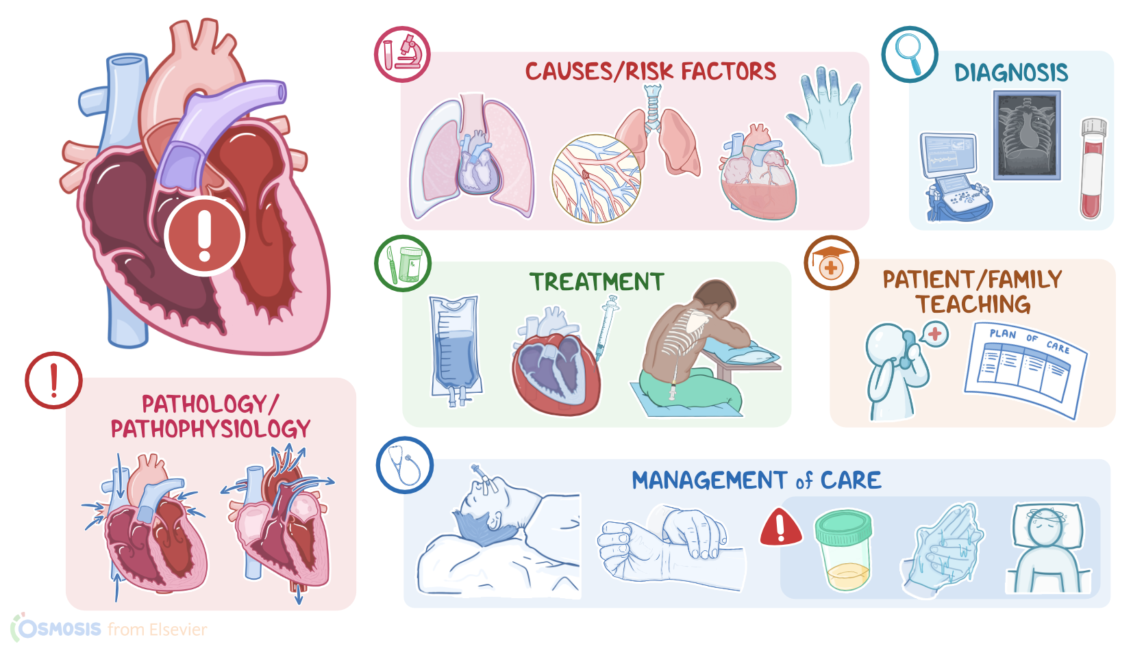
Obstructive shock is a life-threatening condition that develops when a mechanical obstruction prevents the heart from filling properly or pumping enough blood through the cardiovascular system. This reduces the amount of oxygenated blood that reaches the tissues, causing acute hypoperfusion and tissue hypoxia.

Alright, let’s go over the physiology of the cardiovascular system, which consists of the heart and blood vessels. The heart pumps out blood to the body’s organs and tissues with each heartbeat, which consists of two phases: systole, which is when the heart contracts and pumps the blood out; and diastole, which is when the heart relaxes and fills with blood. The stroke volume, meaning the amount of blood pumped out by the heart in a single heartbeat, is influenced by the cardiac contractility, preload, and afterload. Contractility is how strongly the heart is contracting during systole. Preload is how much the heart’s smooth muscle is stretched at the end of diastole, and this is mainly dependent upon how much blood is filling the heart. The more they're stretched the more force they can generate during contraction, kind of like a rubber band. Afterload refers to the resistance that the heart must overcome to pump out blood during systole, and this is affected mainly by peripheral vascular resistance under non-pathological conditions. This is mainly determined by the vasodilation and vasoconstriction of blood vessels. If we multiply the stroke volume by the heart rate we’ll get the cardiac output, which is the amount of blood pumped out by the heart in one minute, and it’s the main measure of the heart’s function.

Okay, now obstructive shock is caused by an obstruction that prevents the heart from filling properly with blood during diastole, thus decreasing preload, or pumping blood out during systole, which increases afterload. One of the most common causes of obstructive shock is cardiac tamponade, where blood or fluid builds up in the pericardial sac, that surrounds the heart, preventing the heart from physically expanding during diastole. Another cause is constrictive pericarditis, where the pericardium becomes thickened and fibrotic, so it prevents the heart from expanding during diastole. Tension pneumothorax, where air builds up within the pleural space, increasing intrathoracic pressure which compresses the veins that return blood to the heart. Other causes that prevent the heart from pumping out blood effectively during systole include a massive pulmonary embolism, or blood clot, which blocks the flow of the blood that is pumped into the lungs, as well as conditions like aortic stenosis or coarctation of the aorta, which prevents blood from being pumped out during systole.

Now, the pathology of obstructive shock develops when a mechanical obstruction reduces blood flow into or out of the heart. As a result, there’s a decrease in stroke volume, which in turn decreases cardiac output, causing blood pressure to drop, and ultimately causing hypoperfusion of organs and tissues. So, in order to increase cardiac output, the body releases vasoconstrictor molecules into the bloodstream. These include catecholamines like epinephrine and norepinephrine which cause vasoconstriction, increase heart rate, and contractility to maintain blood pressure and blood flow; as well as ADH which acts on the kidneys to increase fluid retention, and angiotensin II, which also constricts blood vessels and causes sodium retention in the kidneys, in turn, raising blood pressure.

If the cause of shock is not managed in time, these compensatory mechanisms may begin to fail, leading to severe tissue hypoxia. Vital organs like the heart, brain, and kidneys may begin to shut down, leading to multiple organ failure. At the same time, since blood isn’t being pumped forward into the systemic circulation, it starts backing up into the pulmonary and systemic blood vessels. Eventually, fluid can be be pushed out of the circulation and into the lungs and tissues, leading to complications like pulmonary and peripheral edema.

Okay, during the initial stage, compensatory mechanisms such as increased heart rate and vascular constriction are sufficient to maintain cardiac output within the normal range. Cardiac output can best be seen by obtaining the mean arterial pressure, or the average pressure in one cardiac cycle, because it is a more accurate indicator of perfusion than a normal blood pressure. In this stage, the MAP is decreased by less than 10 mmHg from baseline. Clients may experience tachycardia, but compensatory mechanisms keep blood pressure at around the normal range, so it may be difficult to detect shock at this stage. During the compensation stage, the compensatory mechanisms are fully active but the MAP is decreased and is about 10-15 mmHg below baseline. The client’s skin can become cold and clammy, indicating that blood flow is being redirected to vital organs like the brain and heart. Other symptoms include pallor, severe hypotension and tachycardia, decreased peripheral pulses, and oliguria. Other important signs include pulsus paradoxus, where blood pressure during inspiration drops more than 10 mm Hg; as well as jugular venous distention, where the jugular vein in the neck becomes enlarged and distended. Lastly, some clients may develop pulmonary edema, which typically presents with dyspnea and crackles; as well as peripheral edema, causing the limbs to swell. In the progressive stage, clients can develop organ failure because the compensatory mechanisms can no longer guarantee adequate blood flow to vital organs. The MAP is sustained at more than 20 mmHg below baseline. The client may experience anxiety, altered level of consciousness from decreased perfusion to the brain, cyanosis, increased respirations, decreased oxygen saturation from lung failure, profound hypotension, bradycardia, and irregular heart rhythm from heart failure, as well as anuria, which occurs due to kidney failure. In the refractory stage, cell death occurs in the vital organs due to the lack of oxygen reaching the tissues. The MAP is still sustained at more than 20 mmHg from baseline. At this point, the damaged organs cannot respond to treatment, leading to multiple organ dysfunction. The client may experience a sudden loss of consciousness, shallow respirations, unmeasurable oxygen saturation, non-palpable pulses, or death.

The diagnosis of obstructive shock starts with the client’s history and physical assessment. The client should be monitored closely to detect signs of hypoperfusion, including a drop in oxygen saturation, low or declining blood pressure, sudden changes in the ECG, or decreased urinary output. This is usually followed by additional tests, including an echocardiogram, and a chest X-ray to identify the precipitating cause of obstructive shock. Routine diagnostic investigations also include laboratory tests that can reveal decreased oxygen levels, electrolyte abnormalities, and signs of end-organ damage like elevated lactate levels and metabolic acidosis.

Initial treatment of obstructive shock involves supportive measures, including supplemental oxygen, intravenous fluids, and medications like norepinephrine or dopamine to improve the cardiac output and blood pressure. However, definitive treatment is aimed at addressing the underlying cause of obstruction, when possible. This may include performing a pericardiocentesis to drain fluid from the pericardial space in cases of cardiac tamponade; draining excess air from the pleural space in tension pneumothorax; or treating a pulmonary embolism with anticoagulants and fibrinolytics, or removing a blood clot surgically via an embolectomy.