Adenomyosis

Adenomyosis: What Is It, Causes, Diagnosis, Treatment, and More

Author: Georgina Tiarks, MD
Editor: Alyssa Haag, MD
Editor: Emily Miao, MD, PharmD
Editor: Nikol Natalia Armata, MD
Editor: Kelsey LaFayette, DNP, ARNP, FNP-C
Illustrator: Jillian Dunbar
Copyeditor: David G. Walker
Modified: Dec 06, 2024

What is adenomyosis?

Adenomyosis occurs when endometrial tissue is found within the myometrial, or muscular, layer of the uterine walls, resulting in growth, or hyperplasia, of the surrounding myometrium.  

There are three different layers of tissue in the wall of the uterus. The innermost layer is termed the endometrium, which contains endometrial glands, columnar epithelium, and endometrial stroma. The middle layer is referred to as the myometrium and consists mainly of uterine muscle. The outermost layer is a thin layer of tissue called the perimetrium. Adenomyosis occurs when ectopic, or abnormally positioned, endometrial tissue is found within the myometrium. This occurrence can be classified into either diffuse or focal. Diffuse adenomyosis is characterized by endometrial tissue scattered diffusely within the myometrium. Conversely, focal adenomyosis occurs when the endometrial tissue is only found in a localized area of the myometrium. The ectopic endometrial tissue induces growth within the surrounding myometrium, which causes enlargement of the uterus. Adenomyosis is a common condition among individuals who have given birth. 

An infographic detailing the background, signs and symptoms, diagnosis, and treatment of adenomyosis.

Is adenomyosis serious?

Adenomyosis is most often a benign condition, meaning that it is non-cancerous, does not invade other organs, and grows slowly. In rare circumstances, adenomyosis may become malignant, or cancerous—in which case, the condition may be considered more serious.  

What causes adenomyosis?

Adenomyosis is caused by the invasion of the endometrial basal layer into the myometrium, likely triggered by mechanisms involving tissue injury and repair at the junctional zone between these layers. There are two layers of endometrial tissue: the basal layer and the functional layer. The basal layer, or deepest portion of tissue that connects to the myometrium, is responsible for regeneration after menstruation. This layer does not shed during menstruation. In contrast, the functional layer is the layer of tissue that lines the uterus. The functional layer proliferates throughout the menstrual cycle and then sheds during menstruation if the egg is not fertilized. 

Several factors have been studied that may increase the likelihood of developing adenomyosis. Although the mechanism is unknown, it’s thought that estrogen, progesterone, and prior uterine surgery may contribute to the development of adenomyosis. It has also been linked to elevated aromatase levels, an enzyme responsible for synthesizing estrogen. Additionally, studies have indicated that age and parity may play a role. The risk of adenomyosis has been found to decrease after menopause, likely due to change in the levels of various hormones, such as progesterone, estrogen, prolactin, and follicle stimulating hormone (FSH). 

What are the signs and symptoms of adenomyosis?

The signs and symptoms of adenomyosis often include changes with menstruation, which may include menorrhagia, or heavy menstrual bleeding, or dysmenorrhea, or painful menstrual cycles. Some individuals with adenomyosis may also complain of dyspareunia, which is pain during intercourse, or chronic pelvic pain. Due to excessive tissue proliferation, many individuals experience uterine enlargement and discomfort. Adenomyosis can also impair fertility by disrupting the transport of the fertilized egg from the fallopian tube to the uterus, and by altering the endometrial function and receptivity, thereby reducing the likelihood of successful implantation. In some cases, however, individuals may experience no symptoms at all. 

How is adenomyosis diagnosed?

Diagnosis of adenomyosis begins with a thorough review of symptoms and medical history. A physical exam may also reveal an enlarged and tender uterus. Imaging tests may also be used while diagnosing adenomyosis. Most commonly, a transvaginal ultrasound can reveal an enlarged, round uterus, with myometrial thickening, diverse myometrial texture, cystic glands within the myometrium, and projections from endometrium into the myometrium with increased vascularity. A transabdominal ultrasound may also be performed that show signs of changes to the myometrium. If leiomyomas, or uterine fibroids, are suspected, an MRI may be performed to distinguish between adenomyosis and leiomyomas. A definitive diagnosis can be made through a needle biopsy of the myometrial layer. However, due to the invasive nature of biopsies, they are rarely performed in cases of adenomyosis. 

Additional lab work may be used to rule out other possible diagnoses. If the individual is of childbearing age, a pregnancy test should also be performed. 

How is adenomyosis treated?

Treatment of adenomyosis is primarily focused on relieving the individual's symptoms, which may include pain and irregular menstruation. Non-steroidal anti-inflammatory medications (NSAIDs), such as ibuprofen, may be recommended for pain relief, while hormonal treatment options may be used to prevent irregular bleeding. Studies have shown that combined oral contraceptives, which include both estrogen and progesterone, as well as progestin-only medications, may be used to promote a regular menstrual cycle. In some cases, gonadotropin-releasing hormone (GnRH) agonists may also be used to prevent excess estrogen production. 

If necessary, other treatment options, such as endometrial ablation or resection, may be used to remove the endometrium. Emerging treatments for adenomyosis focus on addressing underlying molecular mechanisms with a particular emphasis on targeting epigenetic modifications that regulate gene expression involved in the disease’s progression, without altering the DNA sequence. Definitive treatment would require a hysterectomy (i.e., removal of the uterus), but this is primarily reserved for individuals who are no longer of childbearing age. 

If necessary, other treatment options, such as endometrial ablation or resection, may be used to remove the endometrium. Definitive treatment would require a hysterectomy (i.e., removal of the uterus), but this is primarily reserved for individuals who are no longer of childbearing age.

What are the most important facts to know about adenomyosis?

Adenomyosis is a benign uterine condition caused by the growth of endometrial tissue into the myometrial layer of the uterus. It’s caused by an invasion of the basal layer of the endometrium into the myometrium. There are several risk factors, including age, parity, and hormone levels. Symptoms often include irregular bleeding and abnormal pain during the menstrual cycle; however, some individuals do not experience any symptoms. Diagnosis relies primarily on imaging modalities, such as ultrasound or MRI. There are a variety of treatment options that range from symptom control to surgical removal. In some cases, prescription of hormonal contraceptives and pain medication can relieve the symptoms of adenomyosis. Alternatively, surgical resection may offer long-term relief in individuals who have completed childbearing. 

References


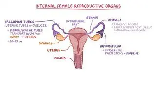
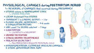
Karavelic A. Atrophic Endometrium. My Pathology Report. Accessed June 29, 2021. https://www.mypathologyreport.ca/atrophic-endometrium/ 


Kho KA, Chen JS, Halvorson LM. Diagnosis, evaluation, and treatment of adenomyosis. JAMA. 2021;326(2):177. doi:https://doi.org/10.1001/jama.2020.26436  


Lukies M. Endometrium. Radiopeadia. Accessed June 29, 2021. https://radiopaedia.org/articles/endometrium?lang=us 


Moawad G, Fruscalzo A, Youssef Y, et al. Adenomyosis: An updated review on diagnosis and classification. J Clin Med. 2023;12(14):4828. Published 2023 Jul 21. doi:10.3390/jcm12144828 


Schrager S, Yogendran L, Marquez CM, Sadowski EA. Adenomyosis: Diagnosis and management. American Family Physician. 2022;105(1):33-38. https://www.aafp.org/pubs/afp/issues/2022/0100/p33.html 


Zhang H, Li C, Li W, Xin W, Qin T. Research advances in adenomyosis-related signaling pathways and promising targets. Biomolecules. 2024; 14(11):1402. https://doi.org/10.3390/biom14111402