Arrhythmias - Atrial flutter (Aflutter): Nursing
Arrhythmias - Atrial flutter (Aflutter): Nursing
NUR 395 Cardiovascular Dysrhythmias
NUR 395 Cardiovascular Dysrhythmias
Notes
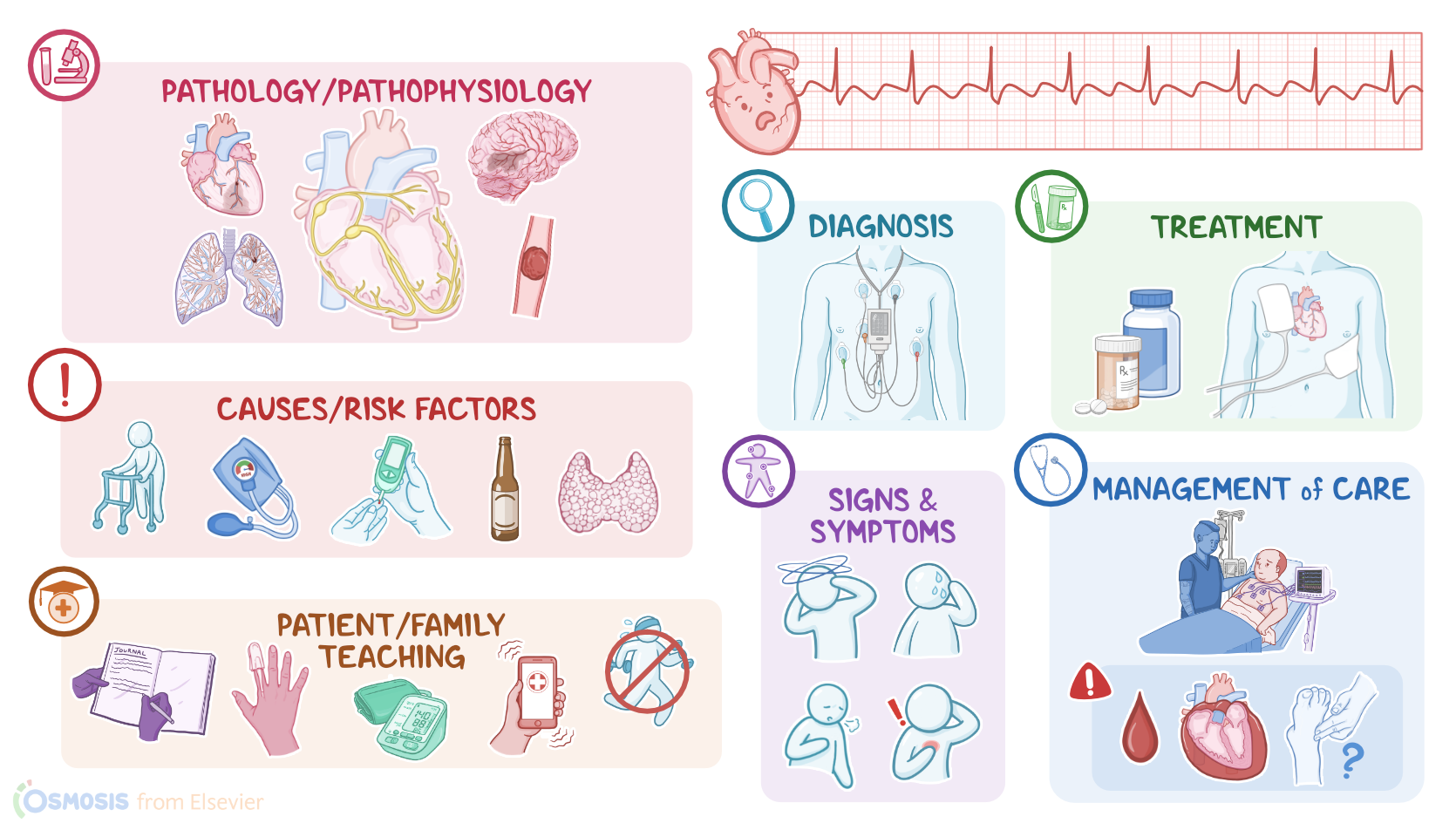
| ARRHYTHMIAS - ATRIAL FLUTTER (AFLUTTER) | ||
| KEY POINTS | NOTES | |
| DEFINITION |
| |
| PHYSIOLOGY |
| |
| CAUSES AND RISK FACTORS |
| |
| PATHOPHYSIOLOGY |
| |
| SIGNS AND SYMPTOMS |
| |
| DIAGNOSIS |
| |
| TREATMENT |
| |
| MANAGEMENT OF CARE |
| |
| PATIENT AND FAMILY TEACHING |
| |
Transcript
Atrial flutter, or AF for short, is a type of tachyarrhythmia, where tachy means fast, and arrhythmia means irregular rhythm. So, AF is a fast, abnormal heart rhythm that causes poor, inefficient atrial contractions, which can affect the heart’s ability to pump blood. It’s called flutter because of its identifiable recurring, regular, sawtooth-shaped flutter waves that can be seen on an ECG.
Now, the cardiac conduction system is made up of specialized myocardial cells that can create and transport electrical potential, also called an action potential. These cells have many special features, including automaticity, meaning that they can generate an impulse, excitability, which is the ability to respond to a stimulus by creating an electrical impulse, conductivity meaning they can carry the impulse to other cells, and contractility, which is the ability to shorten the length of their fibers, causing a contraction.
Alright, now let’s look at the normal electrical conduction pathway in the heart on an ECG, which shows how the depolarization wave flows through the heart during each heartbeat. The normal electrical activity of the heart starts in the sinoatrial or SA node, which is considered the pacemaker of the heart. Then, the impulse is conducted through the atria, causing depolarization and creating the P wave on an ECG. When the atrial muscle cells get depolarized, they contract, pushing blood from the atria into the ventricles. From the atria, the impulse goes to the atrioventricular, or AV node, where the impulse propagation speed slows way down. The interval from the atrial depolarization to just before ventricular depolarization is the PR interval on an ECG. This delay allows the atria to contract while the ventricles fill with blood. From the AV node, the impulse goes through the Bundle of His, then to the right and left bundle branches, and finally through the Purkinje fibers, which deliver the impulse to the right and left ventricles, causing them to depolarize, and is represented by the QRS complex on an ECG. This triggers simultaneous contraction of both ventricles, pushing blood into the systemic and pulmonary circulations. Finally, the ventricles repolarize to prepare for the next cycle, which allows them to relax and fill with blood, called diastole. And on ECG, ventricular repolarization will create a T wave, while the phase between ventricular depolarization and repolarization is represented by the ST segment. Sometimes, immediately after the T wave, there’s a U wave, which represents late repolarization of the ventricles.
Now, the main cause of an atrial flutter is the occurrence of a reentry mechanism, which means an abnormal electrical circuit that forms in the atria, which makes the electrical impulse travel in a loop within the atria, instead of going to the AV node like it normally would.
Risk factors include advanced age; underlying cardiovascular disease like hypertension or cardiac surgery; diabetes; or a history of alcohol use, pulmonary embolism or hyperthyroidism. Interestingly, atrial flutter can also occur following the conversion of atrial fibrillation to normal sinus rhythm. In fact clients taking antiarrhythmics for other conditions can also develop atrial flutter.
Now, the pathology of atrial flutter starts with a reentrant loop that can originate in either the right or left atrium. Reentrant signals loop back on themselves, overriding the sinus node and setting up an endless cycle that causes the atria to contract again and again and again—at really fast rates.
There are actually two types of atrial flutter, depending on how this reentrant circuit arises. Type 1, or typical atrial flutter, is more common and is caused by a single reentrant circuit that moves around the annulus, or the ring of the tricuspid valve of the right atrium. See, around this area, there’s a stretch of tissue called the cavotricuspid isthmus, that propagates the signal more slowly than the surrounding tissue. Tissue that was just activated can’t be activated again until a certain amount of time has passed, which is called the refractory period; so that slow propagation gives the tissue enough time to be out of refractory, and therefore the circuit can loop on itself.
Type 2 or atypical atrial flutter is where a re-entrant circuit develops in either the right or left atrium, but the exact location is less clearly defined. Again, though we’ve got a similar setup where a wave of activated tissue, or depolarization hits a bit of tissue in such a way that it creates a loop of depolarization that keeps going around and around.
Regardless of type, with atrial flutter, the atria contract inefficiently. This means that the ventricles get less blood from the atria, and they also contract at a lesser rate, which decreases cardiac output. Eventually, the inefficient emptying of the atria into the ventricles can cause the blood to stay in the atria for longer than normal, so it can form atrial clots. So, with atrial flutter, there’s a chance that one of these clots can get pumped from the atria into the ventricles, and from there, into the circulation. From there, the clot can get lodged in a cerebral artery, causing a stroke; in a pulmonary artery branch, causing a pulmonary embolism; or in a coronary artery that supplies the myocardial muscle, causing a myocardial infarction.
Clients with atrial flutter may be asymptomatic, but when a client is symptomatic, clinical manifestations include palpitations, fatigue, mild dyspnea, syncope or lightheadedness, tachycardia, and diaphoresis.
Additionally, if a clot leaves the heart and gets lodged in another artery, manifestations depend on the localization. With a stroke, there could be confusion, slurred speech, and unilateral weakness or paralysis of the face or limbs; with a pulmonary embolism, there might be dyspnea and chest pain; and with a myocardial infarction, there could be severe chest pain that radiates, most commonly to the left arm.
The diagnosis of atrial flutter starts with the client's history and physical assessment, followed by cardiac monitoring using an electrocardiogram or a Holter monitor, which is basically a portable ECG that records for a 24-hour period.
So, on the ECG, the heart rate is often between 200 to 350 beats per min with characteristic recurring, regular, sawtooth-shaped flutter waves. Typically, the atrial and ventricular rhythms are regular, at a ratio of 2:1, 3:1, or 4:1. More rarely, ventricular conduction can be variable. The ventricular rate may vary based upon the conduction ratio. For instance, in a 2:1 conduction ratio the ventricular rate is around 150 beats per minute. Also note, P waves are rarely seen and, as a consequence, PR intervals are variable or can’t be measured. The QRS complex is usually normal but contains some AV blocks within flutter waves.
Additionally, a transthoracic echocardiogram can be done to evaluate the overall morphology of the heart, while a transesophageal echocardiogram can be done to rule out the presence of clots in the atrium.
Alright, now, treatment for atrial flutter is geared at slowing the ventricular rate. This often begins with medications including calcium channel blockers like diltiazem or beta blockers like metoprolol. To convert back to a normal sinus rhythm, antiarrhythmics like ibutilide can be used. Or, to maintain the sinus rhythm, antiarrhythmics like amiodarone or flecainide are often used.
Sources
- "Critical care nursing: Diagnosis and management (10th ed.). ISBN: 978-0-443-11581-3 " Elsevier (2026)
- "Lewis’s Medical-Surgical Nursing: Assessment and Management of Clinical Problems. 12th Edition. ISBN:978-0-323-78961-5 " Elsevier
- "Heart failure and atrial flutter: a systematic review of current knowledge and practices. 8(6):4484-4496." ESC Heart Fail. (2021)
- "Stroke and thromboembolism prevention in atrial fibrillation. 106(1):10-17." Heart. (2020)
- "Prevention and Treatment of Atrial Fibrillation via Risk Factor Modification. 160:46-52." Am J Cardiol. (2021)
- "Fundamentals of nursing (10th ed.). ISBN 978-0323810340 " Elsevier (2021)
- "Saunders Comprehensive Review for the NCLEX-RN® Examination. ISBN 978-0323795302 " Saunders (2022)
- "2021 CAEP Acute Atrial Fibrillation/Flutter Best Practices Checklist. 23(5):604-610." CJEM. (2021)