Arrhythmias - Heart blocks: Nursing
Introduction0:00–0:44
Arrhythmias are irregular heartbeats that occur due to any disturbance in the rate, rhythm, site of origin, or conduction of the cardiac electrical impulse, which can affect the heart’s ability to effectively pump blood throughout the body.
Now a heart block, or atrio-ventricular block, occurs when there is delay or disturbance in the conduction of cardiac electrical impulse from the atria to the ventricles.
There are three types of atrio-ventricular block or AV block, for short: first degree AV block, second degree AV block which is further divided into Mobitz type I and Mobitz type II, and lastly, third degree AV block.The cardiac conduction system is made of specialized myocardial cells that create and transport electrical potential which play a role in regulating heart rate and rhythm.
Physiology0:44–2:59
They possess automaticity which is the ability to generate an impulse, excitability or the ability to respond to a stimulus by initiating stimulus, conductivity or the ability to send that impulse, and contractility or the ability to shorten fiber length.Now let’s look at the normal electrical conduction pathway in the heart on an ECG, which shows how the depolarization wave flows through the heart during each heartbeat.
The normal electrical activity of the heart starts in the sinoatrial or SA node, which is considered the pacemaker of the heart.
Then, the impulse is conducted through the atrium, creating the P wave on an ECG. And when the atrial muscle cells get depolarized, they contract, pushing blood from the atria into the ventricles.
From the atrium, electrical activity goes to the atrioventricular, or AV node, where the impulse propagation speed slows way down; this is the PR interval on an ECG.
This pause allows the atria to contract while the ventricles fill with blood.From the AV node, the depolarization wave goes through the Bundle of His, then the right and left branches of the Bundle, and finally through the Purkinje fibers, which deliver the current to the right and left ventricles, causing them to depolarize.
This triggers simultaneous contraction of both ventricles, pushing blood into the systemic and pulmonary circulations, and it’s represented by the QRS complex on an ECG.
Finally, the ventricles repolarize to prepare for the next cycle, which allows them to relax and fill with blood, called diastole.
And on an ECG, ventricular repolarization will create a T wave, while the pause between ventricular depolarization and repolarization is represented by the ST segment.
Sometimes, immediately after the T wave, there’s a U wave, which represents late repolarization of the ventricles.Okay, now let’s look at AV blocks, which are typically caused by any kind of structural damage or fibrosis to the electrical conduction system.
Causes and risk factors2:59–3:45
This damage is strongly associated with several risk factors, particularly cardiac surgery or underlying heart conditions.
These include ischemic heart disease, previous or recent myocardial infarction, congenital heart disease, myocarditis, as well as rheumatic fever.
Other risk factors include drug toxicity, typically with digoxin, calcium channel blockers or beta blockers. Finally, elderly clients, as well as athletes are at a higher risk for developing AV blocks.Okay, the pathology of an AV block starts when any of these factors impair the function of the electrical conduction system.
Pathology3:45–5:38
Now, a first degree AV block is technically not really a block, because it’s more of a delay as the cardiac electrical impulses still make it to the ventricles, but result in a delayed ventricular contraction.On the other hand, second degree AV block has two subtypes: Mobitz type I and Mobitz type II.
In Mobitz I, each atrial impulse encounters a longer and longer delay until one of them does not make it through to the ventricles, and you get what’s called a “dropped beat.” When a signal doesn’t make it from the atria to the ventricles, and if a long enough time passes, then the ventricles’ pacemaker cells kick in, sort of as a fail-safe mechanism, called a ventricular escape beat.
Like Mobitz I, the heart also drops a beat in Mobitz II, except this time, conduction through the AV node is all-or-nothing.
Either the atrial impulse goes through with no delay, or it doesn’t at all. A lot of times, a ratio for the overall number of beats conducted to not-conducted is given, like 2:1 Mobitz II AV block, but it’s important to remember that the dropped beats happen fairly randomly so we can’t really predict exactly when the next beat will get dropped.
Mobitz II can be dangerous and may result in severe bradycardia and decreased cardiac output.Lastly, there is third degree or complete AV block, where the signal is completely blocked when moving from the atria to ventricles, every time.
Because the atria and the ventricles each have their own pacemakers, they now contract independent of one another, which is called AV dissociation.
So, in terms of clinical manifestations, most clients with a first degree AV block and Mobitz type I don’t have symptoms, but occasionally patients feel lightheadedness, dizziness, and syncope.
Clinical Manifestations5:38–6:12
In contrast, most clients with Mobitz type II have symptoms like fatigue, confusion, dyspnea, chest pain, and syncope, though the severity can vary from client to client.
In third degree AV block, these symptoms become more severe and without treatment may lead to cardiac arrest and even death.The diagnosis of an AV block starts with the client's history and physical assessment, followed by a 12-lead electrocardiogram.
Diagnosis6:12–7:31
In first degree AV block, the PR interval is greater than 200 milliseconds. In Mobitz type I AV block, also called Wenckebach block, the PR interval gets progressively longer with each beat until a P wave is blocked completely.
So, maybe the first PR interval is 200 milliseconds, then the next is 260 milliseconds, then 300 milliseconds, and finally the next one doesn’t make it to the ventricles.
In Mobitz type II AV block, there is no progressive prolongation of the PR interval. Instead, there are a couple of normal PR intervals followed by a dropped beat.
Lastly, in third degree AV block, the P-waves and QRS complexes have nothing to do with each other, each appearing at their own rates.
The atrial rate is 60 to 100 beats per minute, whereas the ventricular rate usually ranges between 30 to 45 beats per minute.
The QRS complex is normal if the escape rhythm starts at the bundle of His or above. It is widened if the escape rhythm starts below the bundle of His.Now, treatment of AV block may vary depending on the cause, types and severity of symptoms.
Treatment7:31–9:00
No treatment is required if the client is asymptomatic like in the first degree or Mobitz type I AV block. Now, in a symptomatic client, the underlying cause of the AV block should be identified and treated, when possible.
Additional treatment depends on the type of block as well as whether or not the client is hemodynamically stable. Unstable clients with Mobitz type I second degree AV block could benefit from atropine.
If that doesn’t help stabilize the client, other options include beta-adrenergic agonists like dopamine, dobutamine or isoproterenol; as well temporary pacing options, like transcutaneous or transvenous pacing.
In clients with Mobitz type II second degree AV block that are hemodynamically stable, atropine can also be used. In unstable clients, though, atropine is contraindicated, and beta-adrenergic agonists are preferred instead.
Finally, in unstable clients with a third degree or complete heart block, options include atropine, beta adrenergic agonists as well as temporary pacing options.
Ultimately, implantation of a permanent pacemaker can be a solution for clients with complete heart block, as well as for those with Mobitz type I AV blocks, after they’ve been stabilized.
Alright, let’s look at the nursing care you’ll provide to a client with an AV block. Your priority goal of care is to assist with improving cardiac output.
Management of care9:00–10:22
Begin by initiating continuous cardiac monitoring, and assessing your client to determine if they are hemodynamically stable.
Assess their vital signs and check for symptoms of decreased cardiac output, and report dizziness, decreased level of consciousness (LOC), a heart rate less than 40 beats per minute, hypotension, chest pain, tachypnea, or shortness of breath to the healthcare provider.
If they continue to be unstable, hypotensive, or have signs of heart failure, administer the prescribed beta-adrenergic agonist medications to support heart rate and blood pressure, and prepare your client for transvenous pacing.
Once they are stabilized, assist the healthcare team with identifying a reversible cause, provide the prescribed treatments, and continue to monitor your client.
If no treatable cause is identified, prepare your client for the insertion of a permanent pacemaker. Okay, let’s move onto client and family teaching.
General client & family teaching10:22–13:25
First, teach your client that an AV block causes a delay in the conduction of cardiac electrical impulse from the atria to the ventricles.
Review their treatment plan, including any new medications; and instruct your client to take them exactly as prescribed.Next, talk to them about lifestyle modifications that can help keep their heart healthy.
Encourage them to maintain a healthy weight, and to engage in moderate physical exercise regularly, as prescribed by their healthcare provider.
Instruct them to eat plenty of fruits, vegetables, and whole grains, as well as lean meats, and low-fat or fat-free dairy products.
If they need help quitting smoking, provide them with counseling and refer them to resources for support. Then, teach them how to check their pulse and blood pressure, and instruct them to sit or lie down if they begin to feel dizzy to avoid injuries.
Now, if your client has a permanent pacemaker, let them know that the device has been implanted just beneath the skin, and that there are leads that go directly to the heart muscle.
Explain that when it senses their heart is beating too slow, it will send a signal to their heart to help it beat at the correct pace.
Review with them how to care for the incision site and to let their healthcare provider know right away if they experience any signs of infection, including fever, redness, swelling, tenderness, or discharge at the insertion site.
Next, advise them to avoid raising their arm on the incision side above shoulder level or stretching their arm behind their back, to avoid dislodging the leads and to give them a chance to become more secure inside their heart.
Also let them know that their pacemaker could be affected by electromagnetic fields, so emphasize the importance of keeping their cell phone away from their pacemaker, and review the kinds of electrical devices and medical procedures that are safe as well as those that could interfere with their particular type of pacemaker.
Lastly, stress the importance of always carrying their pacemaker ID card with them at all times; to let all healthcare providers know they have a pacemaker; and to keep all of their follow-up appointments so their pacemaker can be regularly checked.Finally, advise them to notify their healthcare provider immediately if they have a heart rate less than 60 beats per minute, or a systolic blood pressure of less than 90 mmHg; stress the importance of seeking emergency medical care if they experience dizziness, chest pain, shortness of breath, confusion, loss of consciousness, or prolonged hiccupping.Alright, as a quick recap… The three types of AV blocks are first degree AV block; second degree AV block, which is further divided into Mobitz type I and Mobitz type II; and third degree AV block.
Review13:25–15:40
During a first degree AV block, the electrical impulse is delayed and causes a delayed ventricular contraction. A Mobitz type I AV block is when each atrial impulse has a longer delay until one doesn’t make it to the ventricles which causes a dropped beat.
On the other hand, in a Mobitz type II AV block the atrial impulses either go to the ventricles without delay or not at all, and the number of conducted to not-conducted beats can occur either as a ratio or in an unpredictable pattern.
If a third degree AV block occurs, the atrial impulse is completely blocked from reaching the ventricles, causing the atria and ventricles to contract independently of one another.
Clinically, clients with first degree AV block or Mobitz type I AV block can be asymptomatic, while clients with Mobitz type II AV block or third degree AV block typically have more severe symptoms.
Diagnosis involves a 12-lead ECG, and treatment depends on the underlying cause, type of AV block, and severity of symptoms.
Treatment isn’t required for asymptomatic clients, whereas symptomatic clients may benefit from medications to increase their heart rate such as atropine or beta-adrenergic agonists; temporary pacing; or a permanent pacemaker.
Nursing management is focused on improving cardiac output while closely assessing and monitoring the client and assessing for pacemaker dysfunction.
Client and family teaching includes explaining their condition, treatment plan, and medications; lifestyle modifications; pacemaker considerations; as well as symptoms to report to their healthcare provider.
| ARRHYTHMIAS - HEART BLOCKS | ||
| KEY POINTS | NOTES | |
| DEFINITION |
| |
| PHYSIOLOGY |
| |
| CAUSES AND RISK FACTORS |
| |
| PATHOPHYSIOLOGY |
| |
| SIGNS AND SYMPTOMS |
| |
| DIAGNOSIS |
| |
| TREATMENT |
| |
| MANAGEMENT OF CARE |
| |
| PATIENT AND FAMILY TEACHING |
| |
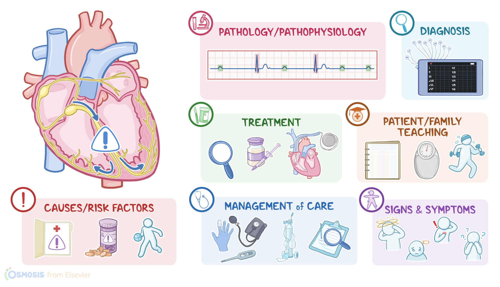
- "Medical-Surgical Nursing: Concepts for Interprofessional Collaborative Care" Elsevier (2020)
- "Lewis’s medical-surgical nursing: Assessment and management of clinical problems" Elsevier (2020)
- "Saunders Comprehensive Review for the NCLEX-RN Examination" Elsevier (2017)
- "Fundamentals of Nursing" Elsevier (2021)
- "Contralateral pneumothorax and pneumopericardium after dual-chamber pacemaker implantation: Mechanism, diagnosis, and treatment" HeartRhythm Case Reports (2018)
- "Right Bundle Branch Block: Current Considerations" Current Cardiology Reviews (2021)
No notes for this video yet
Try adding a note below