Arrhythmias - Sinus tachycardia and sinus bradycardia: Nursing

Arrhythmias - Sinus tachycardia and sinus bradycardia: Nursing

Acute Final

Acute Final

Endocrine system anatomy and physiology
Antepartum assessment - Fetus: Nursing
Assessment of gestational age: Nursing
Fetal circulation: Nursing
Fetal development: Nursing
Group B streptococcus (GBS) infection in pregnancy: Nursing
Hepatitis B virus (HBV) infection in pregnancy: Nursing
Hyperemesis gravidarum: Nursing
Large for gestational age (LGA) infant: Nursing
Preeclampsia and eclampsia: Nursing
Prenatal screening: Nursing
Placenta previa: Nursing process (ADPIE)
Placental abruption: Nursing process (ADPIE)
Birth-related procedures: Nursing
Cesarean birth: Nursing
Intrapartum assessment - Fetal heart rate patterns: Nursing
Intrapartum assessment - Uterine activity: Nursing
Premature rupture of membranes (PROM): Nursing
Shoulder dystocia: Nursing
Prolapsed umbilical cord: Nursing process (ADPIE)
Stages of labor: Nursing
Assessment - Postpartum: Nursing
Perinatal depression: Nursing
Physiology of lactation: Nursing
Postpartum infections: Nursing
Postpartum hemorrhage: Nursing
Biliary atresia: Nursing
Cleft lip and palate: Nursing
Congenital diaphragmatic hernia: Nursing
Congenital heart defects - Acyanotic: Nursing
Congenital heart defects - Cyanotic: Nursing
Esophageal atresia and tracheoesophageal fistula: Nursing
Craniosynostosis: Nursing
Hemolytic disease of the fetus and newborn: Nursing
Hyperbilirubinemia: Nursing process (ADPIE)
Infant of a diabetic mother (IDM): Nursing
Meconium aspiration syndrome: Nursing
Neonatal respiratory distress syndrome (NRDS): Nursing
Neonatal sepsis: Nursing
Neural tube defects: Nursing
Newborn adaptation to extrauterine life: Nursing
Persistent pulmonary hypertension of the newborn (PPHN): Nursing
Physical assessment - Neonate: Nursing
Small for gestational age (SGA) infant: Nursing
Postterm infant: Nursing
Thermoregulation - Neonate: Nursing
Arterial blood gas (ABG) - Overview: Nursing
Arterial blood gas (ABG) - Metabolic acidosis: Nursing
Arterial blood gas (ABG) - Metabolic alkalosis: Nursing
Arterial blood gas (ABG) - Respiratory acidosis: Nursing
Arterial blood gas (ABG) - Respiratory alkalosis: Nursing
Adrenal insufficiency (Addison disease): Nursing
Anemia - Iron-deficiency: Nursing
Anemia - Aplastic: Nursing
Anemia - Macrocytic: Nursing
Case study - Hypothyroidism: Nursing
Case study - Iron-deficiency anemia: Nursing
Case study - Sickle cell anemia: Nursing
Complete blood count (CBC) - Hemoglobin and hematocrit: Nursing
Complete blood count (CBC) - Red blood cells (RBC): Nursing
Complete blood count (CBC) - Platelets: Nursing
Complete metabolic panel (CMP) - Blood urea nitrogen (BUN) and creatinine (Cr): Nursing
Complete metabolic panel (CMP) - Estimated glomerular filtration rate (eGFR): Nursing
Complete metabolic panel (CMP) - Liver function tests (LFT): Nursing
Cushing syndrome and Cushing disease: Nursing
Hematopoietic growth factors: Nursing pharmacology
Hyperparathyroidism: Nursing
Hyperthyroidism: Nursing process (ADPIE)
Hypoparathyroidism: Nursing
Hyperpituitarism: Nursing
Hypopituitarism: Nursing
Hypothyroidism: Nursing process (ADPIE)
Medications affecting the parathyroid glands: Nursing pharmacology
Medications for growth hormone disorders: Nursing pharmacology
Medications for thyroid disorders: Nursing pharmacology
Neutropenia: Nursing
Polycythemia: Nursing
Thrombocytopenia: Nursing
Acute kidney injury (AKI): Nursing process (ADPIE)
Benign prostatic hyperplasia (BPH): Nursing process (ADPIE)
Case study - Cholecystitis: Nursing
Case study - Cirrhosis: Nursing
Case study - Chronic kidney disease (CKD): Nursing
Case study - Benign prostatic hyperplasia (BPH): Nursing
Case study - Gastroesophageal reflux disease (GERD): Nursing
Case study - Pediatric appendicitis: Nursing
Case study - Pyelonephritis: Nursing
Cholecystitis: Nursing
Cholelithiasis: Nursing
Chronic kidney disease (CKD): Nursing
Cirrhosis: Nursing process (ADPIE)
Diverticular disease: Nursing
Gastroesophageal reflux disease (GERD): Nursing process (ADPIE)
Hemolytic uremic syndrome: Nursing
Hirschsprung disease: Nursing
Intestinal obstruction: Nursing
Irritable bowel syndrome (IBS): Nursing
Nephrotic syndrome: Nursing
Pyloric stenosis: Nursing process (ADPIE)
Renal and urinary calculi: Nursing
Urinary incontinence - Stress: Nursing process (ADPIE)
Diabetes insipidus: Nursing process (ADPIE)
Dialysis care: Nursing
Case study - Diabetic ketoacidosis (DKA): Nursing
Case study - Pediatric diabetes mellitus type 1: Nursing
Diabetes mellitus (DM): Nursing process (ADPIE)
Hyperosmolar hyperglycemic state (HHS): Nursing process (ADPIE)
Diabetic ketoacidosis (DKA): Nursing process (ADPIE)
Case study - Epilepsy: Nursing
Case study - Head injury: Nursing
Epidural and subdural hematoma: Nursing
Case study - Stroke: Nursing
Hemorrhagic stroke - Intracranial hemorrhage (ICH) and subarachnoid hemorrhage (SAH): Nursing
Increased intracranial pressure (ICP): Nursing
Hydrocephalus: Nursing process (ADPIE)
Intracranial aneurysm: Nursing
Seizure disorder: Nursing process (ADPIE)
Stroke: Nursing process (ADPIE)
Jaundice: Nursing
Nutrition - Enteral: Nursing skills
Nutrition - Newborn: Nursing
Nutrition - Parenteral: Nursing skills
Phenylketonuria (PKU): Nursing
Arterial embolism: Nursing
Disseminated intravascular coagulation (DIC): Nursing
Hemophilia: Nursing process (ADPIE)
Acute respiratory distress syndrome (ARDS): Nursing
Asthma: Nursing process (ADPIE)
Atelectasis: Nursing
Bacterial pneumonia: Nursing process (ADPIE)
Bronchiolitis and respiratory syncytial virus (RSV): Nursing process (ADPIE)
Case study - Acute respiratory distress syndrome (ARDS): Nursing
Care of an intubated client: Nursing skills
Case study - Chronic obstructive pulmonary disease (COPD): Nursing
Case study - Impaired gas exchange: Nursing
Case study - Pediatric asthma: Nursing
Chest tube care: Nursing
Chronic obstructive pulmonary disease (COPD): Nursing process (ADPIE)
Cystic fibrosis: Nursing
Epiglottitis: Nursing process (ADPIE)
Flail chest: Nursing
Intraoperative care: Nursing
Pleural effusion: Nursing
Pneumothorax and hemothorax: Nursing
Pulmonary edema: Nursing
Smoke inhalation injury: Nursing process (ADPIE)
Tracheostomy: Nursing
Venous thromboembolism (VTE): Nursing process (ADPIE)
Arrhythmias - Asystole: Nursing
Arrhythmias - Atrial flutter (Aflutter): Nursing
Arrhythmias - Premature atrial contractions (PACs): Nursing
Arrhythmias - Heart blocks: Nursing
Arrhythmias - Atrial fibrillation (Afib): Nursing
Arrhythmias - Premature ventricular contractions (PVCs): Nursing
Arrhythmias - Sinus tachycardia and sinus bradycardia: Nursing
Arrhythmias - Supraventricular tachycardia (SVT): Nursing
Arrhythmias - Ventricular fibrillation (Vfib): Nursing
Arrhythmias - Ventricular tachycardia (Vtach): Nursing
Cardiac biomarkers - Troponin: Nursing
Case study - Acute coronary syndrome (ACS): Nursing
Case study - Atrial fibrillation (Afib): Nursing
Case study - Heart failure with reduced ejection fraction (HFrEF): Nursing
Case study - Deep vein thrombosis (DVT): Nursing
Case study - Hypertension: Nursing
Case study - Hypovolemic shock: Nursing
Coronary artery disease (CAD) and angina pectoris: Nursing process (ADPIE)
Electrocardiogram (ECG) - Normal sinus rhythm (NSR): Nursing
Heart defects that decrease pulmonary blood flow - Nursing considerations & client education: Nursing
Hypertension: Nursing process (ADPIE)
Left-sided heart failure: Nursing process (ADPIE)
Myocardial infarction (MI): Nursing process (ADPIE)
Pericardial effusion and cardiac tamponade: Nursing process (ADPIE)
Peripheral arterial disease (PAD): Nursing process (ADPIE)
Rheumatic heart disease: Nursing process (ADPIE)
Shock - Cardiogenic: Nursing
Shock - Neurogenic: Nursing
Shock - Obstructive: Nursing
Shock - Septic: Nursing
Sickle cell disease: Nursing process (ADPIE)
Valvular heart disease: Nursing

Notes

ARRHYTHMIAS - SINUS TACHYCARDIA AND SINUS BRADYCARDIA

KEY POINTS
NOTES
DEFINITION
  • Types of sinus arrhythmias
  • Sinus tachycardia: impulses too fast
  • Sinus bradycardia: impulses too slow

PHYSIOLOGY
  • Impulse begins in sinoatrial (SA) node
  • Conducts through atrium: P wave
  • Through atrioventricular (AV) node, propagation slows: PR interval
  • Bundle of His
  • Right and left bundle branches
  • Purkinje fibers
  • Right and left ventricles contract: QRS complex
  • Ventricles depolarize: T wave
  • Late ventricular depolarization: U wave

CAUSES AND RISK FACTORS
  • Causes
    • Sinus tachycardia
      • Sympathetic nervous system (SNS) stimulation
      • Parasympathetic nervous system (PNS) inhibition
    • Sinus bradycardia
      • PNS stimulation
      • SNS inhibition

PATHOPHYSIOLOGY
  • Sinus tachycardia
    • Increased firing of impulses from SA node
    • Cardiac output (CO) increases
    • Diastolic filling time decreases
    • Stroke volume (SV) decreases
    • Oxygen demand increases
    • Myocardial perfusion decreases
  • Sinus bradycardia
    • Decreased firing of impulses from SA node
    • Decreased heart rate (HR)
    • Increased diastolic filling time
    • Increased coronary perfusion time, decreased CO
    • Decreased myocardial perfusion
    • Increased demand for oxygen
  • Complications
    • Myocardial ischemia or infarction
    • Heart failure
    • Cardiomyopathy

SIGNS AND SYMPTOMS
  • Depends on degree of tachycardia or bradycardia and patient tolerance
  • Asymptomatic
  • Symptomatic
    • Hypotension
    • Dizziness
    • Dyspnea
    • Restlessness
    • Confusion
    • Palpitations
    • Chest pain

DIAGNOSIS
  • History
  • Physical assessment
  • Electrocardiogram (ECG)

TREATMENT
  • Sinus tachycardia
    • Treat underlying cause
  • Sinus bradycardia
    • No treatment if stable
    • Unstable
      • Medications
      • Pacing

MANAGEMENT OF CARE
  • Goals of care
    • Restore normal sinus rhythm
  • Sinus tachycardia
    • Assess vital signs and symptoms of decreased CO
      • Report to HCP
        • Decreased LOC
        • Hypotension
        • Chest pain
        • Tachypnea
        • Shortness of breath
        • Decreased urinary output
    • Administer oxygen, IV fluids, and medications
    • Prepare for synchronized cardioversion
  • Sinus bradycardia
    • Administer medications
    • Prepare for pacing

PATIENT AND FAMILY TEACHING
  • Explain condition, plan of care, and how to safely self-administer medications
  • Instruct how to take their HR and BP
  • Lifestyle modifications
    • Healthy weight
    • Moderate physical exercise
    • Diet high in fruits, vegetables, whole grains, lean meats, low-fat or fat-free dairy products; low in sodium
    • Relaxation techniques
  • Seek emergency care
    • Dizziness
    • Shortness of breath
    • Chest pain

Transcript

Watch video only

Arrhythmias are irregular heartbeats that occur due to any disturbance in the rate, rhythm, site of origin, or conduction of the cardiac electrical impulse, which can affect the heart’s ability to effectively pump blood throughout the body.

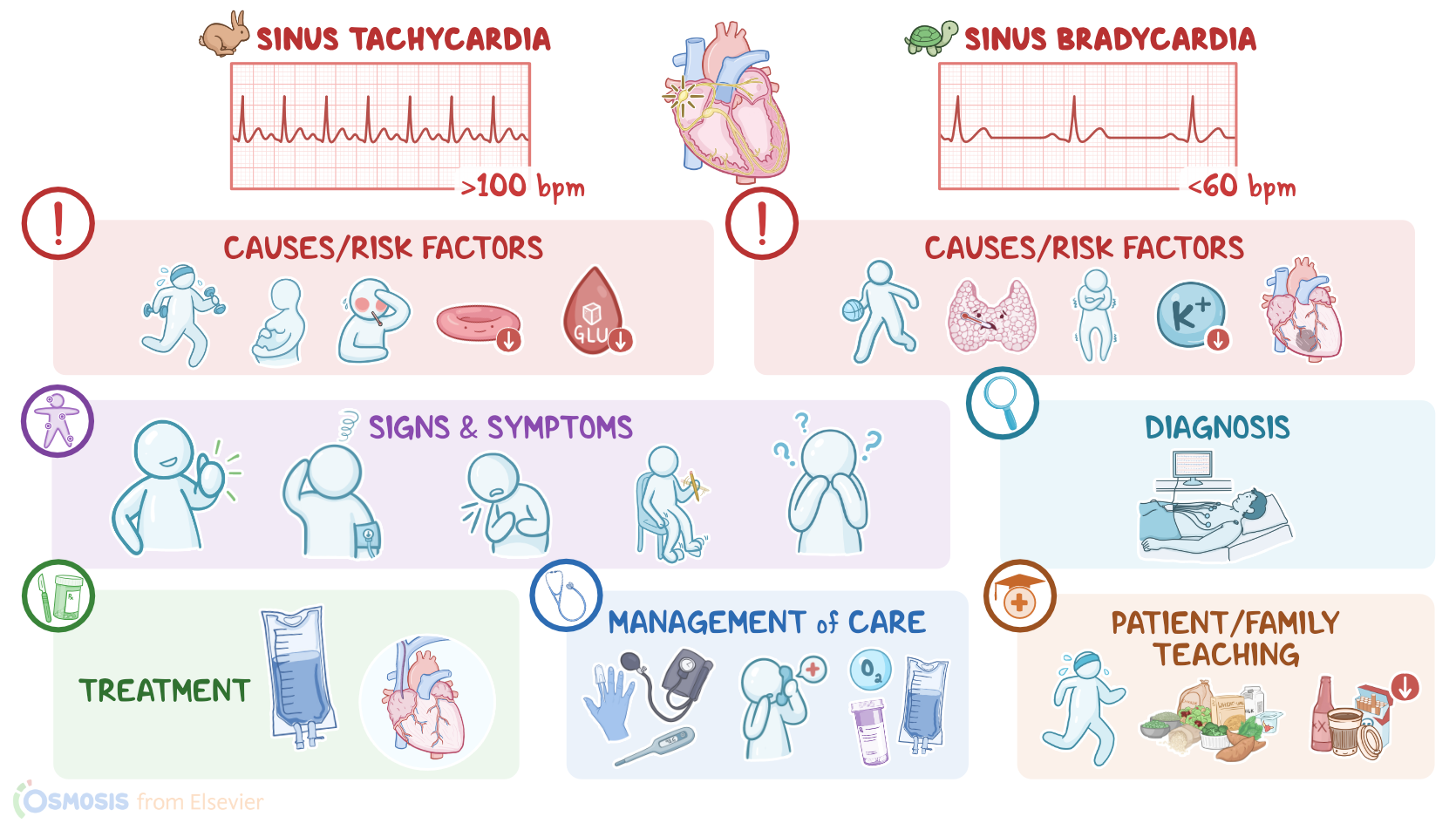
In sinus tachycardia and sinus bradycardia, the electrical impulse originates where it should, in the sinoatrial node, but the node fires either too fast or too slow. The cardiac rhythm is normal, meaning the beats are spaced evenly apart, but in sinus tachycardia the heart rate is more than 100 beats per minute, while in sinus bradycardia, it is less than 60 beats per minute.

Now, the cardiac conduction system is made up of specialized myocardial cells that can create and transport electrical potential, also called an action potential. These cells have many special features, including automaticity, meaning that they can generate an impulse; excitability, which is the ability to respond to a stimulus by creating an electrical impulse; conductivity meaning they can carry the impulse to other cells; and contractility, which is the ability to shorten the length of their fibers, causing a contraction.

Let’s look at the normal electrical conduction pathway in the heart on an ECG, which shows how the depolarization wave flows through the heart during each heartbeat.

The normal electrical activity of the heart starts in the sinoatrial node or SA node, which is considered the pacemaker of the heart.

Then, the impulse is conducted through the atria, causing depolarization and creating the P wave on an ECG. When the atrial muscle cells get depolarized, they contract, pushing blood from the atria into the ventricles.

From the atria, the impulse goes to the atrioventricular node, or AV node, where the impulse propagation speed slows way down. The interval from the atrial depolarization to just before ventricular depolarization is the PR interval on an ECG. This delay allows the atria to contract while the ventricles fill with blood.

From the AV node, the impulse goes through the Bundle of His, then to the right and left bundle branches, and finally through the Purkinje fibers, which deliver the impulse to the right and left ventricles, causing them to depolarize, and is represented by the QRS complex on an ECG. This triggers simultaneous contraction of both ventricles, pushing blood into the systemic and pulmonary circulations.

Finally, the ventricles repolarize to prepare for the next cycle, which allows them to relax and fill with blood, called diastole. And on ECG, ventricular repolarization will create a T wave, while the phase between ventricular depolarization and repolarization is represented by the ST segment. Sometimes, immediately after the T wave, there’s a U wave, which represents late repolarization of the ventricles.

Finally, to figure out the heart rate on an ECG, we can count the number of QRS complexes in six seconds and multiply that by 10. This normally lies between 60 and 100 beats per minute. The heart rate is mainly regulated by the autonomic nervous system. Specifically, the sympathetic nervous system increases the heart rate, while the parasympathetic nervous system decreases it. These two systems oppose each other’s actions so that a normal heart rate can be maintained under regular circumstances.

Now, let’s look at sinus tachycardia, which is typically caused by sympathetic nervous system stimulation, or parasympathetic nervous system inhibition. Sinus tachycardia can be normal during exercise and pregnancy and can also occur as a physiological response to fear or pain.

Pathological causes include fever, anemia, electrolyte abnormalities, as well as hypoglycemia, hyperthyroidism, hypotension, hypovolemia, and hypoxia.

In addition, caffeine, cocaine, and amphetamine, as well as various medications can also lead to sinus tachycardia. These include epinephrine, norepinephrine, atropine, theophylline, hydralazine, and some over the counter cold medications that contain pseudoephedrine.

Finally, withdrawal from alcohol or medications like benzodiazepines can also cause sinus tachycardia.

Okay, on the other side of the spectrum we have sinus bradycardia, which can be caused by parasympathetic nervous system stimulation, or sympathetic nervous system inhibition. Of note, sinus bradycardia, can be completely normal in athletes, and it’s also normal during sleep.

Pathological causes of sinus bradycardia include hypothyroidism, hypothermia, hypokalemia, and inferior wall myocardial infarctions, as well as increased intracranial pressure, which can be due to head trauma, strokes, and brain tumors. In addition, medications like beta blockers, calcium channel blockers, and opiates can cause sinus bradycardia.

Now, the pathology of sinus tachycardia starts with stimulation of the sympathetic system or inhibition of the parasympathetic system, which causes increased firing of impulses from the SA node. This increases the heart rate above 100 beats per minute, which initially increases cardiac output.

Over time, though, a fast heart rate decreases diastolic filling time, which, in turn, decreases the stroke volume or the volume of blood the left ventricle pumps with each heartbeat. At the same time, there’s an increased demand for oxygen by cardiac muscle cells. But since the perfusion of the myocardium by the coronary arteries occurs during diastole, sinus tachycardia also causes decreased myocardial perfusion.

On the other hand, the pathology of sinus bradycardia often starts with excessive parasympathetic stimulation, which causes decreased firing of impulses from the SA node. This leads to decreased heart rate, which increases diastolic filling time, which translates to increased coronary perfusion time. However, since the cardiac output is decreased, there’s also decreased coronary perfusion pressure, which once again decreases myocardial perfusion. In this case, though, there’s also a decreased demand for oxygen by cardiac muscle cells.

Complications linked to both sinus tachycardia and bradycardia include myocardial ischemia or infarction, heart failure, and cardiomyopathy.

Now, in terms of clinical manifestations, these can depend on the degree of tachycardia or bradycardia, and how well the client tolerates the altered heart rate, so some clients with these arrhythmias are asymptomatic. On the other hand, some can develop hypotension, dizziness, dyspnea, restlessness, confusion, palpitations, and even angina, or chest pain.

Sources

  1. "Critical care nursing: Diagnosis and management (10th ed.). ISBN: 978-0-443-11581-3 " Elsevier (2026)
  2. "Lewis’s Medical-Surgical Nursing: Assessment and Management of Clinical Problems. 12th Edition. ISBN:978-0-323-78961-5 " Elsevier
  3. "Guyton and Hall Textbook of Medical Physiology. ISBN 978-0323597128 " Elsevier (2020)
  4. "Mosby’s® Diagnostic and Laboratory Test Reference. 15th edition. ISBN: 978-0-323-67519-2 " Mosby (2020)
  5. "Saunders Comprehensive Review for the NCLEX-RN. 9th Edition. ISBN: 978-0-323-79530-2 " Saunders (2022)
  6. "Fundamentals of Nursing. 11th edition. ISBN: 978-0-323-81034-0 " Elsevier (2022)
  7. "Lewis’s Medical-Surgical Nursing: Assessment and Management of Clinical Problems. 11th Edition. ISBN: 978-0-323-55149-6 " Mosby (2019)
  8. "Continuous heart rate dynamics preceding in-hospital pulseless electrical activity or asystolic cardiac arrest of respiratory etiology. 179:1-8. " Resuscitation (2022)