Brachial plexus injury: Nursing
Introduction0:00–0:12
Brachial plexus injury describes damage to the brachial plexus, which is a network of nerves that supply motor and sensory innervation to the upper limbs.
Physiology0:12–3:00
First, let’s go over some anatomy and physiology. The brachial plexus is divided into roots, trunks, divisions, cords, and branches.
The roots are the ventral rami coming from spinal nerves C5 to T1. These roots combine to form the next part of the brachial plexus, called the superior or upper, middle, and inferior or lower trunks.
Then, these trunks give off divisions, which in turn recombine to form the lateral, posterior, and medial cords. Finally, these cords give off branches, which give rise to the musculocutaneous, axillary, radial, median, and ulnar nerves, which supply motor and sensory innervation to different areas of the upper limb.
The musculocutaneous nerve gives motor innervation to the muscles of the anterior arm, as well as sensory innervation to the lateral side of the forearm.
On the other hand, the axillary nerve carries motor information to the teres minor and deltoid muscles. It also carries sensory information from the shoulder joint, and the skin covering the inferior portion of the deltoid.
The radial nerve gives sensory and motor innervation to the posterior arm and forearm. In the hand, it only supplies sensory innervation on the dorsal side.
Here, it innervates the lateral hand, the thumb, and the proximal 2nd to 4th digits. Next, the median nerve provides motor innervation to most muscles of the anterior forearm.
In the hand, it innervates the thenar muscles, which act on the thumb, and the lateral lumbrical muscles of the other digits.
The median nerve also carries sensory information from the skin of the hand; on the palmar side, it innervates the lateral 3 ½ digits and the adjacent palm.
On the dorsal side, it innervates the distal aspects of the lateral 3 ½ digits. Finally, the ulnar nerve carries motor information to the flexor carpi ulnaris, and the ulnar half of flexor digitorum profundus in the forearm.
In the hand, it innervates most of the intrinsic muscles, except for a couple that are innervated by the median nerve. Just like the median and radial nerves, the ulnar nerve also carries sensory information from the skin of the hand; on the palmar side, it supplies the medial 1 ½ digits and the adjacent palm.
On the dorsal side, it also innervates the medial 1 ½ digits and the adjacent skin of the hand.Now, the most common cause of brachial plexus injury is birth trauma from traction on the arm when the infant is passing through the birth canal.
Causes & risk factors3:00–3:54
Other causes of brachial plexus injury include trauma like sport injuries, falls, surgical procedures, or stab wounds. Lastly, Pancoast tumors, which are tumors that develop at the lung’s apex, can compress the brachial plexus.
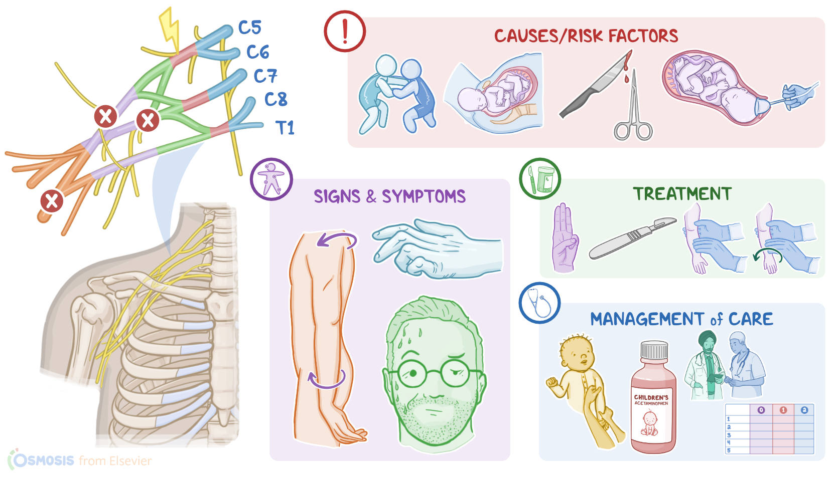
Risk factors for brachial plexus injury include large babies; prolonged labor; precipitous labor, which is labor that lasts less than three hours, resulting in a rapid delivery; and shoulder dystocia, which can happen when one of the baby’s shoulders get stuck during delivery; as well as the use of forceps or vacuum to extract the baby during delivery; and breech position.
Now, the pathology of brachial plexus injury starts with a stretch, tear or hemorrhage of one or more nerves arising from the brachial plexus.
Pathology3:54–5:02
Depending on the injured nerve and the location of the injury, the nerve won’t be able to transmit motor signals or receive sensory signals from the innervated areas, which causes impaired sensation or movement in the affected limb.
Sometimes, there can be paralysis of the affected limb, which is called palsy. The two main palsies linked to brachial plexus injury include Erb and Klumpke.
In Erb palsy, there’s an injury to the upper trunk of the brachial plexus, which is typically caused by an increase in the angle between the neck and the shoulder.
This causes loss of sensation in the upper arm, in addition to paralysis to the deltoid, biceps, and brachialis muscles.
On the other hand, Klumpke palsy happens with an injury to the lower trunk of the brachial plexus, which is typically caused by excessive upward pulling of the upper limb.
This causes paralysis of the intrinsic muscles of the hand. The clinical manifestations of brachial plexus injury typically include impaired sensation, movement, or strength of the affected upper limb, or, in some cases, palsy.
Clinical manifestations5:02–6:52
With Erb palsy, the main clinical finding is the so-called waiter’s tip sign, in which there’s internal rotation at the shoulder joint, adduction of the upper limb, extension and pronation of the forearm, in addition to flexion of the wrist and extension of the fingers.
On the other hand, Klumpke palsy causes the claw hand sign, in which there’s supination of the forearm, extension of the wrist, and flexion of the fingers.
Klumpke palsy can also be associated with Horner syndrome, which affects the sympathetic nerves of the T1 nerve root. Clients with Horner’s syndrome typically present with unilateral drooping eyelids, called ptosis; smaller pupils, called miosis; and decreased sweat production in the face, called anhidrosis.
Finally, both Erb and Klumpke palsies can cause loss of reflexes, including the Moro reflex in newborns, as well as the biceps, and brachioradialis reflexes in both newborns and adults.
Now, clients with brachial plexus injury can experience pain, stiff joints, numbness, muscle atrophy, and permanent disability.
In addition, some clients may present with injuries to nearby structures, such as fractures of the clavicle or humerus, dislocations of the shoulder joint, and even spinal cord injuries.
The diagnosis of brachial plexus injury starts with the client’s history and physical assessment. This can include assessment of reflexes, which are usually absent in those with injury.
Diagnosis6:52–7:13
Additional diagnostic tests include X-rays, CT scan, and MRI, which can be used to observe the brachial plexus and the extent of injury.
Treatment7:13–8:00
The treatment of brachial plexus injury typically involves monitoring, in addition to physical therapy that includes daily range of motion exercises of the shoulder, arms, wrist, and fingers.
More severe injuries may require surgical procedures, such as nerve surgery; osteotomy, which is most commonly done with humerus fractures, and it involves cutting and reshaping the bones; capsulorrhaphy, which involves repairing and tightening the shoulder capsule; tendon transfers, where the muscle is left in place, but it’s tendon insertion is detached and sewn into a different bone or tendon to restore its function; and finally, free functional muscle transfers, where a muscle from the client’s inner thigh is transplanted to the injured area.
Management of care8:00–9:09
Alright, let’s look at the nursing care you’ll be providing for a newborn with a brachial plexus injury. Your priority goals are to prevent further injury to the affected arm, manage pain, and maintain joint mobility.Begin by gently stabilizing the affected extremity and preventing any unnecessary movement.
When the infant needs to be repositioned or held, protect the extremity from stretching or dangling free. Next, assess the infant’s pain using an appropriate neonatal pain assessment scale, and provide comfort measures along with non-nutritive sucking with 24% sucrose solution, or the prescribed acetaminophen, as needed to manage the infant’s pain.Then, after swelling of the soft tissue has resolved, collaborate with the physical therapist to provide passive range of motion exercises to maintain mobility and prevent joint contractures.
Lastly, arrange for follow-up appointments after discharge for continued therapy.Okay, moving on to the education you’ll provide for the baby’s parents or caregivers.
General client & family teaching9:09–10:59
Begin by explaining how a brachial plexus injury involves damage to the nerves that supply their baby’s upper limb, and that this can cause a lack of muscle control and sensation in the arm, hand, wrist, and fingers.Then, provide them with a schedule of appointments with the physical therapist, pediatrician, and pediatric neurologist, and stress the importance of bringing their baby to each appointment for continued therapy, monitoring, and support.Next, teach them how to care for their baby’s injured arm.
Demonstrate how to position their baby for sleep and for rides in their car seat to prevent injury to the arm. Also caution them to avoid pulling on their baby’s arms or lifting them up under the arms; instead, they should pick their baby up by the baby’s trunk.
Show them how to perform each of the exercises, including shoulder rotation; flexion and extension of the elbow, wrist, and each of the fingers; and thumb abduction, adduction, and opposition, and explain how the exercises can help improve function of the arm and prevent formation of joint contractures.
Suggest they perform one set of these exercises at each diaper change to ensure the exercises are done several times each day.
Provide an opportunity for them to practice each of the physical therapy exercises, and ensure they demonstrate confidence in providing therapy to their baby.
Lastly, let them know that recovery can take several months, and provide reassurance that most babies will have a return of function with continued therapy.
Review10:59–13:07
Alright, as a quick recap… A brachial plexus injury involves damage to the brachial plexus, which is a network of nerves that supply motor and sensory innervation to the upper limbs.
Brachial plexus injuries most commonly occur due to birth trauma, resulting in a tear, stretch, or hemorrhage of one of more nerves.
In adults, causes of brachial plexus injury include trauma like sport injuries, falls, surgical procedures, or stab wounds.
The clinical manifestations of brachial plexus injury typically include impaired sensation, movement, or strength of the affected upper limb, or, in some cases, palsy.
With Erb palsy, the main clinical finding is the waiter's tip sign; while with Klumpke palsy, there’s the claw hand sign.
Additional findings can include stiff joints, pain, numbness, muscle atrophy, and permanent disability.Brachial plexus injuries are diagnosed by physical examination and imaging studies.
Non-surgical treatments include regular monitoring, in addition to physical therapy with frequent daily range of motion exercises of the shoulder, arms, wrist and fingers.
If no significant improvement occurs with physical therapy, surgical interventions can be done. Goals of nursing care include preventing further injury to the affected arm, managing pain, and maintaining joint mobility.
Education is focused on understanding the disorder, safe positioning, and learning about how to confidently perform range
| BRACHIAL PLEXUS INJURY | ||
| KEY POINTS | NOTES | |
| DEFINITION |
| |
| PHYSIOLOGY |
| |
| CAUSES AND RISK FACTORS |
| |
| PATHOPHYSIOLOGY |
| |
| SIGNS AND SYMPTOMS |
| |
| DIAGNOSIS |
| |
| TREATMENT |
| |
| MANAGEMENT OF CARE |
| |
| PATIENT AND FAMILY TEACHING |
| |
No notes for this video yet
Try adding a note below