Cardiomyopathy: Nursing
Introduction0:00–0:18
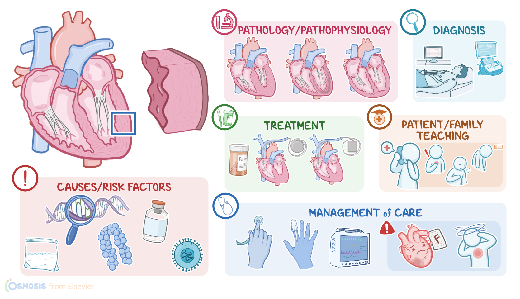
Cardiomyopathies are a group of diseases of the myocardium, which is the muscle layer of the heart wall. The three main types are dilated cardiomyopathy, which is the most common one; as well as hypertrophic, and restrictive cardiomyopathy.
Now, let’s quickly review some anatomy and physiology. The heart wall is made of three layers: the outer layer is the epicardium; the middle and thickest layer is the myocardium, which contracts in a rhythmic way to pump blood; and the inner layer is the endocardium.
Physiology0:18–1:15
These layers line the four heart chambers, so the two atria and two ventricles, which are separated by the heart valves at the end of each chamber.
First, there are two atrioventricular valves, the mitral or bicuspid valve on the left, and the tricuspid valve on the right.
The atrioventricular valves prevent blood from returning to the atria after filling the ventricles. And second, there are two semilunar valves called the aortic valve at the left, and the pulmonary valve at the right.
Causes & risk factors1:15–2:57
The semilunar valves prevent blood from returning to the ventricles after being pumped out. Okay, when it comes to the causes of dilated cardiomyopathy, there are many potential causes, which include genetic mutations; cardiotoxic agents, such as alcohol, cocaine, lead, as well as chemotherapeutic medications like doxorubicin and trastuzumab.
Dilated cardiomyopathy can also be caused by infections such as human herpesvirus 6, cytomegalovirus, or Lyme disease; nutritional deficiencies, like lack of thiamine; and metabolic disorders, like hyperthyroidism.
Sometimes, dilated cardiomyopathy can also be related to pregnancy, called peripartum cardiomyopathy.Next up is hypertrophic cardiomyopathy, which is most commonly caused by mutations in genes coding for components of the cardiac muscle cells.
These mutations are typically inherited in an autosomal dominant fashion. On a side note, it’s important to distinguish hypertrophic cardiomyopathy from “athlete’s heart”, which refers to benign left ventricular hypertrophy in professional athletes.
Lastly, restrictive cardiomyopathy can be idiopathic, or it can be due to toxic; genetic; infiltrative or inflammatory causes.
Some conditions that can cause restrictive cardiomyopathy include amyloidosis or hemochromatosis, where misfolded amyloid proteins, or excess iron, respectively, end up depositing in the myocardium.
Pathology2:57–5:45
Other causes include sarcoidosis, as well as medications like hydroxychloroquine, and radiation therapy.Now, let’s look at the pathology of each type of cardiomyopathy.
With dilated cardiomyopathy, there’s an enlarged left ventricle chamber. As a result, the dilated heart muscle becomes really weak, so its ability to contract and pump blood becomes significantly impaired.
On the flip side, in hypertrophic cardiomyopathy, the heart wall gets thicker, stiffer, and less compliant, which reduces the chamber size and ability to stretch.
This most frequently affects the left ventricle, impairing the heart’s ability to fill up with blood, so less blood is pumped out with each contraction.
Additionally, the interventricular septum is also often thickened, which can obstruct blood flow from the left ventricle into the aorta, causing obstructive hypertrophic cardiomyopathy.
Additionally, the mitral valve, found between the left atrium and the left ventricle, also moves abnormally, meaning that during ventricular contraction, it moves slightly forward.
This further impairs adequate blood flow through the left ventricle. Finally, with restrictive cardiomyopathy, the heart muscle becomes stiffer and less compliant.
Now, the heart wall and chambers generally stay about the same size or get slightly enlarged. However, when blood fills into restricted ventricles, they can’t expand, so this again impairs the heart’s ability to fill up with blood and less blood is pumped out with each contraction.Regardless of the type of cardiomyopathy, clients with a poorly functioning myocardium are more prone to develop several complications.
First off, blood that’s not efficiently pumped out becomes stagnant in the heart, which can increase the risk of clot formation.
This clot can then dislodge, and travel into the systemic circulation, and potentially to the brain, cutting off blood supply, and causing a stroke.
The abnormal myocardium may also impair the function of the heart valves, ultimately leading to valvular heart disease. Over time, as cardiomyopathy progresses, the client may develop heart failure, where the heart becomes unable to pump enough blood to meet the body’s requirements.
Lastly, because cardiomyopathies affect the cardiac muscle as well as the pacemaker cells that run through the cardiac muscle, they can cause arrhythmias like atrial fibrillation, and even cardiac arrest and sudden cardiac death.
Clinical manifestations5:45–7:16
In fact, hypertrophic obstructive cardiomyopathy is the most common cause of sudden cardiac death in clients less than 35 years old, and is especially more common in athletes.Alright, now, regarding clinical manifestations, in all three types of cardiomyopathy, clients are typically asymptomatic at first.
Over time, as heart failure develops, clients may experience fatigue, nausea, vomiting, and anorexia, as well as signs of left sided heart failure, like dyspnea, particularly when lying flat; fatigue and exercise intolerance; and signs of right sided heart failure, like jugular venous distention, swelling of the feet, abdominal bloating, ascites, and hepatomegaly.
And because cardiomyopathies can lead to arrhythmias, clients may experience palpitations. Clients with hypertrophic obstructive cardiomyopathy can also present with syncope, which is a transient loss of consciousness; and sometimes, the presentation of hypertrophic cardiomyopathy can be sudden cardiac death.
Diagnosis7:16–8:06
Now, during auscultation of the heart, dilated and restrictive cardiomyopathies cause an S3 heart sound, which is the result of blood rushing and slamming into the dilated ventricular wall during diastole.
On the other hand, hypertrophic cardiomyopathy produces an S4 sound in the apex of the heart. This sound indicates that the atrium is contracting against a thickened left ventricle.
If there’s also valvular heart disease, heart murmurs might also be found.The diagnosis of cardiomyopathy starts with the client's history and physical assessment, followed by an echocardiogram, which makes it possible to measure the thickness of the heart wall and dimensions of the heart chambers, as well as cardiac output, meaning how much blood the heart pumps out with each stroke.
Treatment8:06–9:31
An ECG should be also performed to look for arrhythmias. In some cases, an exercise stress test might be done, to assess the heart’s ability to perform under stress.
Finally, additional diagnostic tests can be also helpful, including a chest X-ray, cardiac CT and MRI scans, to visualize the heart’s shape and size, genetic testing, to look for associated gene mutations, as well as cardiac catheterization, which is used to evaluate the extent of the disease.
The goal of treatment in cardiomyopathies is aimed at symptom relief and ensuring that the heart continues to function. This involves treating the associated heart failure, and arrhythmias with medications like beta blockers and ACE inhibitors, as well as vasodilators, such as dobutamine, and milrinone, which help decrease the heart’s workload, and diuretics to reduce fluid overload.
Anticoagulants can also be administered to prevent thrombus formation. Additionally, clients prone to arrhythmias need an implanted pacemaker or a cardioverter-defibrillator device or ICD.
Management and care9:31–11:48
Some clients also need a left ventricular assist device or an LVAD, which is a mechanical pump that assists the heart in distributing blood.
In extreme cases, a client with cardiomyopathy might require a heart transplant.Now, some clients may require additional treatment, which depends on the type of cardiomyopathy.
So, those with hypertrophic cardiomyopathy might require procedures to reduce the wall thickness, such as alcohol septal ablation, or ventriculomyotomy, and septal myectomy, where a part of the thickened septal wall is removed.
For clients with restrictive cardiomyopathy, cessation of high-intensity exercise is recommended to avoid sudden cardiac death.
Alright, now let’s look at the nursing care you’ll provide for a client with hypertrophic cardiomyopathy and heart failure.
Your priority goals of care are focused on improving cardiac output and managing symptoms. Begin by assessing your client’s baseline weight, vital signs, and lung and heart sounds.
Institute continuous pulse oximetry, and keep the SpO2 above 95%; and cardiac monitoring, making note of their current rhythm.
Report signs and symptoms of worsening heart failure, including chest pain, pulmonary crackles, dyspnea, decreased SpO2, or tachypnea.
Provide high flow supplemental oxygen and place your client in a high Fowler position. Then administer the prescribed medications to slow the heart rate, increase ventricular filling, and cardiac output; as well as medications to promote diuresis and decrease cardiac workload.
General client and family teaching11:48–16:43
Prepare to assist with catheter placement for monitoring of pulmonary and arterial pressures.Now, if your client is at high risk for sudden cardiac death, and is prescribed an ICD, prepare them for the procedure by administering the prescribed prophylactic IV antibiotics, and sedatives to decrease anxiety.
Then, following placement of an ICD, provide routine post-operative care. Closely monitor the implantation site, and immediately report if you note signs of bleeding, hematoma formation, or wound dehiscence.
Institute continuous cardiac monitoring, and closely monitor them for arrhythmias, hemodynamic instability, and device malfunction.
Immediately report if your client experiences dizziness, syncope, or palpitations, or if there’s an inappropriate delivery of a shock.Finally, collaborate with the case manager to coordinate your client’s care after discharge.
If they are being discharged with an ICD, be sure you provide them with a medical device ID card that names the type of device, model and serial number, and the date the ICD was implanted.Alright, moving on to client and family teaching.
Begin by explaining that hypertrophic cardiomyopathy is a disease where the muscle layer of the heart wall gets thicker and stiffer, which makes it hard for the heart to fill up with blood and pump it out to the body; and explain how the diseased heart muscle interferes with the normal rhythm of the heart, resulting in abnormal and potentially life-threatening heart rhythms.
Regarding their prescribed medications, review how and when to take each of their prescribed medications, how each medication works, and the importance of adhering to their medication regimen.
Lastly, remind them of the importance of maintaining their regular appointments with their healthcare provider for continued management and support.
Next, review lifestyle modifications to help decrease symptoms of heart failure, including maintaining a healthy weight; limiting dietary sodium and eating a low fat, high fiber diet; engaging in physical activity as tolerated; limiting alcohol; and avoiding nicotine and tobacco smoke.
Provide smoking cessation counseling and referrals, as needed. Then, instruct them to call their healthcare provider for worsening symptoms of heart failure, including shortness of breath, chest pain, or increased fatigue.
Now, if your client has an ICD, explain that their ICD has been implanted in a pocket beneath their skin and that the device's leads are threaded down to their heart.
Help them to understand that the ICD is an important part of their treatment, but it does not cure their heart failure. Then, teach them about important postoperative activity limitations to prevent dislodging the ICD leads.
For the first several weeks after implantation, instruct them to avoid lifting the arm on the ICD’s side of the body above the shoulder or extending backwards.
Teach them that they should avoid excessive pushing, pulling, or twisting; and they should not lift anything weighing more than 10 pounds.
Also instruct them to keep the area around your insertion site dry for the next 7-10 days. Let them know they should not take a shower during this time, and that they should keep the insertion site completely dry while bathing.
Reassure them that mild discomfort at the insertion site is normal, and that their healthcare provider has recommended treating any pain with acetaminophen.
Lastly, emphasize the importance of checking the insertion site each day, and to call their healthcare provider right away if their pain can’t be controlled with acetaminophen; if there’s redness, tenderness, warmth around the incision; if there is pus or other fluid seeping from the incision; or if they develop a fever or chills.Next, explain that the ICD device will deliver a shock if it detects a life threatening heart rhythm, and that the shock may startle them the first time it happens.
Let them know that some clients with an ICD only feel a mild sensation, while others describe the shock like a kick in the chest.
Remind them that after a shock occurs, they should sit down and take a few minutes to recover; and as a safety measure, they should not drive for a period of six months afterwards, since the abnormal rhythm may cause them to lose consciousness.
Review16:43–18:37
Stress the importance of contacting their healthcare provider immediately whenever a shock does occur, especially if they experience repeated shocks, if they lose consciousness, or if they have symptoms like chest pain or shortness of breath after a shock.
Lastly, instruct them to immediately contact their healthcare provider for signs that their ICD needs to be checked, like if their device emits a beeping sound.Finally, review some important points about day-to-day living with an ICD.
Teach them that strong electromagnetic fields, like those from heavy industrial equipment, can interfere with the ICD’s functioning, but that regular household equipment like microwave ovens and most power tools will not affect the ICD.
Let them know that using a cell phone is also safe, but that they should always keep the phone on the opposite side of their body than their ICD,
| CARDIOMYOPATHY | ||
| KEY POINTS | NOTES | |
| DEFINITION |
| |
| PHYSIOLOGY |
| |
| CAUSES AND RISK FACTORS |
| |
| PATHOPHYSIOLOGY |
| |
| SIGNS AND SYMPTOMS |
| |
| DIAGNOSIS |
| |
| TREATMENT |
| |
| MANAGEMENT OF CARE |
| |
| PATIENT AND FAMILY TEACHING |
| |
No notes for this video yet
Try adding a note below