Esophageal atresia and tracheoesophageal fistula: Nursing
Introduction0:00–0:22
Esophageal atresia and tracheoesophageal fistula are congenital gastrointestinal anomalies where the esophagus and trachea don’t separate normally during development.
Physiology0:22–0:54
They both form from the primitive foregut between weeks 4 and 6 of intrauterine development. Afterwards, they are separated by the tracheoesophageal septum, which divides the foregut into the laryngotracheal tube at the anterior side and the esophagus at the posterior side.
Eventually, the laryngotracheal tube develops into the larynx and the trachea, while the esophagus elongates and connects to the stomach.Now, the exact cause of both esophageal atresia and tracheoesophageal fistula isn’t known, but it has been suggested that environmental factors, along with genetic factors play an important role in their development.However, there are some risk factors associated with both these conditions.
Causes & risk factors0:54–1:49
They include prenatal factors, such as maternal use of alcohol and smoking, uncontrolled diabetes mellitus, increased maternal age or exposure to drugs like methimazole and diethylstilbestrol.
Risk factors unrelated to maternal ones include chromosomal anomalies, like trisomy 13, 18 or 21 and VACTERL syndrome, which is associated with spinal, anal, heart, tracheoesophageal fistulas, kidneys and limb anomalies.Now let’s switch gears and look at the pathology of these conditions.
Pathology1:49–3:38
With esophageal atresia, the esophagus fails to elongate and connect to the stomach around week 4 of intrauterine development.
So essentially, there’s a proximal esophagus that ends in a blind pouch; and a distal esophagus, which ends right above the diaphragm.
This is commonly associated with a tracheoesophageal fistula, which is a connection between the trachea and either the proximal, the distal or both parts of the esophagus.
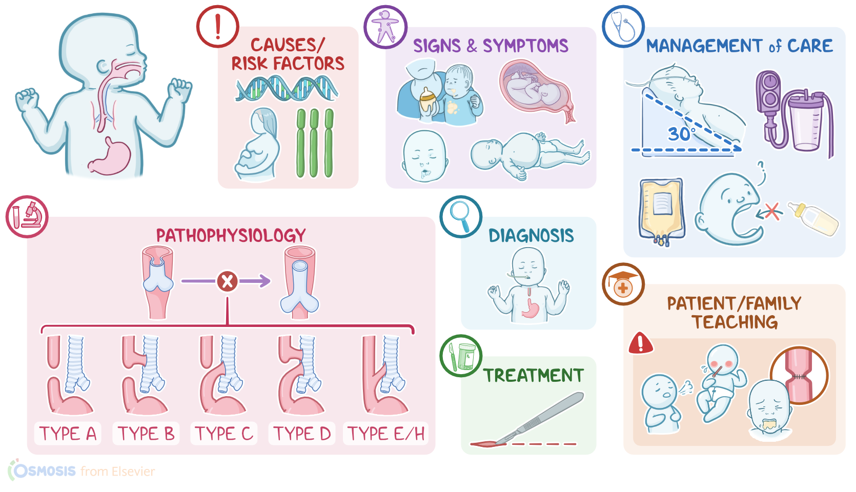
Based on the location and extent of the defect, these two conditions are classified into 5 types. Type A is called isolated esophageal atresia and happens when the middle part of the esophagus is missing.
In type B, the proximal esophagus communicates with the trachea. In type C, which is the most common type, there’s proximal esophagus atresia, but this time the distal esophagus has a communication with the trachea.
Finally, with type E, also referred to as type H, there’s a tracheoesophageal fistula, but no esophageal atresia.Let’s move on and talk about complications.
These include aspiration pneumonia and lung abscesses or bronchiectasis due to recurrent aspiration.Postoperatively, complications include leaking at the site of anastomosis, strictures, or the formation or return of preexisting tracheoesophageal fistula.
Other complications include impaired esophageal motility and resulting gastroesophageal reflux, which increases the risk of recurrent pneumonia and other respiratory problems.
Okay, the most frequent clinical manifestations of esophageal atresia in the prenatal period are polyhydramnios, which happens because the fetus does not swallow amniotic fluid normally; along with the absence of stomach gas on prenatal abdominal echography.
Clinical manifestations3:38–4:55
Postnatal symptoms and signs include frothy saliva in the mouth and nose and excessive drooling, inability to swallow feedings, vomiting, abdominal distention, and in severe cases, respiratory distress.
Keep in mind the three Cs for esophageal atresia: coughing, choking during feeding, and cyanosis. With tracheoesophageal fistula, when the proximal esophagus is connected to the trachea, the above symptoms remain, but there’s a higher chance of developing aspiration pneumonia when content from the esophagus reaches the lungs.
This is associated with respiratory symptoms, like respiratory distress, along with systemic symptoms like malaise and fever.
When the distal esophagus is connected to the trachea, GI symptoms like gastric distention appear, due to the fact that air reaches the stomach through the fistula.
Diagnosis of both conditions start with a full history and clinical exam. Clinically, a nasogastric or an orogastric tube wouldn’t be able to pass all the way from the mouth to the stomach.
Diagnosis4:55–5:40
Imaging exams include abdominal X-rays, where you can see a large gastric bubble in types C, D, and E, but not in type A and B, since in these two there isn’t a communication between the distal esophagus and trachea.
Chest X-rays with a water–soluble contrast solution injected through the orogastric tube shows the anomalies and gives the final diagnosis.
Additionally, anomalies that include the VACTERL syndrome should be assessed. Treatment-wise, surgery is the only effective means to correct these conditions; and in otherwise healthy, term newborns, immediate repair is indicated.
Treatment5:40–6:03
Other options include delayed repair and staged repair, and these can be considered in infants with low or very low birth weight, moderate or severe pneumonia, or other severe comorbidities or defects.
Management of care6:03–7:51
Alright, now let’s talk about nursing management for an infant with esophageal atresia and tracheoesophageal fistula. Your priority goals of care are to prevent complications and provide emotional support.Preoperatively, institute measures to decrease the risk of aspiration.
Initiate NPO status and position the infant’s head at least 30 degrees. Then, place an orogastric tube in the blind esophageal pouch as ordered, ensuring that it is patent and attached to low continuous suction.
Be sure to have suction equipment readily available, and suction secretions as needed.Then, administer the prescribed antireflux medications to decrease the risk of acid-induced pneumonitis.
Also be sure to promote comfort measures to reduce crying, which will reduce the amount of swallowed air and abdominal distention.Postoperatively, provide routine post-operative care and monitor the infant closely for complications related to the procedure.
Regularly assess and intervene for pain, administer the prescribed IV antibiotics, and provide parenteral nutrition.If suctioning is needed, remember that suction catheters should not come in contact with the newly repaired esophagus and trachea, as this can cause an anastomosis leak or even a recurrent fistula.Finally, support the infant’s caregivers by providing emotional and psychological support.
Encourage them to participate in care; and provide clear, concise answers to their questions and concerns.Alright, let's move on to client and family teaching.
General client & family teaching7:51–9:33
Begin by explaining that an esophageal atresia and tracheoesophageal fistula happen when the esophagus and trachea don’t form normally during development, preventing their baby from eating or drinking, and increasing the risk that secretions can get into the lungs, causing damage.
Let them know that after surgery, when the surgical site has healed completely, their baby can start receiving feedings by mouth.Next, teach them how to care for their baby after they are discharged home.
Teach them about each of their prescribed medications, such as antacids and antireflux medications, and teach them how and when to administer the medications to their baby.
Also let them know that sometimes a narrowing of the esophagus, called a stricture, can develop after surgery, so underscore the importance of notifying their healthcare provider immediately if signs and symptoms of a constricted esophagus develop, such as trouble swallowing, coughing during feedings, or spitting up undigested foods; as well as signs of aspiration such as respiratory distress and fever.Also teach them how to take care of the surgical site.
Remind them of the importance of always practicing good hand hygiene, especially when caring for the site. Show them how to cleanse the incision with a clean, damp cloth, and then cover it with a clean dressing.
Instruct them to notify the healthcare provider promptly for signs of a surgical site infection such as increasing redness, swelling, pain or tenderness at the site; if there’s discharge from the site, or if their baby develops a fever.Alright, as a quick recap….
Review9:33–10:45
Esophageal atresia and tracheoesophageal fistula are congenital gastrointestinal anomalies where the esophagus and trachea don’t separate normally during development, and both conditions often occur together.
With esophageal atresia, the esophagus fails to elongate and connect to the stomach; and tracheoesophageal fistula is a connection between the trachea and esophagus.Risk factors can be genetic or environmental.
Clinical manifestations of esophageal atresia in the prenatal period are polyhydramnios and the absence of stomach gas. Postnatal manifestations include frothy saliva, excessive drooling, inability to swallow feedings, and vomiting.
The major complication is aspiration pneumonia.Diagnosis is through history, physical examination, and imaging. Treatment is by surgical correction.
Goals of nursing care are to prevent complications and provide emotional support. Client and family education is centered on home care and when to contact their healthcare provider.
| ESOPHAGEAL ATRESIA AND TRACHEOESOPHAGEAL FISTULA | ||
| KEY POINTS | NOTES | |
| DEFINITION |
| |
| PHYSIOLOGY |
| |
| CAUSES AND RISK FACTORS |
| |
| PATHOPHYSIOLOGY |
| |
| SIGNS AND SYMPTOMS |
| |
| DIAGNOSIS |
| |
| TREATMENT |
| |
| MANAGEMENT OF CARE |
| |
| PATIENT AND FAMILY TEACHING |
| |
- "Foundations of Maternal-Newborn and Women's Health Nursing" Elsevier (2019)
- "Essentials of Pediatric Nursing" LWW (2021)
- "Saunders comprehensive review for the NCLEX-RN examination" Elsevier (2017)
- "Merenstein & Gardner’s handbook of neonatal intensive care: An interprofessional approach" Elsevier (2021)
- " First Aid for the USMLE Step 1 2021" McGraw-Hill Education (2021)
- "Larsen's human embryology" Elsevier (2015)
- "Fetal and Neonatal Physiology (5th ed.)" Elsevier (2017)
- "Tracheoesophageal Fistula" NeoReviews (2021)
- "Esophageal Atresia" Pediatric Surgery Handbook for Residents and Medical Students (2021)
No notes for this video yet
Try adding a note below