Gastric cancer: Nursing
Introduction0:00–0:11
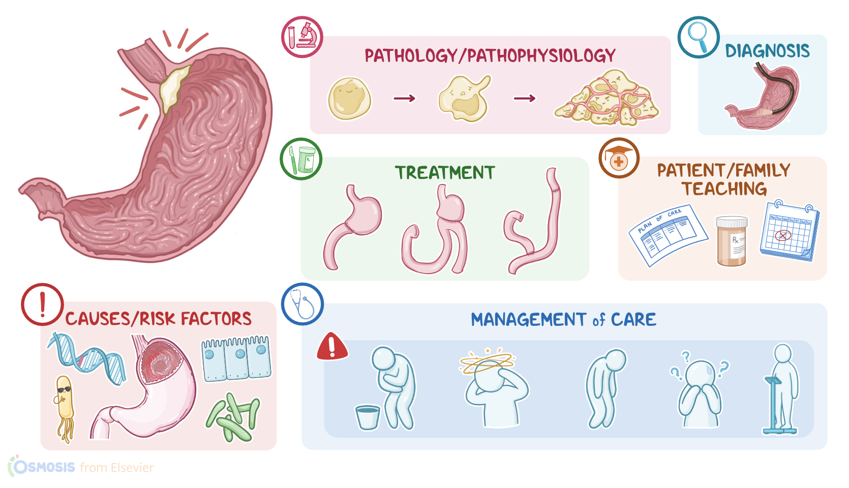
Gastric cancer is a malignant tumor that originates in the stomach, and is one of the most common types of cancer. First, let’s recall the physiology of the stomach.
Physiology0:11–1:39
The stomach is a hollow, J-shaped organ of the digestive system that stores food, mixes it with gastric juices and empties it into the small intestine.
The outermost layer is called serosa or adventitia, and it faces the abdominal or peritoneal cavity. This is the space between the abdominal wall and abdominal organs that is lined with peritoneal membrane and contains a small amount of serous fluid.
Next layer is muscularis, which contracts to stir the food and move it further into the guts. After that is the submucosa, which consists of a dense layer of tissue that contains blood vessels, lymphatics, and nerves.
And finally, there’s the innermost layer, called the mucosa, which consists of a simple columnar epithelium that forms many invaginations, called gastric pits, which connect to gastric glands.
The gastric glands are composed of cells that produce gastric acid, which helps break down food and inactivate ingested bacteria; mucus, which protects the mucosa from the gastric acid; enzymes, which break down proteins; and intrinsic factor, which is a protein necessary for vitamin B12 absorption.Now, the exact cause of gastric cancer is unknown, but there is usually a genetic mutation in a cell of the gastric mucosa.
Causes & risk factors1:39–3:30
These include mutations in a tumor suppressor gene, which results in loss of inhibition of cell division, or a proto-oncogene, which stimulates cell division.
And these mutations can be hereditary, meaning that the client inherits the mutation from one of their parents, or non-hereditary, also known as sporadic, which occur de novo or spontaneously.
Whatever the cause is, the chance of developing gastric cancer increases with certain risk factors. Modifiable risk factors include Helicobacter pylori infection, gastric ulcers, gastroesophageal reflux disease and achlorhydria, which refers to low or absent HCl production by the stomach leading to a more alkaline environment that is suitable for bacterial growth.
Chronic inflammation and atrophic gastritis can lead to intestinal metaplasia, where the epithelial cells type is changed, which is considered a precancerous condition.
Other risk factors include diet high in salted, smoked and processed foods, low vegetables and fruits intake, obesity, contaminated drinking water, exposure to irritants such as smoking and alcohol, and prolonged use of NSAIDs, since they block production of prostaglandins which normally stimulate gastric mucus secretion.
On the other hand, non-modifiable risk factors include age above 40, being assigned male at birth, family history of gastric cancer, previous gastric surgery, radiation exposure, gastric polyps, Epstein-Barr virus infection and lymphoma, such as mucosa associated lymphoid tissue, or MALT lymphoma.Okay, now let’s look at the pathology of gastric cancer.
Pathology3:30–4:33
The most common type of gastric cancer is adenocarcinoma, which occurs when an epithelial cell becomes mutated and cancerous, and starts dividing uncontrollably, forming a tumor mass.
As the tumor keeps growing, new blood vessels also develop via angiogenesis to supply it. Eventually, cancerous cells start invading neighboring tissues, such as the pancreas and transverse colon, and may even spread to nearby or even distal lymph nodes, such as left supraclavicular, or Virchow’s nodes, left axillary, or Irish nodes, and periumbilical, or Sister Mary Joseph nodes.
Gastric cancer can also metastasize to distant organs, such as the liver via portal circulation, and the lungs and bones via systemic circulation.
Finally, it can form seedlings that metastasize to the peritoneum, and even involve both the ovaries, causing a particular type of tumor, called Krukenberg’s tumor.So, the clinical manifestations of gastric cancer vary based on the size and location of the tumor.
Clinical manifestations4:33–5:45
Initially, clients can be completely asymptomatic. Over time, the body mounts an immune response to fight the tumor off, so clients can experience unintentional weight loss, fever, and malaise.
At an early stage, clients might also experience dyspepsia, or indigestion, abdominal discomfort and early satiety. If the tumor grows enough to physically obstruct the stomach, it can cause narrowing of the lumen; dysphagia; nausea; vomiting; hematemesis, or vomiting of blood; or melena where the discolored blood can also appear in the stool, making it black.
Blood loss and impaired gastric function can also cause iron deficiency anemia. In some cases, a palpable epigastric mass may also be present.
Clients can also experience epigastric pain, retrosternal pain, or back pain due to the compression of nearby nerves. Finally, with peritoneal metastases, clients can develop ascites, which is a collection of free fluid in the abdominal cavity.Diagnosis of gastric cancer starts with the client’s history and physical assessment, followed by esophagogastroduodenoscopy with biopsy.
Diagnosis5:45–6:38
Laboratory test results are generally non-specific and may show low hematocrit and hemoglobin levels, as well as elevated blood levels of tumor markers like CEA, and CA 19-9.
Hypoalbuminemia, increased bilirubin and liver enzymes can also be found if there’s liver metastasis, while increased amylase and lipase levels suggest pancreatic involvement.
Once diagnosis has been confirmed, imaging tests like abdominal or pelvic CT scan, MRI, and positron emission tomography, or PET can be used to stage the tumor with the TNM classification, by defining the Tumor location and looking for lymph Node involvement or Metastasis.
Treatment6:38–9:50
Treatment for gastric cancer depends on its aggressiveness and extension. Small, localized tumors can be treated with partial gastrectomy, which is when the affected part of the stomach is surgically removed.
The remaining healthy part of the stomach can be sutured to the duodenum, which is also known as the Billroth I procedure or gastroduodenostomy, or it can be sutured to the jejunum, which is known as the Billroth II procedure or gastrojejunostomy.
On the other hand, for clients with larger tumors, the treatment of choice is total gastrectomy where the whole stomach is removed, while the esophagus is sutured to the jejunum, which is also known as esophagojejunostomy.
Also, any involved nearby lymph nodes should be resected. Metastasectomy or removal of metastatic cancerous tissues in other organs can also be performed.
In addition, many clients may require a gastrostomy, where the stomach contents are diverted into an artificial opening in the abdominal wall called a stoma, and ultimately eliminated into a pouch or bag.
Now, a complication of gastrectomy is dumping syndrome, where impaired gastric motility leads to rapid gastric emptying.
There is early dumping syndrome, where faster emptying of the stomach contents into the bowel draws water into the bowel lumen, leading to bowel distention, and increased intestinal motility.
In contrast, in late dumping syndrome, there is faster emptying of stomach contents into the bowel, increasing the carbohydrate content in the bowel lumen, which leads to rapid glucose absorption, and later on, an insulin surge, followed by hypoglycemia.
In order to treat the dumping syndrome, clients are advised to eat a diet high in protein and fat, and low in carbohydrates, have smaller portions of food and avoid ingesting liquids with meals.
Medications can be also used, including acarbose to decrease carbohydrate absorption, or octreotide, which is a somatostatin analog that slows down stomach emptying.Additionally, chemotherapy and radiation therapy might be performed before surgery to shrink the cancer, or after surgery to kill the remaining cancer cells.
Now, clients can also be given targeted therapy, with medications like trastuzumab, which binds to HER-2 protein, which is the growth-promoting protein on the surface of cancer cells, or ramucirumab, which blocks the receptor for vascular endothelial growth factor, or VEGF.
On the other hand, for clients with unresectable metastatic tumors, as well as clients those who can’t have surgery, treatment can involve a combination of chemotherapy, immunotherapy, radiation therapy, and palliative care, which can include opioid analgesics and antiemetics, in order to decrease their symptoms and improve their quality of life.
Finally, clients may require nutrition therapy, like oral supplements or total parenteral nutrition, nasogastric tube with suction for gastric outlet obstruction, and transfusion of packed red blood cells for anemia.Okay, let’s look at the nursing care you’ll provide for a client with gastric cancer.
Management and care9:50–11:32
Your priority goals of care are to monitor for postoperative complications and provide emotional support. Now, when providing postoperative care for your client after gastric surgery, implement routine post-operative interventions, and monitor them closely for complications related to the procedure.
Assist your client into a semi-Fowler position to minimize any tension on the sutures and anastomosis site. Keep them NPO, or nothing by mouth, as ordered, and administer the prescribed IV fluids.
Then, maintain your client’s nasogastric tube, or NG tube, at low-intermittent suction, and be sure to monitor its patency and the characteristics of the drainage, making note of the color, consistency, and amount.And if you notice clots forming in the drainage, gently irrigate the NG tube, as ordered, to prevent obstruction of the tube.
Immediately report if bright red bleeding occurs after the first few hours postoperatively, as well as tachycardia, tachypnea, dyspnea, fever, or abdominal pain, as these are an indication of anastomotic leak.
Administer pain medication, as needed, and prepare your client for surgical intervention to repair the anastomosis. Finally, be sure to assess your client’s psychosocial needs.
Take time to talk to them about their feelings and provide emotional support. Additionally, collaborate with the oncology case manager to coordinate care and needed services of supplies they will need to take care of themselves at home, as well as resources like palliative and hospice care, as needed.Okay, let’s move on to client and family teaching.
Begin by explaining that gastric cancer is a malignancy of the stomach. Review the plan of care, including all their prescribed medications to ensure they know how and when to take them at home.
General client and family teaching11:32–13:36
Then, provide them with a schedule of their follow-up appointments for ongoing monitoring and care. Then, teach them how to care for their incision site.
Explain that a small amount of drainage is normal, but they should contact their healthcare provider if there’s redness, swelling, bleeding, bad-smelling drainage, or increased pain around their surgical incisions.
Next, talk to them about the importance of maintaining adequate nutrition. Provide them with information about nutrition and the prescribed supplements, as needed to ensure they get the calories, protein, and nutrients they need.
Also explain to your client that they may experience dumping syndrome, which occurs when food passes through the stomach too quickly.
Talk to them about diet modifications they can make to decrease the risk of dumping syndrome from occurring. Instruct them to eat smaller, more frequent meals each day, so instead of 3 large meals, recommend that they eat 6 small meals instead.
Suggest that they take small bites, and to eat slowly. Remind them to limit intake of fluids with meals and to avoid fluids 30 minutes before and after meals, and instruct them to avoid alcohol, milk, milk products, and simple sugars, and to decrease simple carbohydrates, like refined sugar, while increasing complex carbohydrates, fiber, protein, and fat intake.
Also suggest that they lie down for 15 to 30 minutes after meals to help slow the emptying of their stomach and to prevent light-headedness.
Be sure to caution them to get up slowly after lying down to avoid dizziness and falls. Instruct them to contact their healthcare provider if these dietary modifications do not prevent symptoms of dumping syndrome such as nausea, dizziness, weakness, sweating, or confusion; or if they are unable to maintain a healthy weight.
All right, as a quick recap... Gastric cancer is a malignant tumor that originates in the stomach.
Some risk factors for gastric cancer include H. pylori infection, gastric ulcers, GERD, achlorhydria, and chronic inflammation.
The most common type of gastric cancer is adenocarcinoma, which occurs when an epithelial cell mutates and starts dividing uncontrollably, turning into a tumor mass.
Review13:36–15:18
This tumor can then invade nearby tissues and lymph nodes and metastasize to distant organs and the peritoneum. Early clinical manifestations include unexplained weight loss, dyspepsia, abdominal discomfort, and early satiety.
As the tumor progresses, nausea, vomiting, dysphagia, hematemesis or melena can develop. Sometimes there is a palpable epigastric mass or pain in the abdomen, back, or chest.
Diagnosis is made through a history and physical, along with an EGD and tissue biopsy. Lab results can show a low hemoglobin and hematocrit, elevated tumor markers, or abnormal liver or pancreatic function in the case of metastasis.
Imaging tests are used to stage the tumor after diagnosis. Treatment depends on the aggressiveness and extension of the gastric tumor, and options include surgery, chemotherapy, radiotherapy and palliative care.
Treatment also involves addressing post-surgical complications, like dumping syndrome. Priority goals of care are to monitor for postoperative complications and provide emotional support.
Client and family teaching includes learning about their disease and treatment plan, self-care after treatment, and when to contact the healthcare provider.
complications, like dumping syndrome priority goals of care, are to monitor for postoperative, complications and provide emotional support, science, and family, teaching includes learning about their disease and treatment plan, self care after treatment and went to contact the
| GASTRIC CANCER | ||
| KEY POINTS | NOTES | |
| DEFINITION |
| |
| PHYSIOLOGY |
| |
| CAUSES AND RISK FACTORS |
| |
| PATHOPHYSIOLOGY |
| |
| SIGNS AND SYMPTOMS |
| |
| DIAGNOSIS |
| |
| TREATMENT |
| |
| MANAGEMENT OF CARE |
| |
| PATIENT AND FAMILY TEACHING |
| |
No notes for this video yet
Try adding a note below