Glomerulonephritis: Nursing

1,704views

Glomerulonephritis: Nursing

Watch later

Watch later

Infection prevention and control - Precautions and personal protective equipment (PPE): Nursing skills
Urinary tract infections (UTIs): Nursing process (ADPIE)
Modes of infectious disease transmission
Epstein-Barr virus (Infectious mononucleosis)
Pneumonia
Wound healing
Palliative and hospice care: Nursing
Postoperative care: Nursing
Biology of cancer: Nursing
Biliary atresia: Nursing
Cholecystitis: Nursing
Cholelithiasis: Nursing
Colorectal cancer: Nursing
Diarrhea: Nursing
Diverticular disease: Nursing
Esophageal cancer: Nursing
Gastric cancer: Nursing
Hepatitis: Nursing
Inflammatory bowel disease - Crohn disease and ulcerative colitis: Nursing
Intestinal obstruction: Nursing
Irritable bowel syndrome (IBS): Nursing
Jaundice: Nursing
Laryngeal cancer: Nursing
Liver cancer: Nursing
Pancreatic cancer: Nursing
Esophageal atresia and tracheoesophageal fistula: Nursing
Geriatric considerations - Gastrointestinal: Nursing
Hirschsprung disease: Nursing
Hyperemesis gravidarum: Nursing
Necrotizing enterocolitis: Nursing
Omphalocele and gastroschisis: Nursing
Appendicitis: Nursing process (ADPIE)
Celiac disease: Nursing process (ADPIE)
Cirrhosis: Nursing process (ADPIE)
Gastroesophageal reflux disease (GERD): Nursing process (ADPIE)
Hiatal hernia: Nursing process (ADPIE)
Pancreatitis: Nursing process (ADPIE)
Peptic ulcer disease (PUD): Nursing process (ADPIE)
Antacids: Nursing pharmacology
Antidiarrheals: Nursing pharmacology
Antiemetics: Nursing pharmacology
Antispasmodics (GI): Nursing pharmacology
Gallstone-dissolving agents: Nursing pharmacology
Gastric mucosal protective agents: Nursing pharmacology
Histamine H2 antagonists: Nursing pharmacology
Laxatives: Nursing pharmacology
Medications for hepatic encephalopathy: Nursing pharmacology
Pancreatic enzyme replacements: Nursing pharmacology
Proton pump inhibitors (PPIs): Nursing pharmacology
Treatment for Helicobacter pylori: Nursing pharmacology
Weight loss medications: Nursing pharmacology
Bladder tumors: Nursing
Chronic kidney disease (CKD): Nursing
Dialysis care: Nursing
Epididymitis: Nursing
Glomerulonephritis: Nursing
Nephrotic syndrome: Nursing
Polycystic kidney disease (PKD): Nursing
Prostate cancer: Nursing
Pyelonephritis: Nursing
Renal and urinary calculi: Nursing
Renal cancer: Nursing
Testicular cancer: Nursing
Urinary retention: Nursing
Bladder exstrophy: Nursing
Circumcision: Nursing
Cryptorchidism: Nursing
Enuresis: Nursing
Geriatric considerations - Urinary: Nursing
Hypospadias and epispadias: Nursing
Acute kidney injury (AKI): Nursing process (ADPIE)
Benign prostatic hyperplasia (BPH): Nursing process (ADPIE)
Urinary incontinence - Stress: Nursing process (ADPIE)
Antispasmodics (GU): Nursing pharmacology
Cholinergic therapy (GU): Nursing pharmacology
Diuretics - Osmotic and carbonic anhydrase inhibitors: Nursing pharmacology
Diuretics - Thiazide, thiazide-like, loop, and potassium-sparing diuretics: Nursing pharmacology
Anemia - Aplastic: Nursing
Anemia - Iron-deficiency: Nursing
Anemia - Macrocytic: Nursing
Arterial blood gas (ABG) - Overview: Nursing
Arterial blood gas (ABG) - Metabolic acidosis: Nursing
Arterial blood gas (ABG) - Metabolic alkalosis: Nursing
Arterial blood gas (ABG) - Respiratory acidosis: Nursing
Arterial blood gas (ABG) - Respiratory alkalosis: Nursing
Blood cultures: Nursing
Cardiac biomarkers - Creatine kinase (CK): Nursing
Cardiac biomarkers - Troponin: Nursing
Coagulation studies - Partial thromboplastin time (PTT): Nursing
Complete blood count (CBC) - Hemoglobin and hematocrit: Nursing
Complete blood count (CBC) - Platelets: Nursing
Complete blood count (CBC) - Red blood cells (RBC): Nursing
Complete blood count (CBC) - White blood cells (WBC) and differential: Nursing
Complete metabolic panel (CMP) - Blood urea nitrogen (BUN) and creatinine (Cr): Nursing
Complete metabolic panel (CMP) - Chloride: Nursing
Complete metabolic panel (CMP) - Estimated glomerular filtration rate (eGFR): Nursing
Complete metabolic panel (CMP) - Glucose: Nursing
Complete metabolic panel (CMP) - Liver function tests (LFT): Nursing
Complete metabolic panel (CMP) - Potassium: Nursing
Complete metabolic panel (CMP) - Sodium: Nursing
Complete metabolic panel (CMP) - Total protein: Nursing
Disseminated intravascular coagulation (DIC): Nursing
Multiple myeloma: Nursing
Neutropenia: Nursing
Polycythemia: Nursing
Thalassemia: Nursing
Thrombocytopenia: Nursing
Leukemia: Nursing process (ADPIE)
Anticoagulants - Direct thrombin and factor Xa inhibitors: Nursing pharmacology
Anticoagulants - Heparin: Nursing pharmacology
Anticoagulants - Warfarin: Nursing pharmacology
Antihyperlipidemics - Bile acid sequestrants and cholesterol absorption inhibitors: Nursing pharmacology
Antihyperlipidemics - Fibrates: Nursing pharmacology
Antihyperlipidemics - Miscellaneous: Nursing pharmacology
Antihyperlipidemics - Statins: Nursing pharmacology
Antiplatelet agents: Nursing pharmacology
Blood products: Nursing pharmacology
Hematopoietic growth factors: Nursing pharmacology
Hemostatics: Nursing pharmacology
Iron preparations: Nursing pharmacology
Thrombolytics: Nursing pharmacology
Autoimmunity: Nursing
Human immunodeficiency virus (HIV) and acquired immunodeficiency syndrome (AIDS): Nursing
Hypersensitivity reactions - Type I: Nursing
Hypersensitivity reactions - Type II: Nursing
Hypersensitivity reactions - Type III: Nursing
Hypersensitivity reactions - Type IV: Nursing
Immune response - Adaptive: Nursing
Immune response - Innate: Nursing
Immunodeficiency disorders - Primary: Nursing
Immunodeficiency disorders - Secondary: Nursing
Inflammatory process: Nursing
Scleroderma: Nursing
Shock - Anaphylactic: Nursing
Sjögren syndrome: Nursing
Systemic lupus erythematosus (SLE): Nursing
Toxic shock syndrome (TSS): Nursing
Erythema infectiosum (Fifth disease): Nursing
Fever: Nursing
Infectious mononucleosis: Nursing
Mumps (Parotitis): Nursing
Neonatal sepsis: Nursing
Pertussis: Nursing
Poliomyelitis: Nursing
Postpartum infections: Nursing
Roseola (Exanthem subitum): Nursing
Rubella (German measles): Nursing
Rubeola (Measles): Nursing
Smallpox: Nursing
Zika virus: Nursing
Anaphylaxis: Nursing process (ADPIE)
Lyme disease: Nursing process (ADPIE)
Rheumatoid arthritis (RA): Nursing process (ADPIE)
Antirejection immunosuppressants: Nursing pharmacology
Biologic agents: Nursing pharmacology
Disease-modifying therapy for multiple sclerosis: Nursing pharmacology
Immunoglobulins: Nursing pharmacology
Immunomodulators: Nursing pharmacology
Immunosuppressants for autoimmune diseases: Nursing pharmacology
Non-biologic disease-modifying antirheumatic drug (DMARD) therapy: Nursing pharmacology
Vaccines: Nursing pharmacology
Acne: Nursing
Animal and snake bites: Nursing
Burn injury: Nursing
Cutaneous fungal infections: Nursing
Erysipelas and cellulitis: Nursing
Folliculitis, carbuncles, and furuncles: Nursing
Herpes simplex virus (HSV): Nursing
Herpes zoster: Nursing
Impetigo: Nursing
Insect stings and bites: Nursing
Pediculosis and scabies: Nursing
Psoriasis: Nursing
Rocky Mountain spotted fever (RMSF): Nursing
Skin cancer - Basal cell carcinoma, squamous cell carcinoma, and melanoma: Nursing
Urticaria: Nursing
Geriatric considerations - Integumentary: Nursing
Atopic dermatitis: Nursing process (ADPIE)
Frostbite: Nursing process (ADPIE)
Methicillin-resistant Staphylococcus aureus (MRSA): Nursing process (ADPIE)
Pressure injury: Nursing process (ADPIE)
Antibiotics - Topical: Nursing pharmacology
Antifungals - Topical: Nursing pharmacology
Corticosteroids - Topical: Nursing pharmacology
Debridement agents: Nursing pharmacology
Keratolytics: Nursing pharmacology
Medications for acne vulgaris: Nursing pharmacology
Osteoarthritis: Nursing
Osteomyelitis: Nursing
Osteoporosis: Nursing
Hip fractures: Nursing
Developmental dysplasia of the hip: Nursing
Acute compartment syndrome: Nursing process (ADPIE)
Fractures: Nursing process (ADPIE)
Gout: Nursing process (ADPIE)
Musculoskeletal injuries: Nursing process (ADPIE)
Buerger disease: Nursing
Raynaud phenomenon: Nursing
Peripheral arterial disease (PAD): Nursing process (ADPIE)
Peripheral venous disease (PVD): Nursing process (ADPIE)
Atelectasis: Nursing
Geriatric considerations - Respiratory: Nursing
Venous thromboembolism (VTE): Nursing process (ADPIE)
Asthma: Nursing process (ADPIE)
Bacterial pneumonia: Nursing process (ADPIE)
Bronchiolitis and respiratory syncytial virus (RSV): Nursing process (ADPIE)
Carbon monoxide poisoning: Nursing process (ADPIE)
Chronic obstructive pulmonary disease (COPD): Nursing process (ADPIE)
Epiglottitis: Nursing process (ADPIE)
Foreign body aspiration and upper airway obstruction: Nursing process (ADPIE)
Laryngotracheobronchitis (LTB) and croup: Nursing process (ADPIE)
Smoke inhalation injury: Nursing process (ADPIE)
Papulosquamous and inflammatory skin disorders: Pathology review

Notes


GLOMERULONEPHRITIS

KEY POINTS
NOTES
DEFINITION
  • Inflammation of the glomeruli
    • Primary or secondary
    • Acute or chronic

PHYSIOLOGY
  • Nephron is the functional unit of the kidney
    • Renal corpuscle
      • Glomerulus
      • Bowman's capsule
    • Renal tubules
      • Proximal convoluted tubule
      • Loop of Henle
      • Distal convoluted tubule
      • Collecting ducts
  • Flow of blood
    • Enter glomerulus
    • Glomerular filtration
    • Exits glomerulus
    • Only small particles are filtered from the blood
    • Final product of filtration is called filtrate or primary urine
  • Glomerular capillary wall
    • Endothelium
    • Basement membrane
    • Epithelium

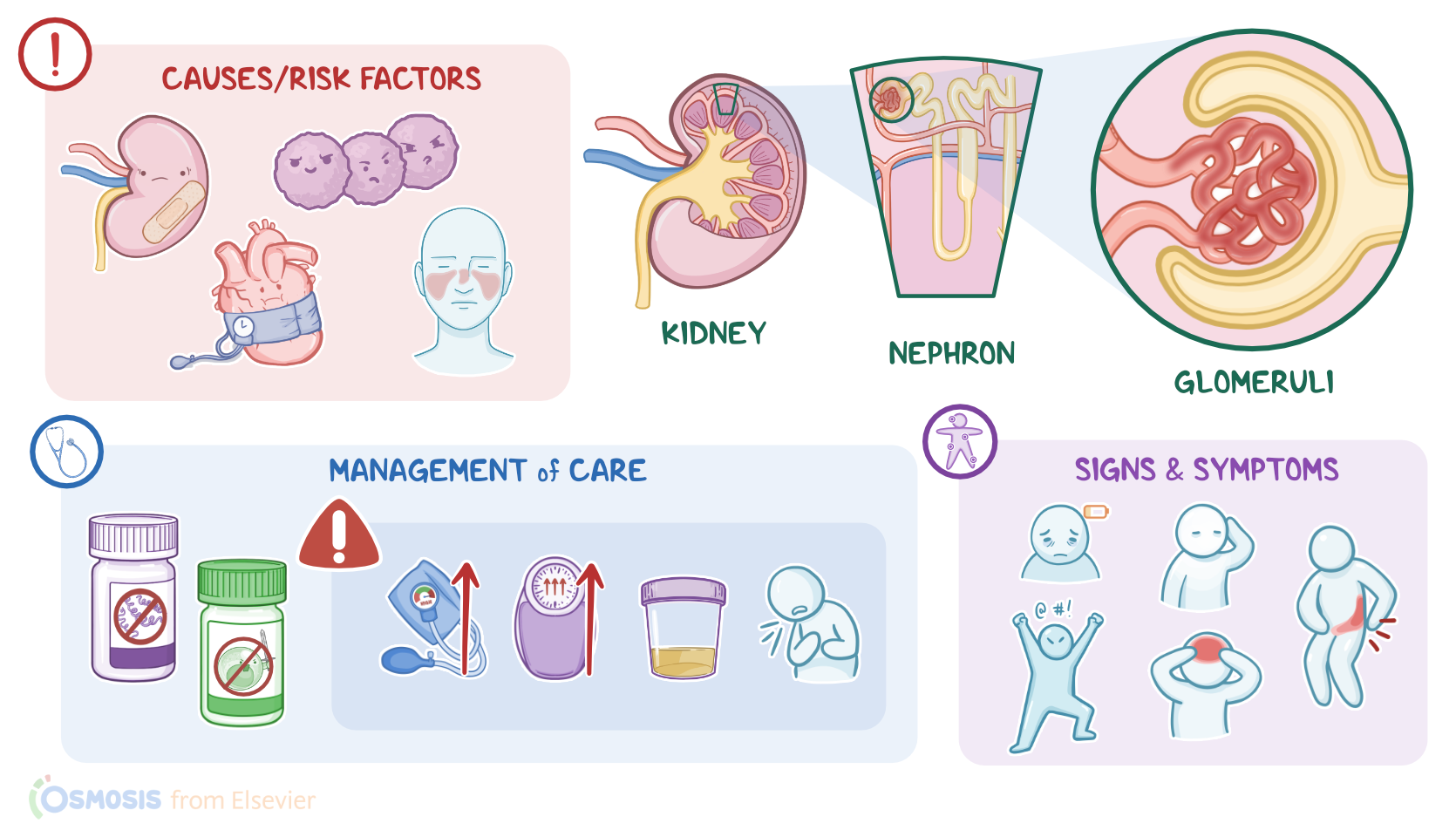
CAUSES AND RISK FACTORS
  • Causes
    • Acute primary
      • Infections
    • Acute secondary
      • Systemic conditions
    • Chronic
      • Unknown
      • Infections
      • Hypertension
      • Immunological injuries
      • Inflammation
      • Impaired renal perfusion

PATHOPHYSIOLOGY
  • Acute glomerulonephritis
    • Excessive immune response
    • Inflammation and injury in the glomerular basement membrane
    • Blood and proteins can make it through the filtration barrier
  • Chronic glomerulonephritis
    • Tissue gradually atrophies
    • Number of nephrons decreases
    • Sclerosis in renal arteries
    • Decline in renal function
    • End-stage renal disease

SIGNS AND SYMPTOMS
  • Lethargy
  • Irritability
  • Pallor
  • Headaches
  • Abdominal or flank pain
  • Nausea, vomiting, anorexia
  • Periorbital edema
  • Generalized edema
  • Dyspnea
  • Urinary manifestations
  • Hypertension

DIAGNOSIS
  • History
  • Physical assessment
  • Urinalysis
  • 24-hour urine  
  • Laboratory tests
  • Kidney biopsy
  • CT

TREATMENT
  • Acute glomerulonephritis
    • Antimicrobials
    • Immunosuppressive therapy
    • Fluid restriction and diuretics
    • Antihypertensives
    • Potassium and protein restriction
    • Dialysis
  • Chronic glomerulonephritis
    • Treat underlying conditions
    • Renal replacement therapy

MANAGEMENT OF CARE
  • Goals of care
    • Address cause of glomerulonephritis
    • Prevent complications
  • Address cause
    • Antimicrobials
    • Immunosuppressants
    • Steroids
  • Prevent complications
    • Vital signs
    • Antihypertensives
    • Daily weights
    • Monitor intake and output
    • Assess for edema
    • Auscultate lungs and heart sounds
    • Diuretics
    • Low sodium diet
    • Fluid restrictions
    • Monitor serum laboratory results
    • Assist with renal replacement therapy
    • Consult with dietician
      • Notify HCP
        • Elevated BP
        • Sudden increase in shortness of breath
        • Decreased urine output
        • Worsening edema
        • Jugular venous distension
        • Elevated BUN, creatinine, potassium
        • Severe fatigue
        • Nausea or vomiting
        • Altered level of consciousness

PATIENT AND FAMILY TEACHING
  • Explain condition, plan of care, and how to safely self-administer medications
  • Track weight
  • Take blood pressure
  • Dietary modifications 
  • Record fluid intake
  • Oral care
  • Check for sodium content
  • Notify HCP
    • Sigs of fluid overload

Transcript

Watch video only

Glomerulonephritis refers to inflammation of the glomeruli. It can be primary when the disease starts in the glomeruli, or secondary, when the glomeruli are affected by systemic disease. It can also be acute, when symptoms develop suddenly and resolve with treatment; or it can be chronic if acute disease is not treated, or when the disease process develops slowly, leading to irreversible failure of the kidneys.

First, let’s quickly review some kidney physiology! Remember that the nephron is the functional unit of the kidney, and its role is to filter waste products and water from the blood. Each nephron is made up of a renal corpuscle and a set of renal tubules, which are, in order: the proximal convoluted tubule, loop of Henle, distal convoluted tubule and finally, collecting ducts which drain urine into the renal papillae and eventually empty into the renal pelvis.

The renal corpuscle is made up of the glomerulus, which is a bundle of capillary loops, and Bowman’s capsule, which surrounds the glomerulus. Now, blood enters the glomerulus through the afferent arteriole, then moves inside the glomerulus, where glomerular filtration occurs. Then, the remaining blood exits the glomerulus through the efferent arteriole. Now, the glomerular capillary wall is a semi-permeable membrane with three layers: the endothelium, which is the inner layer; the basement membrane; and the epithelium, which is the outermost layer.

For filtration to occur, the endothelial and epithelial cells that line the capillary wall are separated by small pores that filter water and small particles from the blood and into the Bowman capsule. Only small particles are filtered from the blood and never proteins, since they are larger molecules. Also, normally, there aren’t any red blood cells in the urine. For glomerular filtration to occur, the hydrostatic pressure created by the blood pressure needs to be greater than opposing forces, such as the tubular filtrate and oncotic pressure, which is the pressure of proteins. The final product of filtration is called filtrate or primary urine, which will flow through the Bowman capsule into the proximal convoluted tubule.

Now let’s look at the causes of glomerulonephritis. Causes and risk factors of acute primary glomerulonephritis include infections with various agents. Some bacteria that can cause acute primary glomerulonephritis include, Group A beta-hemolytic Streptococcus, which is the most frequent in children; but also Streptococcus pneumoniae, Klebsiella pneumoniae, Mycoplasma pneumoniae, and Treponema pallidum, which causes syphilis; as well as staphylococcal or gram negative bacteremia or sepsis. There are also viruses that can cause acute primary glomerulonephritis which include dengue fever virus, hantavirus, varicella, parvovirus, hepatitis B and C viruses, cytomegalovirus, Epstein-Barr virus and human immunodeficiency virus.

Next, acute secondary glomerulonephritis can be caused by systemic conditions, like systemic lupus erythematosus, amyloidosis, chronic liver conditions like hepatitis B and C, autoimmune hepatitis, or cirrhosis, as well as other conditions, like vasculitis, Goodpasture syndrome, diabetic nephropathy and HIV-associated glomerulonephritis. Now, when it comes to chronic glomerulonephritis, sometimes the causative agent isn’t known, but infections, untreated hypertension, immunological injuries to the kidneys, inflammation or impaired renal perfusion can all lead to chronic glomerulonephritis over time.

Now, let’s delve into the pathology of glomerulonephritis. With acute glomerulonephritis, infections or systemic conditions cause an excessive immune response in the kidneys. This leads to inflammation and injury in the glomerular basement membrane, which impairs glomerular filtration, so blood and proteins can make it through the filtration barrier and into the urine.

Acute poststreptococcal glomerulonephritis, develops about 1 to 2 weeks following an infection with nephrotoxic strains of group A beta-hemolytic streptococci. This can be a skin infection like impetigo or a throat infection of the tonsils or pharynx. After the infection, antibodies against the streptococcal antigen are made. Then antigen-antibody complexes are formed and deposited in the glomeruli leading to activation of the complement system. This further leads to inflammation and then acute kidney injury.

With chronic glomerulonephritis, kidney tissue gradually atrophies and the number of nephrons decreases, which leads to reduced glomerular filtration, sclerosis in the renal arteries, and a decline in renal function. If a biopsy is done in the late stages of chronic glomerulonephritis, there’s cell loss, protein and collagen deposits and fibrosis in the kidney tissue. Glomerulonephritis can lead to other problems such as nephrotic syndrome or eventually lead to end-stage renal disease.

Now let’s look at the clinical manifestations of glomerulonephritis. General manifestations include lethargy, irritability, pallor and headaches. The client can also have abdominal or flank pain, as well as nausea, vomiting, and anorexia. There can also be periorbital edema, generalized edema, or even pulmonary edema, which could present as dyspnea. Urinary manifestations can include decreased urine output, dysuria, and hematuria, which could turn the urine tea or cola colored, with a cloudy, smoky aspect. Mild to moderate hypertension is also often present.

Diagnosis of glomerulonephritis starts with the client’s history and physical assessment. Urinalysis shows hematuria and proteinuria. Microscopic examination of the urine usually shows dysmorphic erythrocytes and red blood cell casts, along with epithelial casts. A 24 hour urine collection can be done in order to quantify the protein excretion rate, which is increased, ranging from 500 milligrams to 3 grams per 24 hours.

With both acute and chronic glomerulonephritis, there can be elevated creatinine and BUN levels, which is called uremia. Then an estimated glomerular filtration rate is calculated based on creatinine levels. A serum electrolyte panel, along with an acid-base status can also show abnormalities. With acute poststreptococcal glomerulonephritis, there’s an increased titer of anti-streptolysin O antibodies, and complement fractions like C3 and C4 can be lower than normal. Finally, to identify the cause of glomerulonephritis, an immunological panel, along with a kidney biopsy can be performed. Additionally, a CT-scan of the abdomen can be done to identify structural abnormalities of the kidneys.