Neural tube defects: Nursing
Neural tube defects: Nursing
Medical Surgical
Medical Surgical
Notes
| NEURAL TUBE DEFECTS (NTDs) | ||
| KEY POINTS | NOTES | |
| DEFINITION |
| |
| PHYSIOLOGY |
| |
| CAUSES AND RISK FACTORS |
| |
| PATHOPHYSIOLOGY |
| |
| SIGNS AND SYMPTOMS |
| |
| DIAGNOSIS |
| |
| TREATMENT |
| |
| MANAGEMENT OF CARE |
| |
| PATIENT AND FAMILY TEACHING |
| |
Transcript
Neural tube defects, or NTD for short, refer to developmental defects of the central nervous system secondary to failed closure of the neural tube during the fetus's embryonic development.
Now, let’s quickly look at the physiology of neural tube development. Let’s zoom in on a cross section of the early embryo.
Here we can see it is made up of three main layers: the ectoderm, the mesoderm, and the endoderm.
The ectoderm then goes on and differentiates into three populations of cells.
The first one, located internally, gives rise to the neural tube; the second one, located externally, gives rise to skin cells and the epidermis; and the third population, called the neural crest cells, develop in between the neural tube and epidermis, and give rise to various structures throughout the body.
The mesoderm, on the other hand, develops into a transient midline structure,
called the notochord, which produces various signals that guide the development of other embryonic structures with respect to the midline.
During the third and fourth weeks of development, the notochord signals for the formation of the neural tube, through a process called neurulation, which is highly dependent on adequate folic acid levels. Neurulation progresses in two stages.
Primary neurulation begins when the ectoderm right above the notochord thickens and gives rise to the neural plate.
At both ends of the neural plate, there are neural crest cells, and beyond them, the ectoderm that gives rise to the epidermis.
Soon after, the edges of the neural plate thicken and tilt upward, forming the so-called neural folds.
This allows for a u-shaped neural groove to form, which sets the limit between the right and left sides of the embryo.
During secondary neurulation, the neural folds eventually fuse at the midline, forming the hollow neural tube, separate from the epidermis, above it.
Now let's look at a different view of the developing embryo, this time from above.
At this point, like any hollow tube, the neural tube still has openings at both ends: a large opening at the top end called the cranial neuropore, and a smaller opening at the bottom end called the caudal neuropore. The cranial neuropore seals up around day 25, while the caudal neuropore seals up a few days later, around day 28. The neural tube eventually develops into the brain and spinal cord, while the notochord gives rise to a part of the intervertebral disc called the nucleus pulposus.
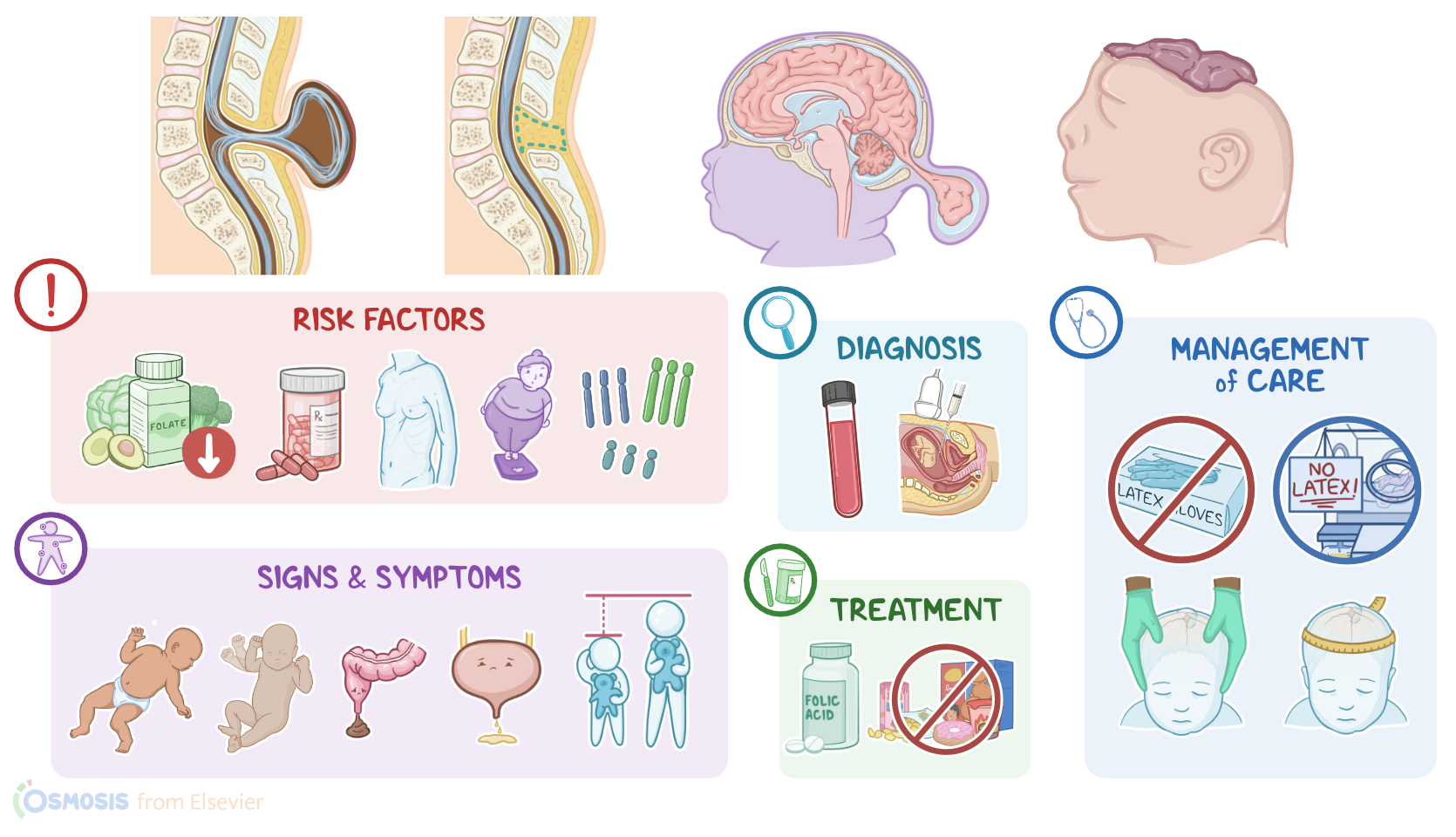
The cause of neural tube defects is not known. However, they are associated with any factor that can interfere with the development of the central nervous system during embryonic development. These include modifiable risk factors such as folate deficiency, malnutrition, maternal obesity, infections, medications such as sodium valproate and methotrexate, and certain chemicals.
There are also non-modifiable risk factors such as advanced maternal age, maternal diabetes, and fetal genetic abnormalities including trisomy 13, 18, and 21.
Pathology-wise, one or more of these risk factors increase the chance of failed neural plate folding or closure during neurulation. More specifically, this can occur either at the cranial neuropore, involving the brain and skull, or at the caudal neuropore, involving the spinal cord.
Now, given the two main locations of neural tube defects, there are two types of defects that can occur. First, spinal defects are more common and can be further divided into two subtypes. Spina bifida occulta is where the posterior vertebral arches fail to close in the lumbosacral area. However, in this case, the spinal cord and the meninges remain in their place and the defect is not usually visible from the outside, hence the name occulta.
Then, spina bifida cystica when parts of the spinal cord herniates through the abnormal gap in the vertebrae, forming a cyst. A meningocele is when only the meninges herniate into the cyst, while a myelomeningocele contains the meninges and spinal cord.
Second, cranial defects are mostly represented by encephalocele, and it is when the brain and the meninges protrude through a defect of the skull. Another cranial defect is anencephaly, which is where the infant has small or missing brain hemispheres, upper skull, and scalp, secondary to failed closure of the cranial neuropore. These infants are typically born without both a forebrain and a cerebrum and the remaining brain tissue may be exposed.
Now, large defects come with a series of complications of their own. For example, a large encephalocele allows the structures in the region to herniate, and, if the defect is not corrected and blood flow compromised, it can cause significant damage to the nervous system. Subsequently, central nervous injury early in the development of the embryo can result in cognitive delay, poor motor skills or cerebral palsy after birth.
In some cases, severe neural tube defects can also cause spontaneous abortion or death in utero, hydrocephalus or buildup of fluid in the brain ventricles, polyhydramnios, and the Chiari malformation.
A Chiari malformation is where the lower part of the brain herniates down into the spinal canal. Sometimes, a spinal NTD can fistulate, causing cerebrospinal fluid to leak out through the skin, increasing the likelihood of a central nervous system infection.
Clinical manifestations of neural tube defects vary depending on the type of defect. Now, spina bifida occulta is usually invisible and asymptomatic. If the defect is visible, it is usually seen only as a dimple on the back, which may be covered in hair. Spina bifida cystica is when the same defect is visible as a protrusion or cyst of the meninges through the vertebrae. With meningocele, the spinal cord remains in the correct position, and these infants often present with minor or no neurologic deficits.
However, a myelomeningocele has a high risk of neurological deficits because the spinal cord herniates out of the vertebral defect, making it easier to damage, thus increasing the risk of developing motor and sensory dysfunction below the defect. Additionally, the infant with such a defect has an increased risk of meningitis, hemorrhage, and hypoxia, hence the infant might present with symptoms suggestive of these complications. With a myelomeningocele, bladder and bowel incontinence can also be present.
Next, an encephalocele is seen as a round protrusion in the occipital area. Generally, the protrusion is covered by skin, but it can also be open. Depending on its location and size, an encephalocele can cause neurological issues, like spastic quadriplegia, ataxia or a lack of muscle control or coordination of voluntary movements, and visual problems. Encephaloceles are also accompanied by craniofacial abnormalities like microcephaly, and by severe cognitive deficits such as developmental delay and intellectual disability, but some children can achieve standard IQs.
Finally, with anencephaly, infants are typically born without both a forebrain and a cerebrum and the remaining brain tissue might be exposed, and it’s incompatible with life. Most infants die in the womb or within hours to weeks after birth.
The diagnosis of neural tube defects can be done prenatally or after birth. Prenatal diagnosis of neural tube defects is usually done by testing the amniotic fluid via amniocentesis and maternal blood for increased levels of alpha-fetoprotein or AFP, and acetylcholinesterase at week 16 and 18.
AFP is a fetal protein produced by the liver and leaks from the fetus into the amniotic fluid through exposed capillaries of the NTD. Acetylcholinesterase, on the other hand, leaks directly from exposed neural tissue into the amniotic fluid.
In cases where AFP levels are increased, high-resolution fetal ultrasonography or fetal MRI can be used to diagnose neural tube defects prenatally.
After birth, diagnosis can also be established by physical examination and imaging such as CT and MRI. Additionally, a cranial ultrasound can help diagnose some neural tube defects and assess for associated complications such as hydrocephalus.
Let’s first look at the prevention of neural tube defects. One of the most efficient ways to prevent a neural tube defect is by ensuring folic acid supplementation. Individuals who plan to give birth should consume 400 mcg of folic acid daily for around 6 months prior to conception, increasing the dose to 600 mcg daily during pregnancy.
Additionally, addressing the risk factors predisposing to neural tube defects, such as obesity through weight loss, diabetes through strict glycaemia control, and discontinuation of drug use will also reduce the risk of a defect occurring.