Pertussis: Nursing

Pertussis: Nursing

NUR243

NUR243

Growth and development - Infant: Nursing
Growth and development - Preschool-age: Nursing
Growth and development - Toddler: Nursing
Growth and development - School-age: Nursing
Growth and development - Adolescent: Nursing
Growth and development - Early and middle adulthood: Nursing
Failure to thrive (FTT): Nursing
Growth and development theories: Nursing
Congenital heart defects - Acyanotic: Nursing
Congenital heart defects - Cyanotic: Nursing
Kawasaki disease: Nursing
Amblyopia: Nursing
Hearing impairment and otosclerosis: Nursing
Pharyngitis: Nursing
Strabismus: Nursing
Otitis media: Nursing
Conjunctivitis: Nursing process (ADPIE)
Eye injury: Nursing process (ADPIE)
Tonsillitis: Nursing process (ADPIE)
Biliary atresia: Nursing
Cleft lip and palate: Nursing
Esophageal atresia and tracheoesophageal fistula: Nursing
Hirschsprung disease: Nursing
Necrotizing enterocolitis: Nursing
Nutrition - Newborn: Nursing
Omphalocele and gastroschisis: Nursing
Appendicitis: Nursing process (ADPIE)
Hyperbilirubinemia: Nursing process (ADPIE)
Poisoning: Nursing process (ADPIE)
Pyloric stenosis: Nursing process (ADPIE)
Bladder exstrophy: Nursing
Circumcision: Nursing
Cryptorchidism: Nursing
Enuresis: Nursing
Hypospadias and epispadias: Nursing
Hemolytic uremic syndrome: Nursing
Sickle cell disease: Nursing process (ADPIE)
Hypersensitivity reactions - Type I: Nursing
Hypersensitivity reactions - Type II: Nursing
Hypersensitivity reactions - Type III: Nursing
Hypersensitivity reactions - Type IV: Nursing
Diphtheria: Nursing
Erythema infectiosum (Fifth disease): Nursing
Fever: Nursing
Mumps (Parotitis): Nursing
Infectious mononucleosis: Nursing
Pertussis: Nursing
Poliomyelitis: Nursing
Rocky Mountain spotted fever (RMSF): Nursing
Roseola (Exanthem subitum): Nursing
Rubella (German measles): Nursing
Rubeola (Measles): Nursing
Smallpox: Nursing
Zika virus: Nursing
Anaphylaxis: Nursing process (ADPIE)
Chickenpox (Varicella): Nursing process (ADPIE)
Lyme disease: Nursing process (ADPIE)
Child maltreatment: Nursing
Crisis intervention: Nursing
Therapeutic communication: Nursing
Autism spectrum disorder (ASD): Nursing
Pediatric psychosocial needs during illness and hospitalization: Nursing
Anxiety disorders: Nursing process (ADPIE)
Eating disorders: Nursing process (ADPIE)
Muscular dystrophies - Duchenne and Becker: Nursing
Clubfoot: Nursing
Craniosynostosis: Nursing
Developmental dysplasia of the hip: Nursing
Juvenile idiopathic arthritis: Nursing
Scoliosis: Nursing
Shoulder dystocia: Nursing
Fractures: Nursing process (ADPIE)
Cerebral palsy: Nursing
Head injury: Nursing
Brachial plexus injury: Nursing
Phenylketonuria (PKU): Nursing
Hydrocephalus: Nursing process (ADPIE)
Reye syndrome: Nursing process (ADPIE)
Cystic fibrosis: Nursing
Influenza: Nursing
Sudden infant death syndrome (SIDS): Nursing
Asthma: Nursing process (ADPIE)
Bronchiolitis and respiratory syncytial virus (RSV): Nursing process (ADPIE)
Epiglottitis: Nursing process (ADPIE)
Foreign body aspiration and upper airway obstruction: Nursing process (ADPIE)
Laryngotracheobronchitis (LTB) and croup: Nursing process (ADPIE)
Smoke inhalation injury: Nursing process (ADPIE)

Notes

PERTUSSIS

KEY POINTS
NOTES
DEFINITION
  • Acute respiratory infection

PHYSIOLOGY
  • Respiratory system
    • Upper airway 
    • Lower airway 
    • Lungs  
    • Protection against pathogens
  • Upper airway and lower airway
    • Lined with ciliated epithelial cells that help expel pathogens out nose or mouth
  • Lungs
    • Immune cells produce cytokines to attract immune cells
    • Immune cells eliminate pathogens

CAUSES AND RISK FACTORS
  • Cause
    • Gram-negative bacillus: Bordetella pertussis
  • Risk factors 
    • Pregnancy
    • Close contact with infected person
    • Lack of or underimmunization

PATHOPHYSIOLOGY
  • Transmitted via airborne droplets via inhalation
    • Bacteria sticks to epithelial cells in upper airway
    • Production of toxin that damages tissues, causing inflammation and edema
      • Increased respiratory secretions
      • Cilia are paralyzed, preventing respiratory clearance
      • Irritated trachea causes coughing
      • Increased vascular histamine (cytokine) sensitivity
        • Blood vessels leak, causing airway edema
        • Swelling obstructs airflow
  • Complications
    • Secondary infections
      • Pneumonia 
      • Otitis media
    • Impaired gas exchange
      • Hypoxia
      • Apnea 
    • CNS complications 
      • Encephalopathy 
      • Seizures

SIGNS AND SYMPTOMS
  • First stage
    • Mild or no fever
    • Rhinorrhea
    • Malaise
    • Mild non-productive cough
  • Second stage
    • Forceful repeated cough; "whoop" sound
  • Third stage
    • Residual cough lasting 2-3 weeks

DIAGNOSIS
  • History
  • Physical assessment
  • Laboratory tests
    • Nasopharyngeal culture
    • Serology antibody testing

TREATMENT
  • Antibiotics
  • Respiratory support
  • DTaP vaccination (<7 years old)
  • Tdap vaccination (>7 years old)

MANAGEMENT OF CARE
  • Goals of care
    • Prevent spread of infection
    • Provide respiratory support
    • Supportive care
  • Institute airborne and droplet precautions
  • Monitor cardiac rhythm and oxygen saturation
  • Auscultate lungs, assess cough
  • Prevent crying to reduce cough
  • Administer oxygen, antibiotics, IV fluids, and antipyretics as prescribed
  • Encourage oral fluids
  • Measure intake and output
  • Report to HCP
    • Decreased SpO2
    • Cyanosis
    • Altered mental status
    • Signs of airway obstruction
    • Low urine output
    • Unable to tolerate oral fluids

PATIENT AND FAMILY TEACHING
  • Explain condition, plan of care, and safe medication administration
  • Prevent future infection through vaccination
  • Avoid environmental triggers like dust and smoke
  • Use a humidifier
  • Encourage rest
  • Decrease spread through handwashing, wearing a mask, and socially distancing 
  • Notify HCP
    • Child has difficulty breathing
    • Change in behavior
    • Refusing to eat or drink

Transcript

Watch video only

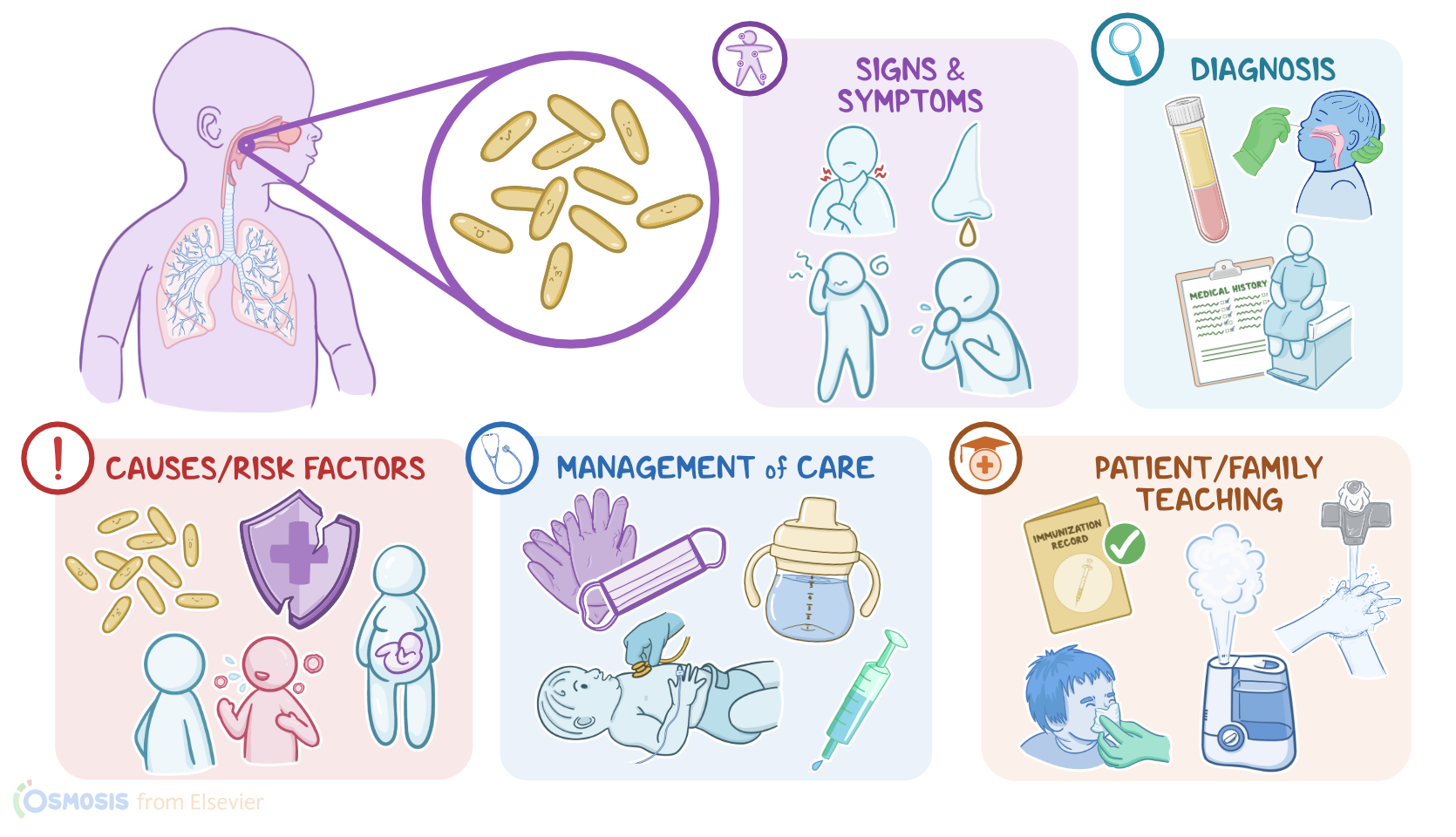
Pertussis, also known as whooping cough, is an acute respiratory infection caused by the bacteria Bordetella pertussis. The disease is characterized by paroxysmal cough, meaning fits of sudden and periodic cough, as well as abundant respiratory secretions. It can affect people of all ages, but it can be particularly severe, and even life threatening in children younger than 6 months.

Now, the respiratory tract consists of the upper and lower airways, as well as the lungs. The tiniest branches of the lower airways, called the bronchioles, end with the alveolar sacs, which are lined by a thin membrane that allows gas exchange to occur.

Now, the respiratory system is equipped with defense mechanisms against potential pathogens. For example, the upper and lower airways are lined with ciliated epithelial cells that sweep pathogens that make their way inside, to be expelled back out through the nose or mouth. At the same time, the lungs have plenty of immune cells, like macrophages, that react when a pathogen makes its way in by producing cytokines that attract more immune cells. These immune cells destroy and dispose of the invading pathogen.

Okay, now, pertussis is an infection caused by a gram-negative bacillus, called Bordetella pertussis. Risk factors for acquiring infection include pregnancy, close contact with an infected individual and lack of immunization or underimmunization.

Pathology-wise, pertussis is transmitted by airborne contagious droplets. After inhalation, the bacteria adheres to the epithelial cells of the upper respiratory tract and the nasopharynx. Once attached, it produces toxins that damage the local tissues, causing inflammation and edema of the respiratory tract. This results in abundant secretions of the respiratory tract. In addition, some of the toxins can also paralyze the cilia, thus preventing the bacteria and the mucus secretion from being cleared. All this mucus irritates the trachea, causing fits of violent coughing.

Furthermore, some toxins will also increase the blood vessels sensitivity to histamine, a cytokine released by immune cells during an infection. This makes the blood vessels leakier, allowing fluid to move out into the surrounding tissues, causing edema in the airway. So the swelling can obstruct air flow, causing shortness of breath, and it also causes the characteristic “whooping” sound when they try to take a deep breath.

Some complications of pertussis include secondary infections, like pneumonia and otitis media, as well as hypoxia and apnea when the physiology of gas exchange is affected. In rare cases, these complications, particularly hypoxia and secondary infections, can cause CNS complications of their own, like encephalopathy and seizures.

Now, the clinical manifestations of pertussis occur following an incubation period of around 7 to 10 days. Then, the disease progresses in three stages. The first stage, called the catarrhal phase, lasts 1 to 2 weeks, and manifests as a mild upper respiratory tract infection with mild or no fever, rhinorrhea, malaise and a mild nonproductive cough. The second stage, called the paroxysmal stage, is characterized by paroxysms of cough, which are severe, forceful and repeated coughs that occur during a single expiration. These bouts are often followed by a prolonged inspiration that produces a characteristic “whoop” sound.

The third and the last stage, called the convalescent phase, is characterized by a residual cough that lasts 2 to 3 weeks.

Diagnosis of pertussis starts with the client’s history and physical assessment. A complete blood count can show leukocytosis and lymphocytosis. The causative bacteria can be identified in nasopharyngeal cultures or with a PCR from nasopharyngeal secretions. Alternatively, the antibodies against Bordetella pertussis can be identified using serology testing.

Treatment of pertussis is focused on treating the bacterial infection with antibiotics and providing respiratory support. The antibiotic of choice is usually a macrolide like erythromycin or azithromycin, or trimethoprim/sulfamethoxazole for those who can’t take macrolides.