Postpartum hemorrhage: Nursing

1,735views

Postpartum hemorrhage: Nursing

Acute Final

Acute Final

Endocrine system anatomy and physiology
Antepartum assessment - Fetus: Nursing
Assessment of gestational age: Nursing
Fetal circulation: Nursing
Fetal development: Nursing
Group B streptococcus (GBS) infection in pregnancy: Nursing
Hepatitis B virus (HBV) infection in pregnancy: Nursing
Hyperemesis gravidarum: Nursing
Large for gestational age (LGA) infant: Nursing
Preeclampsia and eclampsia: Nursing
Prenatal screening: Nursing
Placenta previa: Nursing process (ADPIE)
Placental abruption: Nursing process (ADPIE)
Birth-related procedures: Nursing
Cesarean birth: Nursing
Intrapartum assessment - Fetal heart rate patterns: Nursing
Intrapartum assessment - Uterine activity: Nursing
Premature rupture of membranes (PROM): Nursing
Shoulder dystocia: Nursing
Prolapsed umbilical cord: Nursing process (ADPIE)
Stages of labor: Nursing
Assessment - Postpartum: Nursing
Perinatal depression: Nursing
Physiology of lactation: Nursing
Postpartum infections: Nursing
Postpartum hemorrhage: Nursing
Biliary atresia: Nursing
Cleft lip and palate: Nursing
Congenital diaphragmatic hernia: Nursing
Congenital heart defects - Acyanotic: Nursing
Congenital heart defects - Cyanotic: Nursing
Esophageal atresia and tracheoesophageal fistula: Nursing
Craniosynostosis: Nursing
Hemolytic disease of the fetus and newborn: Nursing
Hyperbilirubinemia: Nursing process (ADPIE)
Infant of a diabetic mother (IDM): Nursing
Meconium aspiration syndrome: Nursing
Neonatal respiratory distress syndrome (NRDS): Nursing
Neonatal sepsis: Nursing
Neural tube defects: Nursing
Newborn adaptation to extrauterine life: Nursing
Persistent pulmonary hypertension of the newborn (PPHN): Nursing
Physical assessment - Neonate: Nursing
Small for gestational age (SGA) infant: Nursing
Postterm infant: Nursing
Thermoregulation - Neonate: Nursing
Arterial blood gas (ABG) - Overview: Nursing
Arterial blood gas (ABG) - Metabolic acidosis: Nursing
Arterial blood gas (ABG) - Metabolic alkalosis: Nursing
Arterial blood gas (ABG) - Respiratory acidosis: Nursing
Arterial blood gas (ABG) - Respiratory alkalosis: Nursing
Adrenal insufficiency (Addison disease): Nursing
Anemia - Iron-deficiency: Nursing
Anemia - Aplastic: Nursing
Anemia - Macrocytic: Nursing
Case study - Hypothyroidism: Nursing
Case study - Iron-deficiency anemia: Nursing
Case study - Sickle cell anemia: Nursing
Complete blood count (CBC) - Hemoglobin and hematocrit: Nursing
Complete blood count (CBC) - Red blood cells (RBC): Nursing
Complete blood count (CBC) - Platelets: Nursing
Complete metabolic panel (CMP) - Blood urea nitrogen (BUN) and creatinine (Cr): Nursing
Complete metabolic panel (CMP) - Estimated glomerular filtration rate (eGFR): Nursing
Complete metabolic panel (CMP) - Liver function tests (LFT): Nursing
Cushing syndrome and Cushing disease: Nursing
Hematopoietic growth factors: Nursing pharmacology
Hyperparathyroidism: Nursing
Hyperthyroidism: Nursing process (ADPIE)
Hypoparathyroidism: Nursing
Hyperpituitarism: Nursing
Hypopituitarism: Nursing
Hypothyroidism: Nursing process (ADPIE)
Medications affecting the parathyroid glands: Nursing pharmacology
Medications for growth hormone disorders: Nursing pharmacology
Medications for thyroid disorders: Nursing pharmacology
Neutropenia: Nursing
Polycythemia: Nursing
Thrombocytopenia: Nursing
Acute kidney injury (AKI): Nursing process (ADPIE)
Benign prostatic hyperplasia (BPH): Nursing process (ADPIE)
Case study - Cholecystitis: Nursing
Case study - Cirrhosis: Nursing
Case study - Chronic kidney disease (CKD): Nursing
Case study - Benign prostatic hyperplasia (BPH): Nursing
Case study - Gastroesophageal reflux disease (GERD): Nursing
Case study - Pediatric appendicitis: Nursing
Case study - Pyelonephritis: Nursing
Cholecystitis: Nursing
Cholelithiasis: Nursing
Chronic kidney disease (CKD): Nursing
Cirrhosis: Nursing process (ADPIE)
Diverticular disease: Nursing
Gastroesophageal reflux disease (GERD): Nursing process (ADPIE)
Hemolytic uremic syndrome: Nursing
Hirschsprung disease: Nursing
Intestinal obstruction: Nursing
Irritable bowel syndrome (IBS): Nursing
Nephrotic syndrome: Nursing
Pyloric stenosis: Nursing process (ADPIE)
Renal and urinary calculi: Nursing
Urinary incontinence - Stress: Nursing process (ADPIE)
Diabetes insipidus: Nursing process (ADPIE)
Dialysis care: Nursing
Case study - Diabetic ketoacidosis (DKA): Nursing
Case study - Pediatric diabetes mellitus type 1: Nursing
Diabetes mellitus (DM): Nursing process (ADPIE)
Hyperosmolar hyperglycemic state (HHS): Nursing process (ADPIE)
Diabetic ketoacidosis (DKA): Nursing process (ADPIE)
Case study - Epilepsy: Nursing
Case study - Head injury: Nursing
Epidural and subdural hematoma: Nursing
Case study - Stroke: Nursing
Hemorrhagic stroke - Intracranial hemorrhage (ICH) and subarachnoid hemorrhage (SAH): Nursing
Increased intracranial pressure (ICP): Nursing
Hydrocephalus: Nursing process (ADPIE)
Intracranial aneurysm: Nursing
Seizure disorder: Nursing process (ADPIE)
Stroke: Nursing process (ADPIE)
Jaundice: Nursing
Nutrition - Enteral: Nursing skills
Nutrition - Newborn: Nursing
Nutrition - Parenteral: Nursing skills
Phenylketonuria (PKU): Nursing
Arterial embolism: Nursing
Disseminated intravascular coagulation (DIC): Nursing
Hemophilia: Nursing process (ADPIE)
Acute respiratory distress syndrome (ARDS): Nursing
Asthma: Nursing process (ADPIE)
Atelectasis: Nursing
Bacterial pneumonia: Nursing process (ADPIE)
Bronchiolitis and respiratory syncytial virus (RSV): Nursing process (ADPIE)
Case study - Acute respiratory distress syndrome (ARDS): Nursing
Care of an intubated client: Nursing skills
Case study - Chronic obstructive pulmonary disease (COPD): Nursing
Case study - Impaired gas exchange: Nursing
Case study - Pediatric asthma: Nursing
Chest tube care: Nursing
Chronic obstructive pulmonary disease (COPD): Nursing process (ADPIE)
Cystic fibrosis: Nursing
Epiglottitis: Nursing process (ADPIE)
Flail chest: Nursing
Intraoperative care: Nursing
Pleural effusion: Nursing
Pneumothorax and hemothorax: Nursing
Pulmonary edema: Nursing
Smoke inhalation injury: Nursing process (ADPIE)
Tracheostomy: Nursing
Venous thromboembolism (VTE): Nursing process (ADPIE)
Arrhythmias - Asystole: Nursing
Arrhythmias - Atrial flutter (Aflutter): Nursing
Arrhythmias - Premature atrial contractions (PACs): Nursing
Arrhythmias - Heart blocks: Nursing
Arrhythmias - Atrial fibrillation (Afib): Nursing
Arrhythmias - Premature ventricular contractions (PVCs): Nursing
Arrhythmias - Sinus tachycardia and sinus bradycardia: Nursing
Arrhythmias - Supraventricular tachycardia (SVT): Nursing
Arrhythmias - Ventricular fibrillation (Vfib): Nursing
Arrhythmias - Ventricular tachycardia (Vtach): Nursing
Cardiac biomarkers - Troponin: Nursing
Case study - Acute coronary syndrome (ACS): Nursing
Case study - Atrial fibrillation (Afib): Nursing
Case study - Heart failure with reduced ejection fraction (HFrEF): Nursing
Case study - Deep vein thrombosis (DVT): Nursing
Case study - Hypertension: Nursing
Case study - Hypovolemic shock: Nursing
Coronary artery disease (CAD) and angina pectoris: Nursing process (ADPIE)
Electrocardiogram (ECG) - Normal sinus rhythm (NSR): Nursing
Heart defects that decrease pulmonary blood flow - Nursing considerations & client education: Nursing
Hypertension: Nursing process (ADPIE)
Left-sided heart failure: Nursing process (ADPIE)
Myocardial infarction (MI): Nursing process (ADPIE)
Pericardial effusion and cardiac tamponade: Nursing process (ADPIE)
Peripheral arterial disease (PAD): Nursing process (ADPIE)
Rheumatic heart disease: Nursing process (ADPIE)
Shock - Cardiogenic: Nursing
Shock - Neurogenic: Nursing
Shock - Obstructive: Nursing
Shock - Septic: Nursing
Sickle cell disease: Nursing process (ADPIE)
Valvular heart disease: Nursing

Notes

POSTPARTUM HEMORRHAGE (PPH)

KEY POINTS
NOTES
DEFINITION
  • Obstetrical emergency where there's significant blood loss after delivery 
    • Blood loss 
      • > 500 mL after vaginal birth 
      • > 1000 mL after C-section 
    • Primary PPH first 24 hours
    • Late/secondary PPH 6-12 weeks after delivery 

PHYSIOLOGY
  • After delivery, uterus regresses back to normal size by the sixth week (involution) 
  • After placenta delivered, smooth muscles contract on placental arteries reducing uterine bleeding
  • Other physiological changes 
    • Blood volume returns to normal
    • Increased diuresis
    • Increased diaphoresis
    • H&H normalize
    • Increased plasma fibrinogen and other factors  

CAUSES AND RISK FACTORS
  • Causes 
    • Tone, Trauma, Tissue, and Thrombin (4 Ts)
  • Risk factors
    • History of PPH in previous pregnancies
    • Placental disorders 
    • Overdistended uterus 
    • Infection
    • Prolonged labor
    • Cesarean section

SIGNS AND SYMPTOMS
  • Overt bleeding
    • Large amount of visible blood
  • Occult bleeding
    • Less visible, blood pools inside uterus or body cavity 
  • Lochia rubra 
    • Abnormal w/ fresh blood and clots 
  • Signs or symptoms of hypovolemic shock 
  • Severe back pain 
  • Abdominal inspection 
    • Fundus above expected level 
    • Uterine atony 
  • Vagina 
    • Bright red discharge w/ clots 
  • Parts of placenta retained 

DIAGNOSIS
  • History
  • Physical assessment
  • Visual inspection
  • Laboratory tests
  • Ultrasound

TREATMENT
  • Administer 
    • IV fluids
    • Blood products
    • Uterotonic medications
  • Bimanual uterine massage
  • Frequent fundal massage
  • Intrauterine balloon tamponade
  • Uterine artery embolization or ligation 
  • Hysterectomy

MANAGEMENT OF CARE
  • Goals of care 
    • Control bleeding
    • Maintain normal fluid volume
    • Provide emotional support
  • Assess fundal 
    • Height
    • Position
    • Consistency
  • Monitor bleeding
    • Count peri-pads
  • Massage fundus until firm
  • Insert two large bore IVs
  • Assess vital signs
  • Insert urinary catheter
  • Monitor intake and output
  • Draw coagulation panel
  • Prepare patient for possible surgery
  • Watch for signs and symptoms of hypovolemic shock
  • Maintain bedrest
  • Elevate legs 30 degrees
  • Notify HCP
    • Boggy uterus
    • Continued hemorrhage
    • Signs or symptoms of shock
  • Keep patient informed about care

PATIENT AND FAMILY TEACHING
  • Explain condition, plan of care, and safe medication administration
  • After discharge
    • Rest
    • Hydrate
    • Eat healthy diet
    • Take iron supplement
    • Monitor lochia
  • Seek emergency care
    • Signs or symptoms of heavy bleeding

Transcript

Watch video only

Postpartum hemorrhage or PPH is an obstetrical emergency which occurs when there is any significant loss of blood after delivery of a baby. Now, some blood loss is expected during childbirth, but PPH is when the blood loss during the first 24 hours exceeds 500 mL after vaginal birth or 1000 mL after cesarean delivery. Significant bleeding in the first 24 hours after delivery is called early or primary postpartum hemorrhage, while late or secondary postpartum hemorrhage occurs 6 weeks to 12 weeks after birth.

Okay, let’s start with some physiology. So, after delivery, the uterus tends to regress back to its normal size and resume its pre-pregnancy position by the sixth week, a process known as involution. Immediately after the delivery of placenta, the smooth muscles of the uterus continue to contract and squeeze down on the placental arteries where they are attached to the uterine wall, causing them to clamp them shut and reduce uterine bleeding.

There are also some physiological changes that occur during pregnancy that begin to change back to pre-pregnancy levels in the postpartum period. For example, during pregnancy there is increased blood plasma volume in relation to red blood cell mass. As a consequence, maternal hemoglobin and hematocrit are usually relatively low during pregnancy, since the same amount of red blood cells are circulating in a higher volume of blood. After delivery, through increased diuresis or urine production, and increased diaphoresis or sweat production, blood volume returns to normal in about 6 to 12 weeks, and hemoglobin, and hematocrit levels normalize in 4 to 6 weeks. Likewise, during pregnancy, plasma fibrinogen and other pro-coagulant factors increase, and they stay elevated until 4 to 6 weeks following delivery.

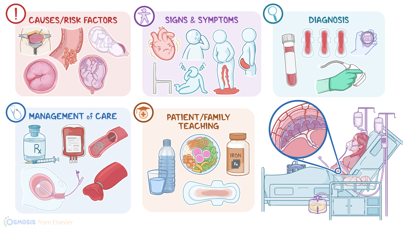
The most common causes of postpartum hemorrhage can be divided into two groups; the early causes and the late causes. The early causes can easily be remembered as the 4 Ts: Tone, Trauma, Tissue, and Thrombin.

Tone refers to a lack of uterine tone, meaning the uterine contraction is weak or absent. This is also known as uterine atony, which is the most common cause of PPH. Repeated distention of the uterus as a result of multiple previous pregnancies or overstretching from multigestational pregnancy can interfere with effective uterine contractions after birth and lead to uterine atony after birth. Uterine atony can also occur when uterine muscles become fatigued after prolonged labor, or if urine retention causes a distended bladder that interferes with uterine involution.

The next T is Trauma, and it refers to damage to any of the reproductive structures, like the uterus, cervix, vagina, or perineum during delivery.

Next is Tissue, which refers to when a part of the placenta is retained in the uterus after birth, interfering with the involution process.

Lastly, Thrombin, refers to the mother having some condition that prevents blood clots from forming normally, for example, a coagulation disorder like von Willebrand disease. These conditions can turn even a tiny bleed into a serious bleed due to the inability to form blood clots.

Finally, some risk factors for PPH which include a history of PPH in previous pregnancies; placental disorders like placenta previa, placenta accreta or placental abruption; an overdistended uterus due to polyhydramnios, multiple gestation, or macrosomic infant; infection, prolonged labor, and lastly delivery by cesarean section.

Clinically, postpartum hemorrhage can be overt, in which a large amount of blood loss can be assessed; or occult, which is when blood pools up somewhere inside the body or inside a hematoma. This can be within the uterus or in the pelvic or abdominal cavity. The lochia rubra, which is the vaginal discharge after delivery, can appear abnormal with large amounts of fresh blood and numerous large blood clots. Severe bleeding can cause signs and symptoms of hypovolemic shock, such as orthostatic hypotension, dizziness, tachycardia, palpitation, shortness of breath, and cold, clammy skin. Some clients can have a severe backache with a feeling of pelvic heaviness due to hematoma formation.

Abdominal inspection may reveal the uterine fundus to be above the expected level, which could indicate internal bleeding and formation of hematoma. With uterine atony, palpation can reveal a boggy, soft and enlarged uterus instead of a firm, contracted one. Vaginal examination can reveal bright red discharge with numerous blood clots, indicating that the uterus is the source of the bleed, and not a cervical or vaginal laceration. Finally, close inspection of the placenta after delivery can show some of the placenta has been left behind.