Pressure injury: Nursing process (ADPIE)
Notes
| PRESSURE INJURY | ||
| KEY POINTS | NOTES | |
| PATIENT REPORT |
| |
| PATHOPHYSIOLOGY |
| |
| DIAGNOSIS AND TREATMENT |
| |
| ASSESSMENT |
| |
| NURSING DIAGNOSES |
| |
| PLANNING |
| |
| IMPLEMENTATION |
| |
| EVALUATION |
| |
Transcript
Joann Mercer is a 78-year-old female client who resides in a skilled nursing facility.
Mrs. Mercer has a history of osteoarthritis and hip fracture.
She needs assistance to walk, and spends most of her time in bed or sitting in her wheelchair.
The certified nursing assistant, or CNA, who is taking care of Mrs. Mercer informs you of redness and a shallow ulcer that developed on her sacrum.
You are concerned that Mrs. Mercer has developed a pressure injury.
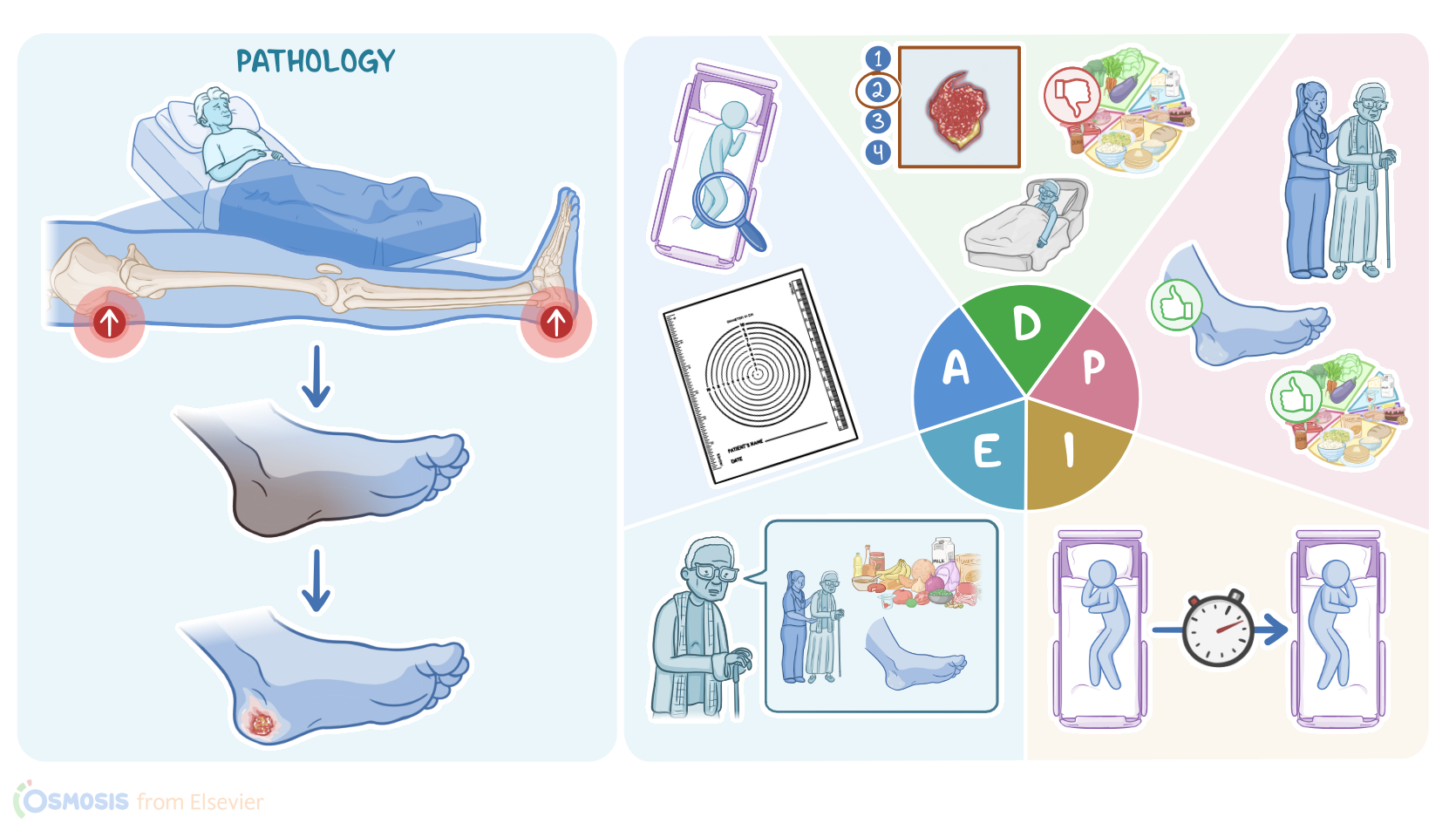
Pressure injuries, also known as decubitus ulcers, involve damage to the skin or underlying tissue that result from prolonged pressure.
Now, pressure injuries usually appear over bony prominences, especially the sacrum, followed by the heels, since these areas have the thinnest subcutaneous tissue between the bone and the skin.
So the prolonged pressure causes a reduced blood flow to that tissue area, resulting in tissue hypoxia and ischemia, and ultimately leading to necrosis and ulceration.
Most often, pressure injuries develop in clients who aren’t moving about, like those on chronic bedrest or consistently in a wheelchair.
Other factors that can increase the risk for skin injury are thinning of skin and subcutaneous tissue due to advanced age as well as dry skin and thin subcutaneous tissue due to inadequate nutrition and hydration; and prolonged contact to skin irritants like sweat, urine, and feces.
Other important risk factors for pressure injuries are conditions that may impair blood flow, such as heart and lung disease and diabetes mellitus.
Clients should be assessed for the risk of developing a pressure injury using a validated assessment tool like the Braden Scale.
This scale looks at six criteria, which include sensory perception, moisture, activity, mobility, nutrition, and friction or shear.
The lower the score, the higher the risk of injury.
Nutritional assessments can be used to assess the likelihood of injury as well as healing.
With non-healing injuries, laboratory tests can be done to assess an underlying cause like diabetes or infection.
Laboratory tests can include a glucose test, which would reveal hyperglycemia in case of diabetes, or a complete blood count showing elevated white blood cells, an elevated erythrocyte sedimentation rate, and elevated C reactive protein or CRP, as well as blood cultures to check for an infectious cause.
Now, pressure injuries can often cause symptoms like pain or pruritus, and can present with purulent drainage or bleeding.
In addition, pressure injuries can lead to complications like local infections of the wound.
In severe cases, the infection may spread and result in bacteremia, which can lead to sepsis, and death.
Pressure injuries are staged according to their level of tissue damage.
There are four stages of injury development and pain can present at any stage.
In stage 1 the skin will appear red, but remain intact.
When pressed, the area will not blanch or turn white.
At stage 2, there is partial-thickness skin loss, and the wound will look like a shallow open wound or blister.
Stage 3 involves full thickness loss of tissue that can present with slough or light-colored dead tissue.
In stage 4, the wound extends deep enough to expose muscle or bone.
Eschar, or dark-colored dead tissue, can be present.
Finally, if a pressure injury is completely covered with slough or eschar, it can be difficult to determine its depth; this is referred to as an unstageable pressure injury.
Treatment for pressure injuries depends on the stage of the wound, but generally involves redistribution of pressure, by regularly repositioning the client, as well as ensuring good nutrition and hydration to encourage wound healing; pain management; and frequent monitoring and wound care to keep the wound clean, moist, and covered.
For stage 1 injuries, either a transparent or hydrocolloid type dressing is used.
Both are useful in acting as a moisture barrier, as well as preventing shear and friction.
For stage 2 injuries, a hydrocolloid or hydrogel dressing can be used to assist with absorption, protection, and debridement of the wound.
An alginate dressing can be used if there is moderate to heavy exudate.
For stage 3 or 4 injuries, a hydrocolloid, hydrogel, foam, or alginate dressing can be used.