Renal and urinary calculi: Nursing

1,474views

Renal and urinary calculi: Nursing

Acute Final

Acute Final

Endocrine system anatomy and physiology
Antepartum assessment - Fetus: Nursing
Assessment of gestational age: Nursing
Fetal circulation: Nursing
Fetal development: Nursing
Group B streptococcus (GBS) infection in pregnancy: Nursing
Hepatitis B virus (HBV) infection in pregnancy: Nursing
Hyperemesis gravidarum: Nursing
Large for gestational age (LGA) infant: Nursing
Preeclampsia and eclampsia: Nursing
Prenatal screening: Nursing
Placenta previa: Nursing process (ADPIE)
Placental abruption: Nursing process (ADPIE)
Birth-related procedures: Nursing
Cesarean birth: Nursing
Intrapartum assessment - Fetal heart rate patterns: Nursing
Intrapartum assessment - Uterine activity: Nursing
Premature rupture of membranes (PROM): Nursing
Shoulder dystocia: Nursing
Prolapsed umbilical cord: Nursing process (ADPIE)
Stages of labor: Nursing
Assessment - Postpartum: Nursing
Perinatal depression: Nursing
Physiology of lactation: Nursing
Postpartum infections: Nursing
Postpartum hemorrhage: Nursing
Biliary atresia: Nursing
Cleft lip and palate: Nursing
Congenital diaphragmatic hernia: Nursing
Congenital heart defects - Acyanotic: Nursing
Congenital heart defects - Cyanotic: Nursing
Esophageal atresia and tracheoesophageal fistula: Nursing
Craniosynostosis: Nursing
Hemolytic disease of the fetus and newborn: Nursing
Hyperbilirubinemia: Nursing process (ADPIE)
Infant of a diabetic mother (IDM): Nursing
Meconium aspiration syndrome: Nursing
Neonatal respiratory distress syndrome (NRDS): Nursing
Neonatal sepsis: Nursing
Neural tube defects: Nursing
Newborn adaptation to extrauterine life: Nursing
Persistent pulmonary hypertension of the newborn (PPHN): Nursing
Physical assessment - Neonate: Nursing
Small for gestational age (SGA) infant: Nursing
Postterm infant: Nursing
Thermoregulation - Neonate: Nursing
Arterial blood gas (ABG) - Overview: Nursing
Arterial blood gas (ABG) - Metabolic acidosis: Nursing
Arterial blood gas (ABG) - Metabolic alkalosis: Nursing
Arterial blood gas (ABG) - Respiratory acidosis: Nursing
Arterial blood gas (ABG) - Respiratory alkalosis: Nursing
Adrenal insufficiency (Addison disease): Nursing
Anemia - Iron-deficiency: Nursing
Anemia - Aplastic: Nursing
Anemia - Macrocytic: Nursing
Case study - Hypothyroidism: Nursing
Case study - Iron-deficiency anemia: Nursing
Case study - Sickle cell anemia: Nursing
Complete blood count (CBC) - Hemoglobin and hematocrit: Nursing
Complete blood count (CBC) - Red blood cells (RBC): Nursing
Complete blood count (CBC) - Platelets: Nursing
Complete metabolic panel (CMP) - Blood urea nitrogen (BUN) and creatinine (Cr): Nursing
Complete metabolic panel (CMP) - Estimated glomerular filtration rate (eGFR): Nursing
Complete metabolic panel (CMP) - Liver function tests (LFT): Nursing
Cushing syndrome and Cushing disease: Nursing
Hematopoietic growth factors: Nursing pharmacology
Hyperparathyroidism: Nursing
Hyperthyroidism: Nursing process (ADPIE)
Hypoparathyroidism: Nursing
Hyperpituitarism: Nursing
Hypopituitarism: Nursing
Hypothyroidism: Nursing process (ADPIE)
Medications affecting the parathyroid glands: Nursing pharmacology
Medications for growth hormone disorders: Nursing pharmacology
Medications for thyroid disorders: Nursing pharmacology
Neutropenia: Nursing
Polycythemia: Nursing
Thrombocytopenia: Nursing
Acute kidney injury (AKI): Nursing process (ADPIE)
Benign prostatic hyperplasia (BPH): Nursing process (ADPIE)
Case study - Cholecystitis: Nursing
Case study - Cirrhosis: Nursing
Case study - Chronic kidney disease (CKD): Nursing
Case study - Benign prostatic hyperplasia (BPH): Nursing
Case study - Gastroesophageal reflux disease (GERD): Nursing
Case study - Pediatric appendicitis: Nursing
Case study - Pyelonephritis: Nursing
Cholecystitis: Nursing
Cholelithiasis: Nursing
Chronic kidney disease (CKD): Nursing
Cirrhosis: Nursing process (ADPIE)
Diverticular disease: Nursing
Gastroesophageal reflux disease (GERD): Nursing process (ADPIE)
Hemolytic uremic syndrome: Nursing
Hirschsprung disease: Nursing
Intestinal obstruction: Nursing
Irritable bowel syndrome (IBS): Nursing
Nephrotic syndrome: Nursing
Pyloric stenosis: Nursing process (ADPIE)
Renal and urinary calculi: Nursing
Urinary incontinence - Stress: Nursing process (ADPIE)
Diabetes insipidus: Nursing process (ADPIE)
Dialysis care: Nursing
Case study - Diabetic ketoacidosis (DKA): Nursing
Case study - Pediatric diabetes mellitus type 1: Nursing
Diabetes mellitus (DM): Nursing process (ADPIE)
Hyperosmolar hyperglycemic state (HHS): Nursing process (ADPIE)
Diabetic ketoacidosis (DKA): Nursing process (ADPIE)
Case study - Epilepsy: Nursing
Case study - Head injury: Nursing
Epidural and subdural hematoma: Nursing
Case study - Stroke: Nursing
Hemorrhagic stroke - Intracranial hemorrhage (ICH) and subarachnoid hemorrhage (SAH): Nursing
Increased intracranial pressure (ICP): Nursing
Hydrocephalus: Nursing process (ADPIE)
Intracranial aneurysm: Nursing
Seizure disorder: Nursing process (ADPIE)
Stroke: Nursing process (ADPIE)
Jaundice: Nursing
Nutrition - Enteral: Nursing skills
Nutrition - Newborn: Nursing
Nutrition - Parenteral: Nursing skills
Phenylketonuria (PKU): Nursing
Arterial embolism: Nursing
Disseminated intravascular coagulation (DIC): Nursing
Hemophilia: Nursing process (ADPIE)
Acute respiratory distress syndrome (ARDS): Nursing
Asthma: Nursing process (ADPIE)
Atelectasis: Nursing
Bacterial pneumonia: Nursing process (ADPIE)
Bronchiolitis and respiratory syncytial virus (RSV): Nursing process (ADPIE)
Case study - Acute respiratory distress syndrome (ARDS): Nursing
Care of an intubated client: Nursing skills
Case study - Chronic obstructive pulmonary disease (COPD): Nursing
Case study - Impaired gas exchange: Nursing
Case study - Pediatric asthma: Nursing
Chest tube care: Nursing
Chronic obstructive pulmonary disease (COPD): Nursing process (ADPIE)
Cystic fibrosis: Nursing
Epiglottitis: Nursing process (ADPIE)
Flail chest: Nursing
Intraoperative care: Nursing
Pleural effusion: Nursing
Pneumothorax and hemothorax: Nursing
Pulmonary edema: Nursing
Smoke inhalation injury: Nursing process (ADPIE)
Tracheostomy: Nursing
Venous thromboembolism (VTE): Nursing process (ADPIE)
Arrhythmias - Asystole: Nursing
Arrhythmias - Atrial flutter (Aflutter): Nursing
Arrhythmias - Premature atrial contractions (PACs): Nursing
Arrhythmias - Heart blocks: Nursing
Arrhythmias - Atrial fibrillation (Afib): Nursing
Arrhythmias - Premature ventricular contractions (PVCs): Nursing
Arrhythmias - Sinus tachycardia and sinus bradycardia: Nursing
Arrhythmias - Supraventricular tachycardia (SVT): Nursing
Arrhythmias - Ventricular fibrillation (Vfib): Nursing
Arrhythmias - Ventricular tachycardia (Vtach): Nursing
Cardiac biomarkers - Troponin: Nursing
Case study - Acute coronary syndrome (ACS): Nursing
Case study - Atrial fibrillation (Afib): Nursing
Case study - Heart failure with reduced ejection fraction (HFrEF): Nursing
Case study - Deep vein thrombosis (DVT): Nursing
Case study - Hypertension: Nursing
Case study - Hypovolemic shock: Nursing
Coronary artery disease (CAD) and angina pectoris: Nursing process (ADPIE)
Electrocardiogram (ECG) - Normal sinus rhythm (NSR): Nursing
Heart defects that decrease pulmonary blood flow - Nursing considerations & client education: Nursing
Hypertension: Nursing process (ADPIE)
Left-sided heart failure: Nursing process (ADPIE)
Myocardial infarction (MI): Nursing process (ADPIE)
Pericardial effusion and cardiac tamponade: Nursing process (ADPIE)
Peripheral arterial disease (PAD): Nursing process (ADPIE)
Rheumatic heart disease: Nursing process (ADPIE)
Shock - Cardiogenic: Nursing
Shock - Neurogenic: Nursing
Shock - Obstructive: Nursing
Shock - Septic: Nursing
Sickle cell disease: Nursing process (ADPIE)
Valvular heart disease: Nursing

Notes


RENAL AND URINARY CALCULI

KEY POINTS
NOTES
DEFINITION
  • Stones in the kidney and urinary tract
  • Nephrolithiasis
    • Calculi in the kidney
  • Urolithiasis
    • Calculi in the urinary tract
  • Five types
    • Calcium oxalate
    • Calcium phosphate
    • Struvite
    • Uric acid
    • Cystine 

PHYSIOLOGY
  • Urinary tract
    • Kidneys
    • Ureter
    • Bladder
    • Urethra
  • Kidneys
    • Produce urine made of water and solutes
  • Process
    • Glomerular filtration
    • Water, electrolytes, and other small particles pass through endothelial lining of the capillaries
    • Large proteins and red blood cells (RBCs) not filtered
    • Tubular reabsorption and secretion
    • Urine drained into renal pelvis, ureter, bladder
    • Urine exits through urethra

CAUSES AND RISK FACTORS
  • Causes
    • Increased concentration of certain solutes in the urine
    • Increase in the solute vs. decrease in the solvent
    • Unknown
  • Risk factors
    • Family history
    • Prolonged immobility
    • Obesity
    • Dehydration
    • Calcium stones
      • Hypercalcemia
      • Hypercalciuria
      • Hypocitraturia
      • Certain medications
      • Fat malabsorption
      • Ingestion of ethylene glycol
      • High levels of vitamin C
      • Diet heavy in oxalate-rich foods
    • Uric acid stones
      • Acidic urine
      • Hyperuricemia
      • Certain medications
      • Purine consumption
    • Cystine stones
      • Cystinuria
    • Struvite stones
      • Urinary tract infection (UTI) with bacteria that increases urine pH

PATHOPHYSIOLOGY
  • Urinary supersaturation of certain solutes
  • Formation of crystals
  • More solutes deposit
  • Kidney stone
  • Obstruction of urinary tract

SIGNS AND SYMPTOMS
  • Depends on location of the stone
  • Asymptomatic
  • Symptomatic
    • Renal colic
    • Stone in ureter
      • Unilateral flank pain that radiates to testicle or labia
      • Nausea, vomiting, pallor, sweating
      • Frank hematuria
    • Distal ureter
      • Symptoms of UTI

DIAGNOSIS
  • History
  • Physical assessment
  • Urinalysis
  • Laboratory tests
  • Ultrasound
  • CT 

TREATMENT
  • Depends on clinical manifestations, location, size, and composition of stones
  • Pain management with NSAIDs or opioids
  • Medical expulsive therapy
  • Lithotripsy
  • Ureteroscopy
  • Percutaneous nephrolithotomy
  • Pyelolithotomy
  • Ureterolithotomy
  • Cystolithotomy
  • Increase fluid intake
  • Dietary changes

MANAGEMENT OF CARE
  • Goals of care
    • Manage pain
    • Promote stone expulsion
    • Monitor for complications
  • Assess pain, nausea, and vomiting
  • Administer analgesics as prescribed
  • Relaxation techniques
  • Warm compresses
  • Report to HCP
    • Pain unrelieved or worsens
  • Administer alpha-adrenergic blocker as prescribed
  • Initiate IV fluids
  • Frequent ambulation
  • Strain urine
  • Assess skin color and temperature
  • Monitor intake and output
  • Check for bladder distention
  • Perform bladder scan
  • Assess vital signs, urine color and odor
  • Review laboratory tests
  • Report to HCP
    • Fever
    • Dysuria
    • Foul smelling urine
    • Elevated WBCs
    • Signs of obstruction

PATIENT AND FAMILY TEACHING
  • Explain condition, plan of care, and how to safely self-administer medications
  • Keep follow-up appointments
  • Ways to prevent stone formation
  • How to strain urine
  • Report to HCP
    • Severe pain
    • Decreased urine output
    • Bladder distention
    • Blood in urine
    • Painful urination
    • Fever

Transcript

Watch video only

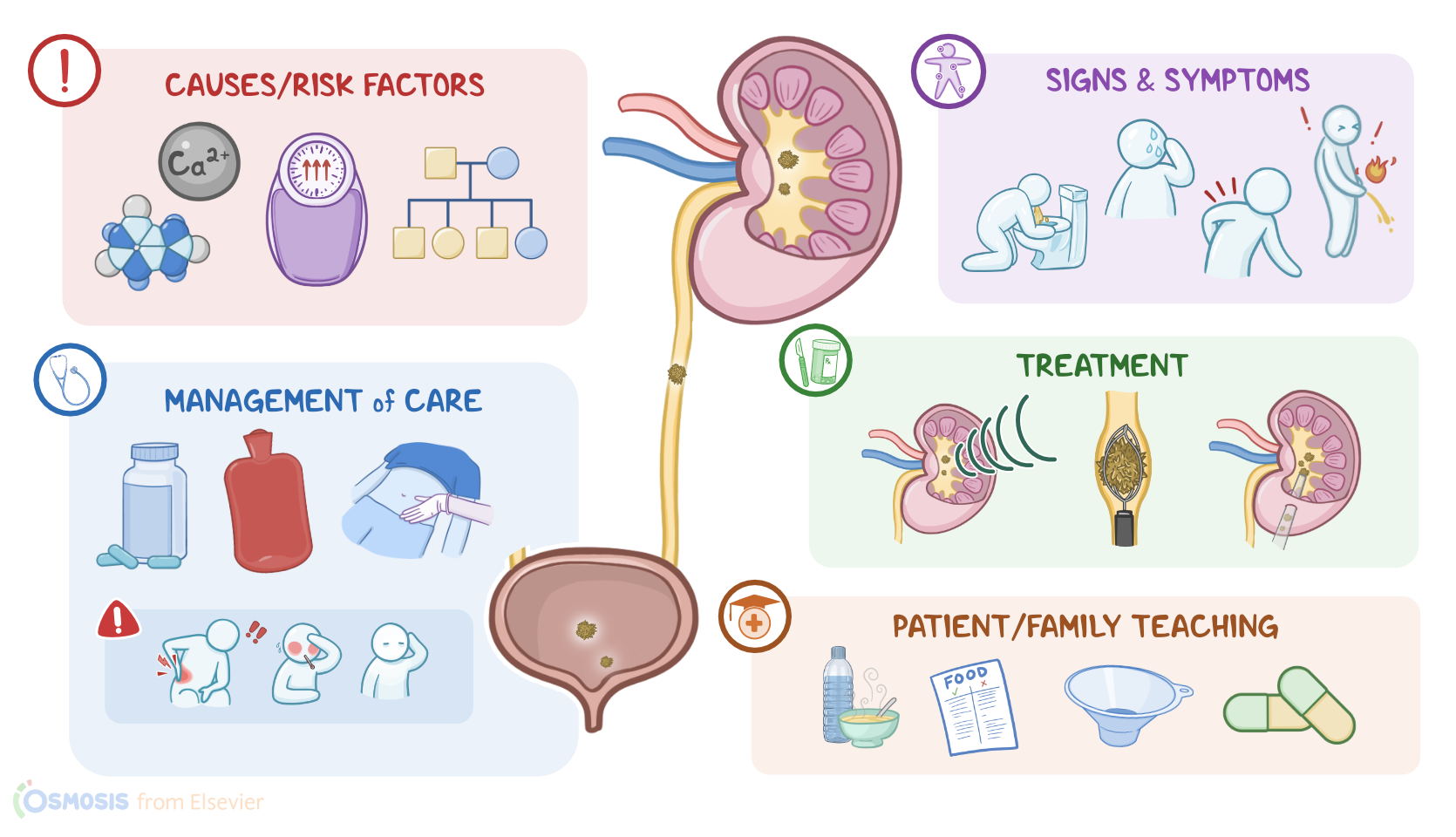
Nephrolithiasis and urolithiasis refer to the presence of stones, also known as calculi, in the kidney and the urinary tract, respectively. Stones form when solutes in the urine precipitate out and crystallize. Depending on which solute precipitates to form the stone, there are five main types of stones: calcium oxalate, calcium phosphate, struvite, uric acid, and cystine stones.

Now, let’s quickly review some anatomy and physiology of the urinary tract, which is made of the kidneys, ureter, bladder, and urethra. The kidneys are in charge of producing urine, which is made up of water and solutes.

The process starts with glomerular filtration in the renal corpuscle, which is made up of the glomerulus and the Bowman’s capsule. As blood flows into the glomerulus, water, electrolytes like sodium, and other small particles in the blood like creatinine, urea nitrogen, and glucose, pass through the endothelial lining of the capillaries and into Bowman's space. Large proteins and red blood cells however, aren’t filtered. Now, the next steps, which include tubular reabsorption and secretion, take place in the renal tubules, which are surrounded by the peritubular capillaries. Here, water and solutes get passed back and forth between the filtrate in the lumen of the renal tubule and the blood in the peritubular capillaries.

Finally, urine is drained into the renal pelvis, which then narrows to form the ureter, and transports urine to the bladder. Ultimately, urine exits the bladder through the urethra.

Now, nephrolithiasis and urolithiasis are caused by the increased concentration of certain solutes in the urine. This can happen if there’s an increase in the solute, which could be calcium with oxalate or phosphate, leading to the formation of calcium stones; magnesium, ammonia, and phosphate, leading to struvite stones, as well as uric acid forming uric acid stones and cystine, forming cystine stones. Alternatively, there could be a decrease in the solvent, or water in the urine.

That being said, the exact cause why these stones form is usually unknown, but they have been associated with several risk factors. Common risk factors for all types of kidney stones include a family history of nephrolithiasis, prolonged immobility, obesity, and dehydration, which is more likely to occur in warm climates or due to low fluid intake.

Now, for calcium stones, risk factors mainly include hypercalcemia and hypercalciuria. This can be a complication of hyperparathyroidism, because parathyroid hormone increases calcium release from the bones and calcium reabsorption from the intestines, while decreasing calcium excretion from the kidneys. Hypocitraturia is another cause, where less citrate is eliminated in the urine. This is because normally, citrate prevents kidney stone formation by inhibiting crystal growth and aggregation.

Additionally, there are medications that can increase the risk of developing calcium stones, like loop diuretics, acetazolamide, theophylline and glucocorticoids. Other risk factors include fat malabsorption, ingestion of ethylene glycol or antifreeze, and high levels of vitamin C, all of which raise oxalate levels in the blood. Finally, a diet heavy in oxalate-rich foods like rhubarb, spinach, chocolate, nuts, and beer increases the risk of calcium stones.

For uric acid stones, risk factors include acidic urine, which can be caused by acidosis, meaning a low blood pH. This can happen with chronic diarrhea, where large amounts of bicarbonate are lost in the stool; or with poorly controlled diabetes, where the body can’t use glucose and instead breaks down fat for fuel, creating acidic ketones as a by-product. Another important risk factor is hyperuricemia, or high levels of uric acid in the blood, which might be due to conditions like gout.

There are also medications that promote the development of uric acid stones, like thiazide diuretics, salicylates, probenecid and allopurinol. Lastly, since uric acid is a breakdown product of purine, consuming lots of purines, which can be found in shellfish, anchovies, red meat or organ meat, increases the risk of uric acid stones.

Next are cystine stones, which occur in the setting of cystinuria, a genetic condition where cystine cannot get reabsorbed from the renal tubules, so instead gets excreted in the urine.

Finally, for struvite stones, the main risk factor is a urinary tract infection with bacteria that produce urease, like Proteus and Klebsiella. These bacteria increase urine pH, making it a favorable environment for magnesium ammonium phosphate to precipitate.

Alright so, pathology of kidney stones begins with urinary supersaturation of certain solutes, which results in precipitation and formation of crystals. Those crystals then act as a nidus, or place where more solutes can deposit, and, over time, this forms a kidney stone. Now, the stone can become stuck and obstruct the urinary tract, which happens most commonly in ureteropelvic junction or in the ureter. When this happens, the ureter becomes dilated and stretched. At the same time, the urothelial lining is damaged, which causes bleeding and consequent hematuria. Also, if the obstruction persists, urine won’t flow, which is called urinary stasis, making the urinary tract prone to infection. Sometimes, kidney stones can completely obstruct the ureter and this way the urine builds up behind the obstruction, causing hydronephrosis, or dilation of the renal pelvis and calyces, which can cause permanent kidney damage.

Key Takeaways

Nephrolithiasis and urolithiasis refer to stones, also called calculi, in the kidney and urinary tract, respectively. They form when solutes in the urine precipitate out and crystallize. Depending on the type of precipitated solute forming the stone, there are five main types of stones: calcium oxalate, calcium phosphate, struvite, uric acid, and cystine stones. Renal and urinary stones can cause urinary tract obstruction, infection, and even hydronephrosis, which can lead to permanent kidney damage.

Even though people with renal and urinary stones may be asymptomatic, common symptoms include renal colic and flank pain. The pain is often sharp, sudden, and severe enough that it may trigger nausea, vomiting, pallor, and sweating. There may also be hematuria, dysuria, urinary urgency and frequency, and signs of infection.

The diagnosis involves a history and physical assessment, followed by lab exams such as urinalysis and blood studies, as well as imaging like CT and ultrasound. Treatment includes medications to treat pain and surgery in some severe cases. There are also other procedures like extracorporeal shock wave lithotripsy (ESWL), which uses high-energy sound waves to break these stones into smaller fragments able to be passed in the urine. Increased fluid intake and dietary changes may be recommended to prevent stone formation in the future.