Shock - Hypovolemic: Nursing
Introduction0:00–0:14
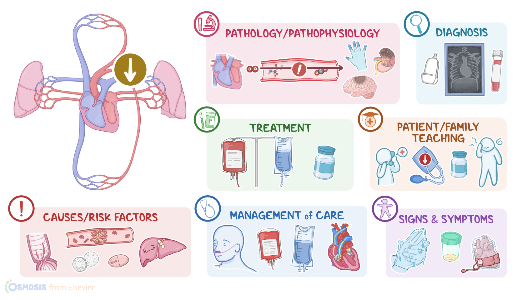
Hypovolemic shock is a life-threatening condition characterized by a decrease in intravascular volume in the cardiovascular system, which becomes insufficient to support adequate perfusion of the body tissues.Now, let’s talk about the physiology of the cardiovascular system, which consists of the heart and blood vessels.
Physiology0:14–1:16
The main job of the heart is to pump oxygenated and nutrient-rich blood through the arteries to the body tissues, and receive deoxygenated blood through the veins and pump it to the lungs to pick up more oxygen.
For this to be possible, there are three key factors to keep in mind: the cardiac output, which is the amount of blood pumped out by the heart per minute; the intravascular volume, which is the amount of blood in the client’s circulation; and the peripheral vascular resistance, which is the resistance of blood flow in peripheral arteries.
Normally, oxygen is delivered to the tissues because there’s enough pressure in the cardiovascular system to push blood through the body; so, blood pressure has a major effect on the amount of blood that reaches tissues and organs.
For the most part, blood pressure is regulated by changes in peripheral vascular resistance and cardiac output, so that if one of them decreases, the other increases to try to compensate.So, hypovolemic shock is caused by a significant decrease of intravascular volume, and can be classified as hemorrhagic and non-hemorrhagic.
Causes & risk factors1:16–2:25
Hemorrhagic shock is the most common, and it’s usually caused by severe bleeding from postpartum hemorrhage, gastrointestinal bleeding, or traumatic injuries.
On the other hand, non-hemorrhagic hypovolemic shock can be caused by anything that results in significant fluid loss, such as vomiting or diarrhea; renal losses, such as excessive diuresis in diabetic ketoacidosis or diabetes insipidus; excessive sweating, like in hyperthermia or hyperthyroidism; or third-spacing, which includes anything that causes intravascular fluid to shift into the interstitial space, like burns or acute pancreatitis.Risk factors to look out for in hypovolemic shock include any conditions that could potentially worsen a hemorrhage, like having a clotting disorder or liver problems, as well as taking antiplatelet medications like aspirin, or anticoagulant medications like warfarin.
Additional risk factors include anything that increases the risk of dehydration, such as persistent vomiting or diarrhea.
Pathology2:25–3:58
Now, the pathology of hypovolemic shock develops from a loss of intravascular volume, which in turn decreases the blood pressure and the venous return to the heart, decreasing cardiac output.
So, in order to increase cardiac output, the body responds by releasing hormones into the bloodstream. This includes catecholamines such as epinephrine and norepinephrine which cause vasoconstriction, increase heart rate and contractility to maintain blood pressure and increasing blood flow; as well as ADH, which acts on the kidneys, increasing fluid retention and angiotensin II, which also constrict the blood vessels and causes sodium retention in the kidneys, raising blood pressure.
As a result, the body may initially compensate by increasing peripheral vascular resistance and blood pressure, as well as the cardiac output.
Additionally, vasoconstriction in the kidneys causes them to produce less urine in an attempt to retain as much fluid as possible, and thus maintain the intravascular volume.
However, if shock isn’t addressed quickly, these compensatory mechanisms may not be able to maintain blood pressure, and as a result, perfusion to the tissues decreases even more.
If this occurs, the organs won’t get enough oxygenated blood, so they’ll need to switch to anaerobic metabolism, which eventually leads to metabolic acidosis, or excess acid in the body, leading to hyperventilation, confusion, or death, if severe.
Finally, if shock persists for too long, vital organs such as the brain, heart, and kidneys may begin to shut down, leading to multiple organ failure.The clinical manifestations of hypovolemic shock depend on the severity of the intravascular volume loss.
Clinical manifestations3:58–6:07
During the initial stage, compensatory mechanisms such as increased heart rate and vascular constriction are sufficient to maintain cardiac output within the normal range.
Cardiac output can best be seen by obtaining the mean arterial pressure, or the average pressure in one cardiac cycle, because it is a more accurate indicator of perfusion than a normal blood pressure measurement.
In this stage, the MAP is decreased by less than 10 mmHg from baseline. Clients may experience tachycardia, but compensatory mechanisms keep blood pressure at around the normal range, so it may be difficult to detect shock at this stage.
During the compensation stage, the compensatory mechanisms are fully active but the MAP is decreased and is about 10-15 mmHg below baseline.
The client’s skin can become cold and clammy, indicating that blood flow is being redirected to vital organs like the brain and heart.
Other symptoms include pallor, severe hypotension and tachycardia, decreased peripheral pulses, and oliguria. In the progressive stage, clients can develop organ failure because the compensatory mechanisms can no longer guarantee adequate blood flow to vital organs.
The MAP is sustained at more than 20 mmHg below baseline. The client can experience anxiety, altered level of consciousness, cyanosis, increased respirations, decreased oxygen saturation from lung failure, profound hypotension, bradycardia, and irregular heart rhythm from heart failure, as well as anuria, which occurs due to kidney failure.
In the refractory stage, cell death occurs in the vital organs due to the lack of oxygen reaching the tissues. The MAP is still sustained at more than 20 mmHg from baseline.
At this point, the damaged organs cannot respond to treatment, leading to multiple organ dysfunction. The client may experience a sudden loss of consciousness, shallow respirations, unmeasurable oxygen saturation, non-palpable pulses, or death.
The diagnosis of hypovolemic shock starts with the client’s history and physical assessment. The assessment identifies signs of hypoperfusion, including a drop in oxygen saturation, low or declining blood pressure, sudden changes in the ECG, or decreased urinary output.
Diagnosis6:07–6:48
Additionally, laboratory tests can reveal decreased oxygen levels, metabolic acidosis, increased lactate levels, and coagulation abnormalities, which can be helpful to determine the evolution of shock and efficacy of treatment.
Finally, imaging techniques, such as an ultrasound, chest X-ray, or CT scan can be performed to look for an internal source of hemorrhage.The main goal of treatment in hypovolemic shock is to stabilize the client’s blood pressure and maintain perfusion.
Treatment6:48–7:37
The initial treatment for hypovolemic shock is fluid resuscitation using colloids or crystalloids, as well as blood transfusions, if needed.
Colloids contain protein molecules and are administered to improve tissue perfusion by increasing osmotic pressure in the plasma volume.
If blood pressure cannot be maintained with fluids alone, vasopressor medications like norepinephrine, phenylephrine, or dopamine can be administered to promote vasoconstriction in an attempt to increase blood pressure.
Definitive treatment of hypovolemic shock involves addressing the underlying cause, when possible.Alright, let’s look at the nursing care you’ll be providing for a client with hypovolemic shock.
Management and care7:37–10:43
Your priority nursing goals are to restore and maintain intravascular volume, monitor for fluid overload and complications of shock, and monitor for improved hemodynamic stability.
Begin by assisting with identifying the source and extent of fluid loss. Perform a thorough assessment, noting their skin color, vital signs, oxygen saturation, and peripheral pulses.
Also be sure to assess their hemoglobin and hematocrit. Administer the prescribed IV fluids, and insert an indwelling urinary catheter to monitor urinary output.
Notify the healthcare provider immediately if your client experiences an altered LOC, cyanosis, cold skin, weak pulses, minimal urinary output, or hypotension despite fluid resuscitation.
Prepare your client for insertion of a central venous pressure catheter to monitor ventricular function of the heart, if indicated.
If hypovolemia is due to blood loss, administer ood products as prescribed. During care, continue monitoring your client closely for fluid overload.
Assess your client’s respiratory rate, lung sounds, oxygen saturation, urine output, and presence of edema. Notify the healthcare provider immediately if your client experiences crackles in the lungs, shortness of breath, bounding pulses, or minimal urinary output.
Standby to assist with pulmonary catheter insertion, and administer the prescribed vasopressor therapy to stabilize your client’s hemodynamic status; and prepare your client for endotracheal intubation and mechanical ventilation, if indicated.
Also, assess your client’s ABG results to assist in identifying acidosis. During your assessment, pay close attention to your client’s level of consciousness, blood pressure, heart rate, and respiratory rate.
Then, prepare to assist in treating the underlying cause. Then, monitor your client closely for indications of improved hemodynamic status and improved perfusion, including capillary refill, increased blood pressure, improving skin color and temperature, regular heart rate and rhythm, and increased urine output.
Also, monitor your client’s CVP level to determine if it is in the normal range of 2 to 5 mmHg. Be sure to assess your client’s MAP as well, which should be within 10 mmHg of their baseline reading.
If your client responds to the therapeutic interventions, prepare to reduce the rate of fluids and begin weaning vasopressor support, as indicated.Now, moving on to client and family teaching.
General client and family teaching10:43–11:32
First, teach your client that hypovolemic shock is a serious condition caused by severe loss of fluid in the blood vessels that leads to decreased oxygen delivery to major organs.
Then, explain the specific cause of the hypovolemic shock they experienced, and allow plenty of time for them to ask questions.
Next, teach them about treatments they received for hypovolemic shock, such as IV fluids and blood products to restore circulation and vasopressors to increase blood pressure.Teach your client who is discharged home how to recognize early signs of hypovolemic shock due to blood loss such as dizziness, low blood pressure, decrease in urine output, and to immediately notify their healthcare provider if they occur.
Review11:32–12:26
Alright, as a quick recap… Hypovolemic shock is a serious condition in which loss of intravascular volume in the cardiovascular system causes hypoperfusion of organs and tissues.
Hypovolemic shock can be caused by blood loss or fluid loss related to certain chronic conditions or therapies. The treatment goal is to identify the cause, correct it and stabilize your client.
Clients at risk include those on certain medications that cause unintended fluid loss, clients who have experienced trauma, and postoperative clients.
Goals of nursing care include restoring and maintaining intravascular volume, monitoring for fluid overload and complications of shock, and assess for improved hemodynamic stability.
Client and family education includes teaching them about treatments for hypovolemic shock, and to seek immediate medical care as soon as possible when early signs of hypovolemic shock occur.
| SHOCK - HYPOVOLEMIC | ||
| KEY POINTS | NOTES | |
| DEFINITION |
| |
| PHYSIOLOGY |
| |
| CAUSES AND RISK FACTORS |
| |
| PATHOPHYSIOLOGY |
| |
| SIGNS AND SYMPTOMS |
| |
| DIAGNOSIS |
| |
| TREATMENT |
| |
| MANAGEMENT OF CARE |
| |
| PATIENT AND FAMILY TEACHING |
| |
No notes for this video yet
Try adding a note below