Venous thromboembolism (VTE): Nursing process (ADPIE)

Last updated: October 29, 2025

Venous thromboembolism (VTE): Nursing process (ADPIE)

Watch later

Watch later

Hypokalemia: Clinical
Movement of water between body compartments
The role of the kidney in acid-base balance
Blood histology
Blood components
Respiratory acidosis
Metabolic acidosis
Respiratory alkalosis
Metabolic alkalosis
Metabolic and respiratory alkalosis: Clinical
Metabolic and respiratory acidosis: Clinical
Bronchodilators: Beta 2-agonists and muscarinic antagonists
Chronic obstructive pulmonary disease (COPD): Clinical
Chronic obstructive pulmonary disease (COPD): Nursing process (ADPIE)
Adrenergic antagonists: Alpha blockers
Sympatholytics: Alpha-2 agonists
Alpha-2 adrenergic agonists: Nursing pharmacology
Adrenergic receptors
Adrenergic antagonists: Beta blockers
Diabetes mellitus: Clinical
Diabetes mellitus (DM): Nursing process (ADPIE)
Diabetes mellitus
Diabetes mellitus: Pathology review
Stroke: Nursing process (ADPIE)
Stroke: Clinical
Ischemic stroke
Intracerebral hemorrhage
Peptic ulcers and stomach cancer: Clinical
GERD, peptic ulcers, gastritis, and stomach cancer: Pathology review
Sickle cell disease (NORD)
Sickle cell disease: Nursing process (ADPIE)
Sickle cell disease: Clinical
Blood groups and transfusions
Erythropoietin
Blood products: Nursing pharmacology
Oxygen binding capacity and oxygen content
Blood products and transfusion: Clinical
Hemophilia
Hemophilia: Nursing process (ADPIE)
Leukemia: Nursing process (ADPIE)
Chronic leukemia
Leukemias: Pathology review
Acute leukemia
Leukemia: Clinical
Lymphomas: Pathology review
Lymphoma: Clinical
Non-Hodgkin lymphoma
Hodgkin lymphoma
Lymphatic system anatomy and physiology
Anatomy of the abdominal viscera: Liver, biliary ducts and gallbladder
Liver anatomy and physiology
Cirrhosis
Cirrhosis: Pathology review
Cirrhosis: Clinical
Cirrhosis: Nursing process (ADPIE)
Hepatitis C virus
Hepatitis A and Hepatitis E virus
Liver cancer: Nursing
Cholestatic liver disease
Non-alcoholic fatty liver disease
HIV (AIDS)
HIV and AIDS: Pathology review
Antiretrovirals for HIV/AIDS - Protease inhibitors: Nursing pharmacology
Antiretrovirals for HIV/AIDS - NRTIs and NNRTIs: Nursing pharmacology
Antiretrovirals for HIV/AIDS - Integrase strand transfer inhibitors: Nursing pharmacology
Anatomy of the abdominal viscera: Pancreas and spleen
Pancreatitis: Nursing process (ADPIE)
Glaucoma: Nursing process (ADPIE)
Glaucoma
Eye conditions: Refractive errors, lens disorders and glaucoma: Pathology review
Acute respiratory distress syndrome (ARDS): Nursing
Anatomy of the coronary circulation
Coronary artery disease: Pathology review
Coronary artery disease: Clinical
ECG basics
ECG normal sinus rhythm
ECG rate and rhythm
ECG axis
ECG intervals
ECG QRS transition
ECG cardiac infarction and ischemia
Cardiac cycle
Arterial disease
Delirium
Dementia and delirium: Clinical
Vascular dementia
Frontotemporal dementia
Dementia with Lewy bodies
Dementia: Pathology review
Tumor lysis syndrome (TLS): Nursing Process (ADPIE)
Cholelithiasis: Nursing
Coronary artery disease (CAD) and angina pectoris: Nursing process (ADPIE)
Breast cancer: Nursing process (ADPIE)
Ovarian cancer: Nursing
Cervical cancer: Nursing
Hormones and hormone modulators for cancer: Nursing pharmacology
Endometriosis: Nursing
Heart failure
Left-sided heart failure: Nursing process (ADPIE)
Heart failure: Pathology review
Pneumonia: Pathology review
Pneumonia
Bacterial pneumonia: Nursing process (ADPIE)
Respiratory stimulants: Nursing pharmacology
Corticosteroids - Inhaled: Nursing pharmacology
Pneumothorax and hemothorax: Nursing
Chest trauma: Clinical
Pleural effusion: Nursing
Tuberculosis (TB): Nursing
Parkinson disease: Nursing process (ADPIE)
Huntington disease
Multiple sclerosis (MS): Nursing
Multiple sclerosis
Guillain-Barré syndrome: Nursing
Guillain-Barre syndrome
Myasthenia gravis: Nursing
Acute kidney injury (AKI): Nursing process (ADPIE)
Acute kidney injury: Clinical
Chronic kidney disease (CKD): Nursing
Chronic kidney disease
Chronic kidney disease: Clinical
Polycystic kidney disease (PKD): Nursing
Renin-angiotensin-aldosterone system
Osteoarthritis: Nursing
Osteoarthritis
Rheumatoid arthritis (RA): Nursing process (ADPIE)
Rheumatoid arthritis: Clinical
Rheumatoid arthritis
Rheumatoid arthritis and osteoarthritis: Pathology review
Systemic lupus erythematosus
Systemic lupus erythematosus (SLE): Pathology review
Systemic lupus erythematosus (SLE): Clinical
Systemic lupus erythematosus (SLE): Nursing
Mycobacterium tuberculosis (Tuberculosis)
Peripheral venous disease (PVD): Nursing process (ADPIE)
Peripheral arterial disease (PAD): Nursing process (ADPIE)
Buerger disease: Nursing
Raynaud phenomenon: Nursing
Aortic dissections and aneurysms: Pathology review
Aortic aneurysms and dissections: Clinical
Aortic dissection
Aortic aneurysm: Nursing process (ADPIE)
Venous thromboembolism (VTE): Nursing process (ADPIE)
Benign prostatic hyperplasia (BPH): Nursing process (ADPIE)
Benign prostatic hyperplasia
Prostate cancer: Nursing
Prostate cancer
Testicular cancer: Nursing
Testicular cancer
Diabetic ketoacidosis (DKA): Nursing process (ADPIE)
Endocrine system anatomy and physiology
Vitamin B12 deficiency
Thalassemia: Nursing
Anemia - Iron-deficiency: Nursing
Anemia - Macrocytic: Nursing
Anemia - Aplastic: Nursing
Thyroid hormones
Medications for thyroid disorders: Nursing pharmacology
Hyperthyroidism: Nursing process (ADPIE)
Hypothyroidism: Nursing process (ADPIE)
Hypoparathyroidism: Nursing
Hyperparathyroidism: Nursing
Anxiety disorders: Nursing process (ADPIE)
Spinal cord injury (SCI): Nursing
Cluster A personality disorders
Smoke inhalation injury: Nursing process (ADPIE)
Shock - Anaphylactic: Nursing
Shock - Obstructive: Nursing
Shock - Neurogenic: Nursing
Shock - Hypovolemic: Nursing
Shock - Cardiogenic: Nursing
Shock - Septic: Nursing
Pulmonary edema: Nursing
Burn injury: Nursing

Notes

VENOUS THROMBOEMBOLISM (VTE)

KEY POINTS
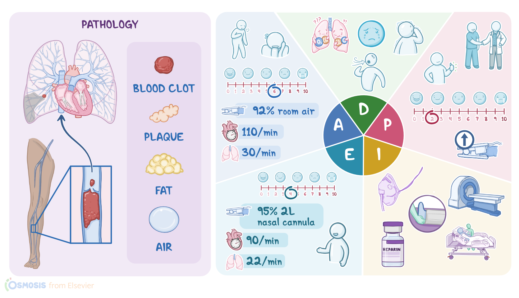
NOTES
PATIENT REPORT
  • 34-year-old woman
  • Shortness of breath, chest pain, blood-tinged sputum
  • Delivered healthy, full-term infant via C-section 1 week ago
  • No other significant medical history

PATHOPHYSIOLOGY
  • Pulmonary embolism
    • Embolus lodges in pulmonary artery
    • Pulmonary circulation obstructed
  • Deep vein thrombosis (DVT)
    • Clot forms in a large vein
  • Venous thromboembolism
    • Blood clot forms in a vein
  • Risk factors
    • Virchow triad
  • Signs and symptoms (PE)
    • Depend on size and location of pulmonary artery blockage
    • Dyspnea
    • Cough
    • Tachypnea
    • Chest pain
    • Disorientation
    • Anxious
    • Apprehension or impending doom
    • Hemoptysis
  • Complications (PE)
    • Pulmonary hypertension
    • Right ventricular failure
    • Shock
    • Death

DIAGNOSIS AND TREATMENT
  • Diagnosis
    • CT
    • V/Q scan
    • Laboratory tests
  • Treatment
    • Small
      • Resolve on own
    • Large
      • Fibrinolytics
      • Anticoagulants
      • Embolectomy
      • IVC filter

ASSESSMENT
  • Short of breath, pain with breathing
  • Anxious
  • Dyspneic with conversation
  • Telemetry: sinus tachycardia
  • Heart rate: 110
  • Respiratory rate: 30, shallow
  • Productive cough with blood-tinged sputum
  • Crackles
  • Blood pressure: 95/72 mmHg
  • Temperature: 98.6 F (37.0 C)
  • Pain: 6/10
  • Oxygen saturation: 90% room air
  • Unilateral calf swelling and warmth
  • Hemoglobin: 12.5 mg/dL (125 g/L)
  • Hematocrit: 40%
  • Red blood cells: 5.0 million/mm3 (5.0 x10¹²/L)
  • White blood cells: 15,000 cells/mm(15 x 109/L)
  • Prothrombin time: 10 seconds
  • Activated partial thromboplastin time: 30 seconds
  • D-dimer: 2300 ng/dL (23 mg/L)
  • Troponin: 0.2 ng/dL (2 μg/L)
  • pH: 7.49
  • PaCO2: 30 mmHg (4.0 kPa)
  • HCO3-: 20 mEq/L (20 mmol/L)

NURSING DIAGNOSES
  • Impaired gas exchange related to decreased lung perfusion
  • Ineffective breathing pattern related to anxiety and dyspnea
  • Acute pain related to verbalization of pain
  • Anxiety related to verbalization and situational crisis 

PLANNING
  • Optimize oxygenation and ventilation
  • Decrease pain
  • Verbalize decrease in anxiety

IMPLEMENTATION
  • Apply face mask with 8L oxygen
  • Transfer to radiology for CT
  • Elevate head of bed
  • Administer medications as prescribed
  • Explain condition
  • Education on hydration, physical activity
  • Review symptoms of VTE
  • Review bleeding precautions 

EVALUATION
  • Respiratory rate: 22
  • Crackles
  • Heart rate: 90
  • Telemetry: normal sinus rhythm
  • Blood pressure: 110/80
  • Temperature: 98.0 F (36.6 C)
  • Pain: 4/10
  • Oxygen saturation: 95% 8L simple mask
  • Verbalizes education
  • Reports less anxiety

Transcript

Watch video only

Renata Gutierrez is a 34-year-old woman who came to the Emergency Department, or ED, with her partner, because of shortness of breath, chest pain, and coughing up blood-tinged sputum. The triage nurse informs you that Ms. Gutierrez delivered a healthy full-term baby by cesarean section one week ago. She has no other significant past medical history.

Venous thromboembolism, or VTE, refers to a clot that starts in a vein.

Specifically, a pulmonary embolism, or PE, occurs when an embolus, which is a small mass that could be a blood clot, fat, or even an air bubble, becomes lodged in the pulmonary artery and obstructs the pulmonary circulation. Most often the embolus is a blood clot associated with deep vein thrombosis, or DVT, which is when a clot forms in a large vein, usually in the leg or pelvis. The clot can break off and travel up the inferior vena cava to the right atrium, into the right ventricle, and finally into the pulmonary artery. This causes decreased blood flow to the lung tissue and impaired oxygenation.

Factors that increase the risk of a pulmonary embolism are summarized in Virchow triad, which include slowed blood flow, or venous stasis, hypercoagulation, meaning the blood is more likely to form clots, and damage to the endothelial lining of a blood vessel. Venous stasis can occur because of prolonged immobility like during a severe illness or after surgery, when an enlarged uterus compresses the nearby veins during pregnancy, or due to long-haul travel. Hypercoagulability can be caused by clotting disorders, use of oral contraceptives, smoking, and it occurs normally during pregnancy. And lastly, damage to the endothelial cell lining of a blood vessel can be the result of trauma or surgery.

Signs and symptoms of a pulmonary embolism depend on the size and location of pulmonary artery blockage. Even a small blockage impairs blood from getting into the lungs to pick up oxygen. Impaired oxygenation may cause a sudden onset of dyspnea, cough, tachypnea and chest pain that is described as pleuritic, meaning a sharp pain felt when inhaling and exhaling.

The patient may become disoriented and anxious from the hypoxemia, or they may report a feeling of apprehension and impending doom. As inflammation sets in, fluid buildup causes crackles which can be heard upon auscultation. Further deprivation of oxygen from the lung tissue leads to infarction of the lung tissue, which may result in hemoptysis. Complications from a PE can include pulmonary hypertension, right ventricular failure, shock and sudden death.

Diagnostics used to identify a PE include computed tomography pulmonary angiogram, or CTPA, where a dye is injected into the blood vessels to locate the blockage. A ventilation-perfusion scan, or V/Q scan, also called lung scintigraphy or ventilation-perfusion scintigraphy, can reveal areas of the lung that are ventilated, but not perfused, called a V/Q mismatch. Lab tests include a D-dimer test to detect fibrin breakdown products, which are usually present when there’s a blood clot.

Small clots may resolve on their own, however, large clots usually need treatment with fibrinolytic medications like tissue plasminogen activator, or tPA, to help break down the clot, and anticoagulants, like low molecular weight heparin to decrease blood coagulation. A

pulmonary embolectomy can also be done, either surgically or percutaneously, to remove the clot. For those at risk of developing further blood clots, anticoagulant medications may be given long term, or an inferior vena cava filter, or IVC filter, can be surgically placed into the inferior vena cava to prevent blood clots from entering the pulmonary artery and lungs. Keep in mind that the treatment course and medications may be influenced by factors such as whether the patient is pregnant or breastfeeding.

So, after you greet Ms. Gutierrez and introduce yourself as her nurse, you wash your hands, confirm her identity, and begin your assessment by asking how she’s doing. She replies she’s feeling short of breath, and it hurts every time she breathes. She goes on to tell you she has never experienced anything like this before and is very anxious. You notice that she has trouble getting the words out between breaths. After attaching a telemetry monitor, you observe sinus tachycardia with a heart rate of 110. Her respiratory rate is 30 per minute and shallow, she has a productive cough with blood-tinged sputum, and crackles are auscultated bilaterally. Her blood pressure is 95/72 mmHg, oral temperature 98.6° F, or 37°C, pain 6/10, and SpO2: 90% on room air.

Key Takeaways

Venous thromboembolism (VTE) is a condition where blood clots form in the veins, typically in the legs or pelvis, and can travel to the lungs, causing a pulmonary embolism (PE).

There are three main factors that lead to abnormal formation of clots, which are known as the Virchow's triad. They include venous stasis; a state of hypercoagulation; and damage to the endothelial cell lining. Venous stasis is said when blood doesn't flow appropriately in the veins due to prolonged periods of physical inactivity. Endothelial injury can occur when there is an injury or trauma to a blood vessel such as in trauma or surgery; and finally, hypercoagulable states can be seen in certain conditions like cancer and pregnancy.

Symptoms of VTE can include pain, swelling, warmth, and redness in the affected area, as well as shortness of breath, chest pain, and coughing up blood in the case of PE. The treatment involves thrombolytics to help break down the clot, or a thrombectomy, which is an intervention that aims to surgically remove the clot. Long-term treatment to prevent future thrombi from forming can include staying physically active, wearing compression stockings, and anticoagulant medications like warfarin or heparin.

Sources

  1. "Inflammation, Infection and Venous Thromboembolism. 128(12):2017-2036. " Circ Res. (2021)
  2. "Management of venous thromboembolism in pregnancy. 211:106-113. " Thromb Res (2022)
  3. "Long-term treatment of venous thromboembolism. 135(5):317-325. " Blood (2020)
  4. "Harrison’s Principles of Internal Medicine. 21st edition. ISBN: 978-1-264-26850-4 " McGraw Hill / Medical (2022)
  5. "Epidemiology and prevention of venous thromboembolism. 20(4):248-262. " Nat Rev Cardiol. (2023)
  6. "American Society of Hematology 2021 guidelines for management of venous thromboembolism: prevention and treatment in patients with cancer. 5(4):927-974. " Blood Advances. (2021)
  7. "Robbins & Kumar Basic Pathology. 11th edition. ISBN: 978-0-323-79018-5 " Elsevier (2022.)