Definitions & Key takeaways

Venous thromboembolism (VTE) is a condition where blood clots form in the veins, typically in the legs or pelvis, and can travel to the lungs, causing a pulmonary embolism (PE).

There are three main factors that lead to abnormal formation of clots, which are known as the Virchow's triad. They include venous stasis; a state of hypercoagulation; and damage to the endothelial cell lining. Venous stasis is said when blood doesn't flow appropriately in the veins due to prolonged periods of physical inactivity. Endothelial injury can occur when there is an injury or trauma to a blood vessel such as in trauma or surgery; and finally, hypercoagulable states can be seen in certain conditions like cancer and pregnancy.

Symptoms of VTE can include pain, swelling, warmth, and redness in the affected area, as well as shortness of breath, chest pain, and coughing up blood in the case of PE. The treatment involves thrombolytics to help break down the clot, or a thrombectomy, which is an intervention that aims to surgically remove the clot. Long-term treatment to prevent future thrombi from forming can include staying physically active, wearing compression stockings, and anticoagulant medications like warfarin or heparin.

Chapters:

Client Report0:00–0:34

Renata Gutierrez is a 34 year old woman who came to the emergency department or EED with her partner because of shortness of breath, chest pain and coughing up blood tinged sputum.
The triage nurse informs you that Miss Gutierrez delivered a healthy full term baby by Cesarean section one week ago. She has no other significant past medical history.
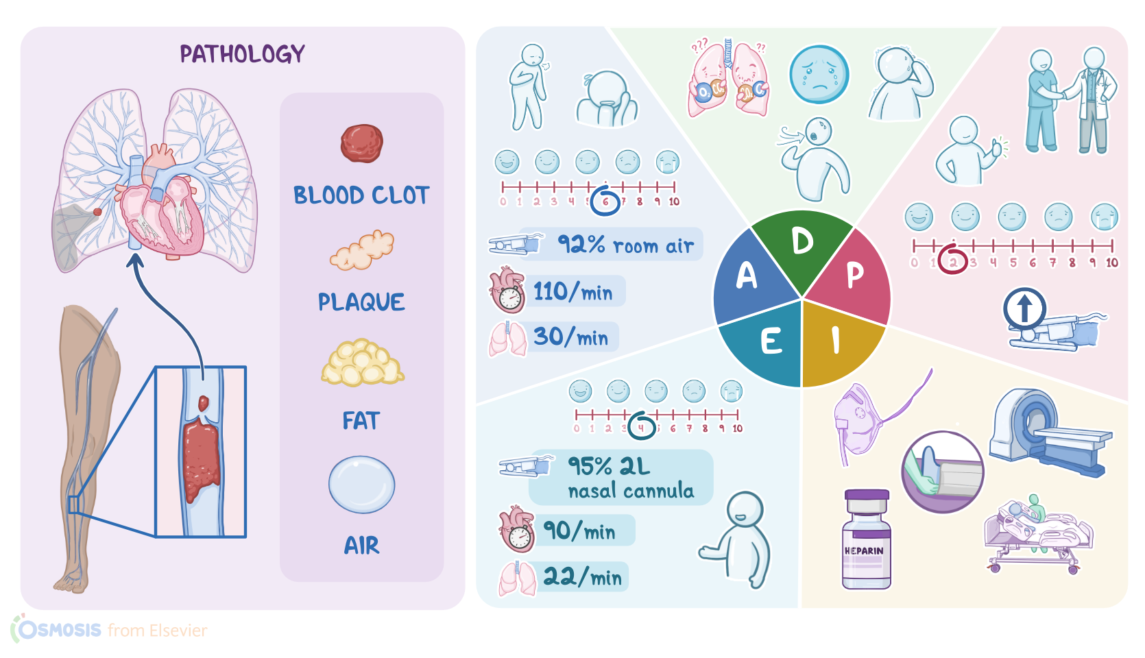
Venous thromboembolism or VTE E refers to a clot that starts in a vein, specifically a pulmonary embolism or pe occurs when an embolism, which is a small mass that could be a blood clot fat or even an air bubble becomes lodged in the pulmonary artery and obstructs the pulmonary circulation.

Pathology0:34–5:04

Most often the embolus is a blood clot associated with deep vein thrombosis or DVT, which is when a clot forms in a large vein.
Usually in the leg or pelvis. The clot can break off and travel up the inferior vena cava to the right atrium into the right ventricle and finally into the pulmonary artery.
This causes decreased blood flow to the lung tissue and impaired oxygenation factors that increase the risk of a pulmonary embolism are summarized in the fco triad, which includes slowed blood flow or venous stasis hypercoagulation, meaning the blood is more likely to form clots and damage to the endothelial lining of a blood vessel.
Venous stasis can occur because of prolonged immobility like during a severe illness or after surgery. When an enlarged uterus compresses the nearby veins during pregnancy or due to long haul travel, hypercoagulability can be caused by clotting disorders, use of oral contraceptives, smoking and it occurs normally during pregnancy and lastly damage to the endothelial cell lining of a blood vessel can be the result of trauma or surgery.
Signs and symptoms of a pulmonary embolism depend on the size and location of pulmonary artery blockage. Even a small blockage impairs blood from getting into the lungs to pick up oxygen, impaired oxygenation may cause a sudden onset of dyspnea, cough, tachypnea, and chest pain.
That is described as pleuritic, meaning a sharp pain felt when inhaling and exhaling. The patient may become disoriented and anxious from the hypoxemia or they may report a feeling of apprehension and impending doom as inflammation sets in fluid build up causes crackles which can be heard upon auscultation.
Further deprivation of oxygen from the lung tissue leads to infarction of the lung tissue which may result in hemoptysis.
Complications from a pe can include pulmonary hypertension, right ventricular failure, shock and sudden death. Diagnostics used to identify a pe include computed tomography, pulmonary angiogram or CTPA where a dye is injected into the blood vessels to locate the blockage, a ventilation perfusion scan or VQ scan, also called lung scintigraphy or ventilation perfusion scintigraphy can reveal areas of the lung that are ventilated but not perfused.
Called A VQ mismatch. Lab tests include ad dimer test to detect fibrin breakdown products which are usually present when there's a blood clot.
Small clots may resolve on their own. However, large clots usually need treatment with fibrinolytic medications like tissue plasminogen activator or T PA to help break down the clot and anticoagulants like low molecular weight heparin to decrease blood coagulation.
A pulmonary embolectomy can also be done either surgically or percutaneously to remove the clot. For those at risk of developing further blood clots.
Anticoagulant medications may be given long term or an inferior vena cava filter or IVC filter can be surgically placed into the inferior vena cava to prevent blood clots from entering the pulmonary artery and lungs.
Keep in mind that the treatment course and medications may be influenced by factors such as whether the patient is pregnant or breastfeeding.

Assessment5:04–7:38

So after you greet Miss Gutierrez and introduce yourself as her nurse, you wash your hands, confirm her identity and begin your assessment by asking how she's doing.
She replies she's feeling short of breath and it hurts every time she breathes. She goes on to tell you that she has never experienced anything like this before and is very anxious.
You notice that she has trouble getting the words out between breasts after attaching a telemetry monitor. You observe sinus tachycardia with a heart rate of 110.
Her respiratory rate is 30 per minute and shallow. She has a productive cough with blood tinged sputum and crackles are auscultated.
Bilaterally. Her BP is 95/72 millimeters of mercury.
Oral temperature, 98.6 °F or 37 °C pain. Six out of 10 and sp 2 90% on room air.
You quickly apply a nasal cannula and administer oxygen at 2 L per minute because she has recently delivered a baby. You inspect her calves and not her left calf is swollen and warm to the touch and dorsiflexion of her left foot is painful indicating a possible DVT.
You quickly review stat labs and note the following hemoglobin. 12.5 g per deciliter hematocrit, 40% RBC 5 million per cubic millimeter W BC, 15,000 per cubic millimeter prothrombin time, 10 seconds activated partial thromboplastin time, 30 seconds D dimer 2300 nanograms per milliliter troponin 0.2 nanograms per milliliter ph 7.49 PC, 230 millimeters of mercury and HC 320 milliequivalents per liter.
You immediately notify the emergency room physician of your assessment findings who arrives to assess her, continue to provide reassurance and document your assessment findings.

Diagnosis7:38–8:08

Now that you've completed your initial nursing assessment, you put together your nursing diagnosis for miss gutierrez impaired gas exchange related to decreased lung perfusion ineffective breathing pattern related to anxiety and hypoxia, acute pain related to verbalization of pain upon inhalation and anxiety related to verbalization of anxiety and experiencing a new situational crisis.

Planning8:08–8:41

Now that you've formulated your nursing diagnoses, it's time to collaborate with the physician and Miss Gutierrez to develop a holistic plan of care.
By the time of her transfer from the ed, your goals are to optimize oxygenation and ventilation resulting in decreased dyspnea and an increased oxygen saturation level.
Her pain rating will decrease to her identified comfort level of two out of 10 and she will verbalize a decrease in anxiety.
Next, you implement the plan of care. First, you assist with the transfer to radiology for a stat CT PA while waiting for her to return.

Implementation8:41–11:04

You note the physician's additional findings of a well score of 10 and prepare to implement additional physician orders.
M Gutierrez returns from the C TPA and you note the diagnosis of acute pulmonary embolism is confirmed. You elevate the head of the bed to reduce her dyspnea and you begin an infusion of the anticoagulant heparin.
You administer morphine, an opioid analgesic to decrease her pain and decrease her anxiety. You continually monitor for any changes to her status that require immediate notification of the physician such as a decrease in her SP O2, increased dyspnea, increased chest pain or signs of right heart failure.
Once Miss Gutierrez is stable and comfortable, you take the opportunity to explain how a recent pregnancy and Cesarean section increase the risk of DVT to help with prevention in the future.
You remind her to drink 6 to 8 cups or 1.5 to 2 L of water each day to avoid sitting for long periods of time and to watch for signs and symptoms of A DVT such as pain, redness, warmth or swelling in her lower legs.
In addition, you review the symptoms of VTE including shortness of breath, chest pain and cough. Tell her that if she experiences any of these, she should call for an ambulance immediately because she will be at increased risk for bleeding on anticoagulation medications.
You review safety precautions when using sharp objects and remind her to watch for bruising and signs of bleeding toward the end of your shift.
It's time to evaluate how Miss Gutierrez is doing. And if she is stable enough for transfer to the medical surgical unit for further care and monitoring her vital signs are respiratory rate 22 per minute with crackles still audible upon auscultation.

Evaluation11:04–11:58

Heart rate 90 with normal sinus rhythm BP. 110/80 temperatures, 98 °F or 36.6 °C pain, four out of 10 sp 2 95% with oxygen at 2 L per minute via nasal cannula.
She goes on to tell you the care and teaching you provided has made her much less anxious about how to care for herself when she is discharged home.
Ok. As a quick recap, your patient, Miss Gutierrez presented to the Ed today reporting dyspnea, chest pain and hemoptysis.
She is one week postpartum after delivering a full term baby via a Cesarean section. Her pregnancy and Cesarean delivery increased her risk for DVT.

Summary11:58–13:00

And when a clot formed in her calf broke off and traveled to her lungs, she developed a pe resulting in impaired perfusion and oxygenation in the lungs.
Your assessment revealed a swollen and tender left calf dyspnea, hemoptysis, chest pain, hypoxia, and sinus tachycardia.
Your nursing diagnoses included impaired gas exchange, ineffective breathing pattern, acute pain and anxiety. The planning goals included decreased dyspnea and a decreased level of pain and anxiety.
Along with the health care team, you work to implement these actions to achieve the goals of the plan of care throughout your shift.
And up until Miss Gutierrez transfers from the ed, you will continue to evaluate and revise your plan of care as needed.