Hemangioma · What Is It, Signs and Symptoms, and More

Published: May 29, 2025
Author: Nikol Natalia Armata
Editor: Alyssa Haag
Editor: Emily Miao, PharmD
Illustrator: Jessica Reynolds, MS
Copyeditor: Sadia Zaman, MBBS, BSc
7-day free trial

Go deeper with Osmosis

Osmosis is a learning platform with videos, questions, and AI tools to help you master topics like this.

4.8 · 12,000+ reviews
Watch quick, visual videos
Practice with Qbank-style questions
Use AI to explain, quiz, and review
Study anytime with the mobile app
Start free trial

No credit card · Cancel anytime

What is a hemangioma?

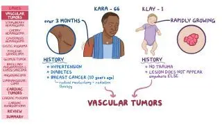
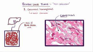
Hemangiomas, also known as strawberry marks, are benign tumors of blood vessels, most commonly seen in children. They have a characteristic clinical appearance of bright red papules, or plaques (i.e., bumps on the skin surface) of a few millimeters to a few centimeters, that may occur anywhere in the body. However, most frequently they are found on the face, scalp, chest, and back. The majority of lesions are solitary, but multiple lesions may occur in up to 20% of infants. Although most hemangiomas are superficial, they can also appear deeper in internal organs, such as the liver.

There are two common types of hemangiomas: congenital hemangiomas, which are present from birth at their maximal size, and infantile hemangiomas, which appear weeks after birth and increase gradually in size until they spontaneously disappear. Congenital hemangiomas can be further subcategorized into RICH (rapidly involuting congenital hemangioma), which regress like infantile hemangiomas, and NICH (non-involuting congenital hemangioma), 

Learn deeper with Osmosis

Master this topic faster with videos, questions, and AI.

Used by 8M+ healthcare learners.

Start free trial

No credit card · Cancel anytime

Are hemangiomas serious?

Hemangiomas are typically benign and self-limited in nature. In rare instances, however, they can be associated with other congenital conditions, like PHACE syndrome, which stands for Posterior fossa anomalies, Hemangiomas, Arterial anomalies, Cardiac anomalies, and Eye anomalies. Therefore, detailed screening is advised for individuals with large or multiple hemangiomas. 

What causes hemangiomas?

Hemangiomas are caused by the rapid growth of endothelial cells (i.e., the single-cell layer that lines all blood vessels) and the construction of pathological blood vessels. Several hypotheses try to explain what triggers such endothelial proliferation. The most dominant one suggests that hypoxic stress regulates the overexpression of several growth factors (e.g., Vascular Endothelial Growth Factor, or VEGF) that act on the endothelial cells. It may also be that angiogenic factors trigger the formation of new vessels (i.e., angiogenesis). Lastly, according to another hypothesis, hemangiomas may originate from stem cells (i.e., cells that have the capacity to evolve in any type of cell) of the trophoblast (i.e., the outermost layer of the blastocyst, which is the fertilized structure formed in early development), that differentiate uncontrollably to endothelial cells. 

Risk factors associated with hemangiomas include premature birth and low birth weight. White infants of non-Hispanic descent are also at an increased risk and infants assigned female at birth are 2 to 3 times as likely to experience hemangiomas. Additionally, advanced maternal age, preeclampsia, placenta previa, and bleeding during the first trimester of pregnancy may be associated with the development of hemangiomas.

What are the signs and symptoms of hemangiomas?

Hemangiomas typically do not cause any discomfort in the affected individuals. However, depending on their size and location, they may result in several signs and symptoms. Hemangiomas can be easily identified by their appearance. Characteristically, hemangiomas blanch (i.e., become white) when pressed as blood vessels empty - whereas, purpura (i.e., blood under the skin) does not. Although most hemangiomas are superficial and bright red, some are deeper in the skin and thus are "blue or skin-colored" (i.e., deep infantile hemangiomas). Large hemangiomas located near the eyes can restrict eye movement and even limit vision. When they are located near the airway (i.e., near the mouth and nose), breathing can be affected. Furthermore, hemangiomas can bleed or break on the skin surface, causing ulceration. When hemangiomas affect internal organs, symptoms may vary depending on the organ. For example, liver hemangiomas may cause loss of appetite, nausea, vomiting, and bloating after eating. Lastly, self-esteem can be negatively affected, especially in children,  by the appearance of large hemangiomas that are in exposed areas of the skin. 

What are the differential diagnoses for hemangiomas?

Differential diagnoses involve considering various possible conditions that could be causing symptoms and then ruling out each one through use of history, clinical evaluation, diagnostic tests, and critical thinking. This process helps to narrow down the list of potential diagnoses to determine the most likely cause of the symptoms.

Differential diagnoses can be broken down into four categories: most likely, less likely, least likely, and can’t miss. Most likely diagnoses are conditions most probable based on symptoms and clinical presentation. Less likely diagnoses are not as probable but should still be considered. On the other hand, least likely diagnoses can be considered if other, more probable conditions are excluded. Finally, can’t miss diagnoses are less common but critical to promptly identify and treat as they can lead to severe consequences.

Differential diagnoses for hemangioma include:
Most likely:
  • Infantile hemangioma: Common benign vascular tumor in infants, typically appearing within the first few weeks of life.
  • Congenital hemangioma: Present at birth, may be rapidly involuting (RICH) or non-involuting (NICH).
  • Cherry angioma: Common in adults, especially with aging; small, red, dome-shaped papules.

Less likely:
  • Pyogenic granuloma: Rapidly growing red lesion, often following trauma.
  • Venous malformation: Soft, compressible, bluish lesion that does not involute over time.
  • Capillary malformation (port-wine stain): Flat, pink to red lesion present at birth, does not regress.

Least likely:
  • Angiosarcoma: Malignant vascular tumor, more common in elderly or in areas of prior radiation.
  • Kaposi sarcoma: Vascular tumor associated with HHV-8, often seen in immunocompromised individuals.

Can’t miss:

How are hemangiomas diagnosed?

Hemangioma diagnoses begins with a history and physical assessment. Most hemangiomas can be diagnosed using observation and requiring no other testing. If diagnosis is uncertain or the hemangioma is atypical or in a critical location (e.g., liver), imaging tests and biopsy can be done. Imaging tests can include ultrasound, MRI, CT, or X-ray, depending on the presentation. 

How are hemangiomas treated?

Most hemangiomas resolve on their own, but careful observation is suggested in all cases. One of the first signs of involution is a change in color, from a bright red color to a darker gray-red color. If hemangiomas persist and become symptomatic, further treatment might be required. More specifically, treatment should be considered if lesions cause life-threatening complications (e.g., hemangiomas in the airway, liver or GI tract); cause functional impairment (e.g., hemangiomas in the periorbital region that cause visual abnormalities); are very large (>5cm) and rapidly growing; or have the potential for permanent damage.

Treatment options include administering beta blockers, such as oral propranolol, or corticosteroids, such as oral prednisone. Both may cause side effects to the user (e.g., bradycardia, hypotension, bronchospasm, and hypoglycemia from beta blockers; and increased appetite, osteoporosis, acne, and lower resistance in infections from corticosteroids) and therefore treatment should be adjusted if side effects arise. Topical beta blockers (e.g., timolol) and topical corticosteroids (e.g., betamethasone) can be used for solitary, small, superficial lesions. Direct intralesional injection of steroids can also be considered.

Laser therapy is a treatment option typically used for persistent or remaining lesions. In such cases, pulse dye laser treatment is commonly used. Surgical excision of hemangiomas is reserved for ulcerative, bleeding, and protruding hemangiomas. Additionally, hepatic hemangiomas may require surgical removal. Minimally invasive surgical techniques for hepatic hemangioma include arterial embolization, radiofrequency ablation, and hepatic radiation. A combination of more than one treatment option is often preferred in larger or resistant lesions.
What are the most important facts to know about hemangiomas?

A hemangioma is a benign tumor of blood vessels, most commonly seen in children. Having a characteristic clinical appearance of mostly bright red papules or plaques all over the skin surface, hemangiomas are also frequently called strawberry marks. They may present anywhere in the body, usually as single lesions, however, are most frequently found on the face, scalp, chest, and back. The rapid growth of endothelial cells (i.e., the single-cell layer that lines all blood vessels) and pathological blood vessels are the main cause of their formation. Hemangiomas are typically benign and are generally asymptomatic in the affected individual. However, signs and symptoms may vary depending on the location. Most hemangiomas regress on their own, therefore observation is suggested in all cases. If hemangiomas persist or become life-threatening, they may require treatment. 
Students say Osmosis is 100% worth it

Because Osmosis saves them time. Lowers stress. And actually helps them remember when it counts.

I used Osmosis to prepare for my first medical school licensing exam! Super helpful and interactive for people who may not do great with just pages of text info!

Cecilia Ruiz

Cecilia Ruiz

MD student

Sayan Misra

I have used Osmosis for about four years. Best thing I have ever used for my medical studies.

Sayan Misra

Sayan Misra

Med student

Osmosis videos are superior because they define simple concepts, tell a story with a clear progression, and provide context.

Jay Pate

Jay Pate

Dental student

References


Boston Children's Hospital. (2021). PHACE syndrome. In Boston Children’s Hospital. Retrieved November 16, 2021, from https://www.childrenshospital.org/conditions-and-treatments/conditions/p/phace-syndrome.   


Chamli A., Aggarwal P., Jamil R. T., et al. (2021, July 18). Hemangioma. In StatPearls [Internet]. Retrieved November 16, 2021, from https://www.ncbi.nlm.nih.gov/books/NBK538232/. 


DeHart, A., & Richter, G. (2019). Hemangioma: Recent advances. F1000Research, 8(F1000 Faculty Rev):1926. DOI: 10.12688/f1000research.20152.1