Nephrolithiasis · What Is It, Types, Signs and Symptoms, Diagnosis, Treatment, and More

Published: Sep 17, 2025
Author: Anna Hernández, MD
Editor: Antonella Melani, MD
Editor: Ian Mannarino, MD, MBA
Illustrator: Abbey Richard, MSc
7-day free trial

Go deeper with Osmosis

Osmosis is a learning platform with videos, questions, and AI tools to help you master topics like this.

4.8 · 12,000+ reviews
Watch quick, visual videos
Practice with Qbank-style questions
Use AI to explain, quiz, and review
Study anytime with the mobile app
Start free trial

No credit card · Cancel anytime

What is nephrolithiasis?

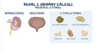
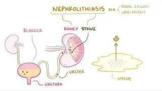
Nephrolithiasis, also known as kidney stones or renal calculi, refers to the presence of stones within the kidneys. It is one of the most common kidney diseases in adultsStone formation occurs when there’s an excess of crystal-forming substances that can’t be dissolved in the urine. High urinary excretion of certain substances—for instance, calcium, oxalate, uric acid, and cysteine—can promote stone formation, whereas the excretion of others, such as citrate, has a protective effect. Disturbances in urinary pH, a low urine volume, and a lack of protective substances that prevent the crystals from sticking together may contribute to stone formation. Additionally, environmental factors such as decreased fluid intake, hot climates, and dietary factors play an important role in the development of nephrolithiasis.  

Learn deeper with Osmosis

Master this topic faster with videos, questions, and AI.

Used by 8M+ healthcare learners.

Start free trial

No credit card · Cancel anytime

What does “nephrolithiasis” mean?

The word nephrolithiasis comes from “nephro,” which is the Latin word for kidneys, and “lithiasis,” which is the medical term used to refer to stones 

Does nephrolithiasis mean the same as kidney stones?

Nephrolithiasis refers specifically to kidney stones, although it is broadly used to refer to stones in the urinary tract. Ureterolithiasis, on the other hand, refers to stones within the ureter, and urolithiasis refers to stones in any part of the urinary tract (kidneys, ureter, bladder, and urethra). 

What is hypocitraturic nephrolithiasis?

The vast majority of kidney stones are made out of calcium salts. Calcium oxalate stones are the most common type, followed by calcium phosphate stones. The main risk factor for developing calcium stones is an increased excretion of calcium (i.e., hypercalciuria) and oxalate (i.e., hyperoxaluria). These abnormalities can be idiopathic, which means their cause is unknown, or can arise due to other systemic conditions, such as hyperparathyroidism, renal tubular acidosis, and malabsorption. 

Citrate has a protective role in stone formation by forming complexes with calcium and inhibiting further aggregation and growth of crystals. For this reason, a low excretion of citrate (hypocitraturia) is associated with an increased risk of developing stones. In most cases, the cause of hypocitraturia is not known. However, it has been associated with the presence of acid-balance disturbances, low levels of potassium in the blood, high-protein diets, and chronic diarrhea.  

What are the other types of nephrolithiasis?

Other less frequent types of stones found in the urinary tract can be struvite stonesuric acid stonescystine stones, or drug-induced stones.  

Struvite stones are sometimes referred to as infectious stones, since they can be associated with infections of the urinary tract, especially those caused by urea-splitting organisms (e.g., Proteus mirabilisKlebsiella, Staphylococcus, Xanthomonas, Pseudomonas, and Mycoplasma)These bacteria split urea molecules into ammonium and CO2, thereby raising the urine’s pH to neutral or alkaline values, and ultimately leading to the precipitation of solutes, to which the bacteria can adhere.  

Moreover, other types of stones can become infected by urea-splitting organisms, leading to secondary struvite stone formationStruvite stones are more common in biologically female individuals. They tend to grow large, filling part of the renal pelvis and calyces, and may adopt a staghorn shape. In the urine, they can be seen as “coffin-lid” shaped crystals. After their removal, if infected fragments of the stone are left behind in the kidney, they can often grow back, causing a persistent urinary tract infection and decreased renal function.  

Uric acid stones generally develop due to increased excretion of uric acid (hyperuricosuria) and low urine pH. Risk factors include high-protein diets, gout, inflammatory bowel disease, genetic diseases, and diabetesUric acid stones are one of the few kidney stones that can be managed successfully through medical treatment that consists of adequate hydration and supplements to raise the urine’s pH.  

Cystine stones are generally caused by cystinuria, a hereditary disease that causes increased excretion of cystine in the urine, as well as low urinary pH. Cystine stones are very hard and can be seen as thin hexagonal crystals in urine analysis 

Finally, drug-induced stones can develop by several mechanisms. Some drugs may alter excretion of urine solutes, contributing to an environment favorable for stone formation. Furthermore, excessive and chronic use of laxatives or diuretics can contribute to stone formation by leading to volume depletion and urine concentration that increase crystallization. . Additionally certain medications (e.g.,  indinavir or ciprofloxacin), or their metabolites, are poorly soluble in urine and can thus crystallize in the urine and create stones. 

What are the signs and symptoms of nephrolithiasis?

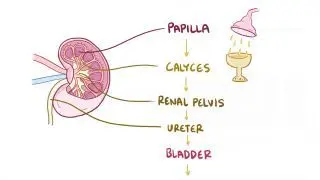
The symptoms of nephrolithiasis depend on stone size, shape, and location within the urinary tract. Initially, the stones are lodged in the renal pelvis, where they may be asymptomatic and cause no pain or obstruction of urine flow.   

Over time, the stones may move from the kidney into the ureter, which is a narrow tube that connects a kidney to the bladder. The passage of the ureteral stone causes increased pressure inside the ureter, followed by spasms of the smooth muscle lining and distension of the walls. This typically presents as renal colicsevere flank pain of sudden onset that can radiate to the inner thigh or the groin. Due to the intense pain, people may find themselves feeling restless or unable to find a comfortable position. Some individuals may even show signs of sweating or have nausea and vomiting as a physiological response to the intense pain. In addition, as the stone moves down, it can damage the ureter, causing bloody urine (i.e., hematuria).   

When stones reach the bladder, they can irritate and obstruct the bladder, leading to frequent or painful urination. Ultimately, small stones may spontaneously be eliminated through urine, relieving the pain. However, if the obstruction isn’t resolved, it can lead to severe complications, including chronic bladder discomfort, hydronephrosis, and a decrease in renal function due to urine build up. Additionally, the obstructed kidney can become infected, causing obstructive pyelonephritis, a life-threatening condition that requires urgent surgery to drain the kidney, as well as intravenous antibiotics 

How is nephrolithiasis diagnosed?

Asymptomatic nephrolithiasis may be detected as an incidental finding on imaging techniques or on urinalysis for hematuria. 

On the other hand, symptomatic individuals with renal colic or flank pain are first assessed through physical examination. A common sign in people with kidney infection or kidney stones is Giordano’s sign, which refers to tenderness upon percussion to the costovertebral angle, formed by the angle between the 12th rib and the lumbar spine  

Next, kidney stones can be identified through imaging techniques. Abdominopelvic X-rays can visualize the majority of calcium stones, determine their location and measure their size, but. small stones may be missed, and some stones such as uric acid and cystine stones are radiolucent, meaning that they can’t be seen on an X-ray due to their composition. A non-contrast CT scan, however, is able to detect most stones, regardless of their size and composition, and can also help determine the type of stone by looking at its density and is thus preferred.  

An ultrasound is an alternative imaging technique that doesn’t require radiation and therefore can be used to assess nephrolithiasis in vulnerable individuals like children or pregnant people. The presence of stones can be seen directly as a dense line with distal shadowing on the kidney or ureter, as well as indirectly by evaluating the dilation of the kidneys (hydronephrosis). For pregnant people with a high clinical suspicion and a negative ultrasound, an MRI can be used to identify kidney stones. 

Once an acute episode has subsided, a metabolic evaluation can be recommended, especially for individuals with recurrent episodes of nephrolithiasis. A metabolic evaluation generally includes assessing the characteristics of a 24-hour urine sample and, when possible, analyzing the stone crystals found in the urine. Additionally, blood tests may be performed to detect metabolic abnormalities, such as high calcium and uric acid levels in the blood. 

How is nephrolithiasis treated?

To treat the acute phase of nephrolithiasis pain control is vital and usually achieved with nonsteroidal anti-inflammatory drugs (NSAIDs) that reduce the inflammation and ureteral spasms. Individuals with persistent pain, contraindications to NSAIDs, or severe renal dysfunction, can be given opioid medications. Additionally, antiemetic medications may be given to help with the associated nausea and vomiting, and adequate fluid intake is encouraged to aid with stone passage and possible dehydration 

Subsequent treatment depends on the size, composition, and location of the stone, as well as the presence of complications 

For small stones (< 1 cm) without complications and effective pain treatment, expectant management (i.e., waiting for the stone to pass spontaneously) is appropriate. Most stones will pass over the course of four weeks. Some cases may be treated with medical expulsive therapy (MET), in which medication such as tamsulosin is given to relax the ureteral smooth muscle and help the stone pass more easily.   

Larger stones or those that do not pass despite MET should be considered for lithotripsy or surgical intervention. Urgent surgery may be required if there’s severe obstruction or infection of the urinary tract, decrease in renal function, uncontrollable pain, or renal obstruction in individuals with a solitary kidney. 

Lithotripsy, also known as extracorporeal shock wave lithotripsy (ESWL), is a non-invasive procedure that uses shock waves to fragment the stone, breaking it into smaller pieces that are excreted in the urine. Shock waves can only fragment low density stones, so hard stones like cysteine or calcium oxalate can’t be treated with lithotripsy. 

When lithotripsy isn’t possible, surgical removal of the stones is the preferred treatment. Stones can be removed either by ureteral endoscopy or through minimally-invasive surgery, as in percutaneous nephrolithotomy (PCNL). PCNL may be necessary for large stones (> 2 cm) or those that can’t be removed endoscopically because of their shape, such as staghorn stones. 

In rare cases in which stones are unable to be removed by endoscopy or minimally invasive surgery, open surgery such as ureterolithotomy or nephrolithotomy may be necessary. 

Finally, many people that develop nephrolithiasis are at risk for future recurrence. Because of that, general dietary modifications such as increased fluid intake and decreased sodium and protein consumption may be recommended to prevent future stone formation. Additionally, individuals who develop hypocitraturic or uric acid stones may be recommended supplements to raise the urine’s pH, such as potassium citrate or bicarbonate. In individuals with struvite or infectious stones, removal of the stone should be followed by antibiotic treatment to eliminate the presence of urea-splitting organisms. 
What are the most important facts to know about nephrolithiasis?

Nephrolithiasis refers to the presence of stones within the kidneys. Calcium oxalate stones are the most common type, followed by calcium phosphate, struvite, and uric acid stonesRisk factors for stone formation include low urine volume, increased excretion of solutes, and pH disturbances. Kidney stones may be asymptomatic for a long time. When the stones pass from the kidney into the ureter, the most common clinical presentation is a renal colic, which can be accompanied by nausea, vomiting, and hematuria. For diagnosis, symptomatic individuals are first assessed through physical examination, followed by imaging techniques —most commonly a non-contrast CT scan, and in selected cases, ultrasound with or without X-ray. Most cases can be treated with pain medications, such as NSAIDs and expectant management (waiting for the stone to pass spontaneously). Larger stones, or those that are not able to pass spontaneously, usually require stone removal with extracorporeal lithotripsy, endoscopy, or surgery. After the first episode, many people will have a recurrence. Because of that, lifestyle modifications such as increased fluid intake and dietary changes might be recommended.  

Key Takeaways

Definition

Nephrolithiasis (i.e., kidney stones or renal calculi) refers to the presence of stones within the kidneys. 

Types of Stones 

Hypocitrauric stones (e.g., calcium oxalate, calcium phosphate) 

- Struvite stones (i.e., infectious stones) 

Uric acid stones 

Cystine stones 

- Drug-induced stones 

Etiopathogenesis 

- Hypocitrauric stones:  

     - Formation of crystals due to ↓ secretion of citrate + ↑ secretion of calcium and oxalate 

     - Idiopathic / Systemic conditions 

- Struvite stones:  

     - UTI infectious organisms split urea into ammonium and CO2 → urine pH ↑ → solute precipitation + bacterial adhesion 

     - Other stones infected by urea-splitting organisms 

- Uric acid stones: 

     - ↑ excretion of uric acid + low urine pH 

     - Drug-induced stones 

     - Drug-induced changes in urine composition 

     - Drug-induced dehydration and volume depletion (e.g., excessive laxative or diuretic use) 

     - Drug crystallization in the urine 

Signs & Symptoms 

- Asymptomatic 

- Renal colic (severe flank pain of sudden onset with radiation to inner thigh or groin) → restlessness, discomfort 

- Sweating, nausea and vomiting 

- Hematuria 

- Frequent or painful urination 

- Unresolved obstruction: chronic bladder discomfort, hydronephrosis, ↓ renal function, obstructive pyelonephritis 

Diagnosis 

- Physical examination (Giordano’s sign)  

- Urinalysis 

- 24-hour urine collection 

- Blood tests 

- Imaging – Abdominopelvic X-rays, non-contrast CT scan, Ultrasound, MRI 

Treatment 

- Pain control via NSAIDs or opioids 

- Antiemetics 

- Adequate fluid intake 

- Small stones 

     - Expectant management 

     - Medical Expulsive Therapy (MET) 

- Large stones or stones refractory to MET 

     - Lithotripsy (ESWL) 

     - Surgical intervention 

     - Infectious stones: 

     - Antibiotics 

- Recurrence prevention 

     - ↑ Fluid intake 

     - ↓ Sodium & protein consumption 

     - Potassium citrate/bicarbonate supplements

Students say Osmosis is 100% worth it

Because Osmosis saves them time. Lowers stress. And actually helps them remember when it counts.

I used Osmosis to prepare for my first medical school licensing exam! Super helpful and interactive for people who may not do great with just pages of text info!

Cecilia Ruiz

Cecilia Ruiz

MD student

Sayan Misra

I have used Osmosis for about four years. Best thing I have ever used for my medical studies.

Sayan Misra

Sayan Misra

Med student

Osmosis videos are superior because they define simple concepts, tell a story with a clear progression, and provide context.

Jay Pate

Jay Pate

Dental student

References


Afshar K, Gill J, Mostafa H, Noparast M. Nonsteroidal anti-inflammatory drugs (NSAIDs) for acute renal colic. Cochrane Database Syst Rev. 2025;3(3):CD006027. Published 2025 Mar 14. doi:10.1002/14651858.CD006027.pub3z 


Gottlieb M, Long B, Koyfman A. The evaluation and management of urolithiasis in the ED: A review of the literature. Am J Emerg Med. 2018;36(4):699-706. doi:10.1016/j.ajem.2018.01.003 


Han H, Segal AM, Seifter JL, Dwyer JT. Nutritional management of kidney stones (nephrolithiasis). Clin Nutr Res. 2015;4(3):137-152. doi:10.7762/cnr.2015.4.3.137 


Matlaga BR, Shah OD, Assimos DG. Drug-induced urinary calculi. Rev Urol. 2003;5(4):227-231. Accessed August 26, 2020. https://www.ncbi.nlm.nih.gov/pmc/articles/PMC1508366/ 


Scales CD Jr, Smith AC, Hanley JM, Saigal CS. Prevalence of kidney stones in the United States. Eur Urol. 2012;62(1):160-165. doi:10.1016/j.eururo.2012.03.052 


Semins MJ, Matlaga BR. Medical evaluation and management of urolithiasis. Ther Adv Urol. 2010;2(1):3-9. doi:10.1177/1756287210369121