Rhabdomyolysis · What Is It, Causes, Treatment, and More

Published: Mar 06, 2026
Author: Anna Hernández, MD
Editor: Alyssa Haag, MD
Editor: Ian Mannarino, MD, MBA
Editor: Kelsey LaFayette, DNP, RN, FNP-C
Editor: Arianna Succi, MD
Editor: Lisa Miklush PhD, RN, CNS
Illustrator: Jessica Reynolds, MS
Copyeditor: Stacy Johnson, LMSW
7-day free trial

Go deeper with Osmosis

Osmosis is a learning platform with videos, questions, and AI tools to help you master topics like this.

4.8 · 12,000+ reviews
Watch quick, visual videos
Practice with Qbank-style questions
Use AI to explain, quiz, and review
Study anytime with the mobile app
Start free trial

No credit card · Cancel anytime

What is rhabdomyolysis?

Rhabdomyolysis is a condition in which skeletal muscle rapidly breaks down, causing the leakage of muscle breakdown products into the bloodstream. Some of these products, such as the protein myoglobin, are harmful to the kidneys and can lead to acute kidney injury. The severity of rhabdomyolysis depends on the extent of muscle damage. It ranges from asymptomatic elevations in serum muscle enzymes to life-threatening diseases associated with extreme enzyme elevations, electrolyte imbalances, and renal injury and impairment.  

Learn deeper with Osmosis

Master this topic faster with videos, questions, and AI.

Used by 8M+ healthcare learners.

Start free trial

No credit card · Cancel anytime

What causes rhabdomyolysis?

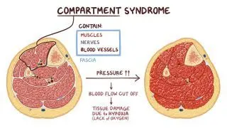
Rhabdomyolysis is caused by extensive muscle damage, which can have multiple causes. Traumatic causes of rhabdomyolysis include a crush injury (e.g., individuals involved in a building collapse); prolonged immobilization that can happen with hospitalized individuals or due to surgical positioning; high-voltage electrical injuries (e.g., from lightning or power supplies); and severe third-degree burns. Another important cause of rhabdomyolysis is compartment syndrome, which occurs when increased pressure inside an enclosed anatomical space reduces blood supply, resulting in tissue necrosis.  

Rhabdomyolysis may also occur after strenuous physical activity, especially in untrained individuals and in hot, humid environments that predispose to heat stroke. Impaired heat dissipation from wearing heavy sports equipment increases the risk of rhabdomyolysis. In addition, numerous metabolic or genetic disorders (e.g., inflammatory and metabolic myopathies, glycogen storage disorders, mitochondrial diseases, etc.) also increase the risk of rhabdomyolysis. Other conditions that may lead to rhabdomyolysis include hyperkinetic states, such as seizures, psychomotor agitation; delirium tremens; hyperthermia; viral or bacterial infections; electrolyte disorders, like hypokalemia and hypophosphatemia; use of amphetamines, cocaine, or alcohol; and several medications, including statins, colchicine, and propofol. Certain toxins, such as snake venom, insect venom from wasp and bee stings, and mushroom poisoning, are other known causes of rhabdomyolysis.  

Regardless of the mechanism, muscle injury results in a cascade of events that lead to the influx of extracellular sodium and calcium into muscle cells. Excessive intracellular sodium causes water influx, distorting the integrity of the intracellular space. Excessive intracellular calcium causes sustained myofibril contraction, resulting in ATP depletion within these cells, which affects normal cellular processes. Additionally, the elevated intracellular calcium activates intracellular proteases and lipases, promoting cell membrane damage and disrupting ion channels. The resulting inflammatory process can cause muscle fiber necrosis and release of muscle breakdown products - such as potassium and myoglobin, creatine kinase (CK), phosphate, uric acid, and various organic acids - into the bloodstream. 
What are the signs and symptoms of rhabdomyolysis?

The severity of rhabdomyolysis depends on the extent of muscle damage. The "classic triad” of rhabdomyolysis signs consists of muscle pain, weakness, and dark urine. Swelling of the affected muscles can also occur. Other symptoms may include confusion, fever, vomiting, and abdominal pain. The kidneys filter the massive amount of myoglobin released from damaged muscle cells, causing renal tubular obstruction, leading to kidney damage 

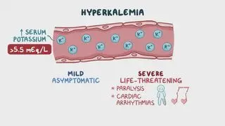
Hypovolemia and other electrolyte imbalances, like hyperkalemia, hypocalcemia, hyperuricemia, and acidosis, are also common in rhabdomyolysis. Hyperkalemia may predispose to cardiac arrhythmias, which may be life-threatening. More rarely, the release of prothrombotic substances from damaged muscle cells may lead to disseminated intravascular coagulation (DIC), another life-threatening complication that causes widespread clot formation and platelet depletion. Finally, compartment syndrome is both a cause of a complication of severe rhabdomyolysis that may arise after fluid resuscitation, due to worsening edema within the affected limb and muscle compartments 

How is rhabdomyolysis diagnosed?

Diagnosis of rhabdomyolysis is based on clinical presentation and laboratory results. A thorough medical history can help identify and treat the underlying cause. Common lab findings include acute elevation in serum creatine kinase (CK); elevated lactate dehydrogenase (LDH); and electrolyte imbalances, such as hyperkalemia, hyperphosphatemia, hyperuricemia, and metabolic acidosis. CK levels are usually greater than 5,000 UI/L, which is about five times higher than the upper limit of the reference range. A urinalysis is also typically performed to determine the presence of myoglobin in the urine. Myoglobin gives urine a red-brownish color and may cause a false-positive result for blood on a urinary dipstick test, which cannot distinguish between myoglobin and hemoglobin. However, no red blood cells are typically observed on urine sediment examination.  

Additional testing like magnetic resonance imaging (MRI), muscle biopsy, or electromyography (EMG) is not required to diagnose rhabdomyolysis, but may be conducted when the underlying diagnosis is uncertain.  

How is rhabdomyolysis treated?

The main goal of rhabdomyolysis treatment is to prevent kidney injury and address any underlying cause when possible. Treatment involves administering intravenous (IV) fluids to maintain an adequate urine output and correct any electrolyte imbalances. In addition, alkalinization of the urine with IV bicarbonate may be done in some instances to decrease myoglobin toxicity to the renal tubules. If compartment syndrome is the cause of rhabdomyolysis, surgical treatment with an emergency fasciotomy should be performed promptly, as IV fluid administration may result in a worsening of the edema within the muscle compartment. Hemodialysis may be considered in individuals with low urine output, even with intensive fluid resuscitation, or who have already developed acute kidney failure 

The prognosis of rhabdomyolysis varies depending on the underlying cause. Nonetheless, most individuals completely recover their kidney function, even if they initially developed acute kidney failure. 

What are the most important facts to know about rhabdomyolysis?

Rhabdomyolysis occurs when skeletal muscle breaks down rapidly, releasing muscle breakdown products into the bloodstream. Some breakdown products, such as myoglobin, are harmful to the kidneys and may lead to acute kidney failure. Signs and symptoms may include muscle pain, muscle weakness, and dark urine, with severity depending on the extent of muscle damage and renal impairment. Rhabdomyolysis is diagnosed based on clinical presentation and acute elevation in creatine kinase (CK) levels. Treatment focuses on preventing kidney injury with intensive fluid resuscitation, correcting any electrolyte abnormalities, and treating the underlying cause when possible.  

Key Takeaways

Definition 
 

A condition in which skeletal muscle rapidly breaks down, causing the leakage of muscle breakdown products into the bloodstream, potentially leading to acute kidney injury 

Causes 
 

- Trauma  

     - Crush injury, high-voltage electrical injury, severe burns 

- Prolonged immobility 

- Compartment syndrome 

- Strenuous physical activity 

- Metabolic or genetic disorders 

- Hyperkinetic states 

- Delirium tremens 

- Hyperthermia 

- Infections 

- Electrolytes disorders 

- Drugs (e.g., statins 

- Toxins (e.g., snake or insect venom) 

Signs and Symptoms 
 

- Classic triad:  

     - Muscle pain 

     - Muscle weakness 

     - Dark urine 

- Muscle swelling 

- Confusion 

- Fever 

- Vomiting 

- Abdominal pain  

- Hypovolemia 

- Electrolyte imbalances 

- Disseminated intravascular coagulation 

- Compartment syndrome 

Diagnosis 
 

- Clinical presentation 

- Labs 

- ↑ ↑ CK >5,000 UI/L 

- ↑  LDH  

- Hyperkalemia, hyperphosphatemia, hyperuricemia 

- Metabolic acidosis 

- Urinalysismyoglobin  

Treatment 

- IV fluids 

- Urine alkalinization 

- Emergency fasciotomy 

- Hemodialysis  

Students say Osmosis is 100% worth it

Because Osmosis saves them time. Lowers stress. And actually helps them remember when it counts.

I used Osmosis to prepare for my first medical school licensing exam! Super helpful and interactive for people who may not do great with just pages of text info!

Cecilia Ruiz

Cecilia Ruiz

MD student

Sayan Misra

I have used Osmosis for about four years. Best thing I have ever used for my medical studies.

Sayan Misra

Sayan Misra

Med student

Osmosis videos are superior because they define simple concepts, tell a story with a clear progression, and provide context.

Jay Pate

Jay Pate

Dental student

References


Giannoglou GD, Chatzizisis YS, Misirli G. The syndrome of rhabdomyolysis: pathophysiology and diagnosis. Eur J Intern Med. 2007;18(2):90-100. https://doi.org/10.1016/j.ejim.2006.09.020 


Khan FY. Rhabdomyolysis: a review of the literature. Neth J Med. 2009;67(9):272-283. 


Sauret JM, Marinides G, Wang GK. Rhabdomyolysis. Am Fam Physician. 2002;65(5):907-913. https://www.aafp.org/pubs/afp/issues/2002/0301/p907.html. Accessed February 22, 2026. 


Torres PA, Helmstetter JA, Kaye AM, Kaye AD. Rhabdomyolysis: pathogenesis, diagnosis, and treatment. Ochsner J. 2015;15(1):58-69.