Antepartum assessment - Fetus: Nursing
Definitions & Key takeaways
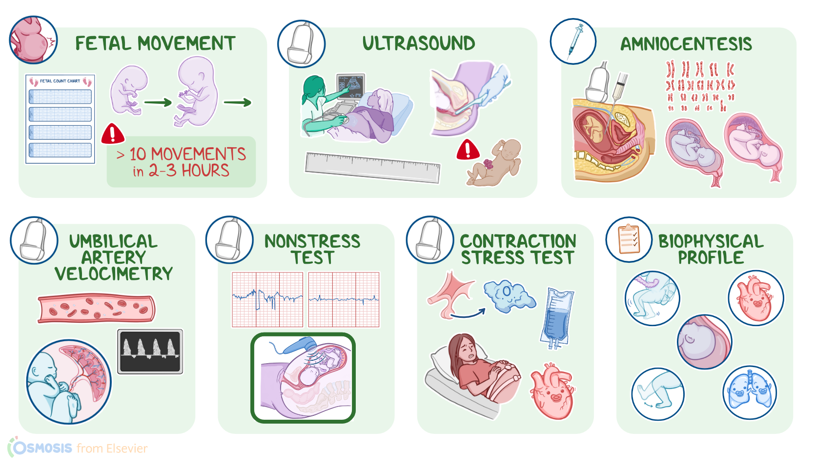
Antepartum means before childbirth �. So, antepartum assessment refers to prenatal medical assessment of a mother and her fetus, aimed at assessing maternal and their health, diagnosing, managing, and counseling about any potential health issue. Antepartum assessment also includes assessing the fetus's size, measuring its head circumference and femur length, listening to its heart rate, and checking for any abnormalities on ultrasound or other various methods.
Introduction0:00–0:10
Antepartum fetal assessment refers to diagnostic procedures used to evaluate the fetal condition before birth.Okay, let’s begin with assessment of fetal movement.
Fetal Movement0:10–1:22
Counting of fetal movements is a simple, cost-effective, and widely used method to monitor the well-being of a fetus. It’s based on the principle that if a fetus is compromised in some way, it will reduce its activity to conserve oxygen and energy.
So, decreases in fetal movements or decreased activity in a previously active fetus might be due to a disturbance of placental function, and may precede an intrauterine fetal demise Now, fetal movements start around the 7th to 8th week of gestation, and increase as gestation progresses.
These movements can be felt by the client, typically starting around the 16th and 18th weeks of gestation. One of the most common methods is the “count to ten” method.
The client is instructed to count fetal movements, including at the same time each day, ideally this will be a time when the client knows her fetus will be active, which is often during the evening hours.
Although there is no well-established evidence of the number of fetal movements that are considered normal, generally, if less than 10 movements in a 2-3 hour period are felt, the client should contact her healthcare provider.
Next up is ultrasound, which is a non-invasive test that uses high-frequency sound waves to visualize the fetus and monitor fetal growth; and can be done either transabdominally or transvaginally.
Ultrasound1:22–2:37
Additionally, fetal anomalies such as open neural tube defects, including microcephaly or anencephaly, as well as ventral wall defects like omphalocele and gastroschisis can be detected.
An ultrasound can also be used to measure nuchal translucency, which is a collection of fluid under the skin behind the fetal neck associated with trisomy 21.
Another important assessment is the amniotic fluid volume. Too much amniotic fluid, called polyhydramnios or sometimes hydramnios, is associated with problems that impair fetal swallowing, such as esophageal atresia.
On the other hand, too little amniotic fluid, called oligohydramnios, is associated with uteroplacental insufficiency or problems that impair fetal urine production, including renal agenesis or polycystic kidneys.
Next, let’s look at amniocentesis, which is an invasive diagnostic procedure, where an ultrasound is used to guide a needle through the abdominal and uterine walls and into the amniotic sac.
Amniocentesis and Chorionic Villus Sampling2:37–3:51
Once inside the amniotic sac, amniotic fluid is drawn, and analyses can be performed, including karyotyping, screening for congenital anomalies, and determining fetal lung maturity.
An amniocentesis can also be used to remove excess amniotic fluid in cases of polyhydramnios.On the other hand, there’s chorionic villus sampling, which involves taking a small sample of the placenta.
A transabdominal approach is often used, where an ultrasound is used to locate the placenta, and a needle is inserted through the abdominal and uterine walls and into the placenta, where a sample is aspirated for chromosomal analysis.
Finally, it's important to note that these procedures can result in complications, including the risk of bleeding, infection, premature rupture of membranes, amniotic emboli, Rh isoimmunization, fetal injury, or spontaneous abortion.
Another technique is umbilical artery doppler velocimetry, which is a non-invasive method of evaluating blood flow in the maternal-fetal circulation, most often of the umbilical artery, using Doppler ultrasound.
Umbilical Artery Doppler Velocimetry3:51–4:50
It assesses the direction of blood flow, as well as the velocity, volume, and resistance to flow from the fetus to the placenta.
Common indications include pregnancies where uteroplacental insufficiency is suspected, like with preeclampsia, which causes placental resistance to increase.
Now, the Doppler captures the movement of red blood cells through the vessels, creating a waveform where the peak represents systole and the lowest represents diastole.
When there is decreased, absent, or even backward blood flow during diastole, this is an indication that there’s high resistance in the placenta, which increases the risk of intrauterine growth restriction and fetal hypoxia.
Now, let’s switch gears and look at the nonstress test, or NST for short, which assesses fetal heart rate changes in response to fetal movements.
Non-Stress Test4:50–6:34
This diagnostic method does not require external factors to trigger the changes in the fetal heart rate, thus the name “non-stress.” Common indications for the non-stress test include decreased fetal movements, intrauterine growth restriction, postterm pregnancy, and maternal cardiovascular conditions, such as chronic hypertension, and preeclampsia.
The ultrasound transducer is placed over the fetal back to record the fetal heart rate; while the tocotransducer is placed over the uterine fundus to track uterine activity.
The client might be asked to press a button every time they feel fetal movement, over a 20 minute period. Each press is recorded on the tracing as a mark, which helps evaluate the fetal heart rate in response to the fetal movement.
Now, fetal heart rate accelerations refer to an increase in the fetal heart rate of at least 15 beats per minute with a duration of at least 15 seconds.
When there are at least 2 accelerations over 20 minutes, the nonstress test is considered reactive or normal. On the flip side, if there are no accelerations during a 40 minute period, the nonstress test is nonreactive or abnormal.
But, it's important to note that a nonreactive result may happen if the fetus is sleeping, so vibroacoustic stimulation can be used to wake the fetus up!
This is where a device is placed on the maternal abdomen and sound is emitted, resulting in a startle reflex in the fetus and subsequent fetal heart rate acceleration.
Now, switching gears and moving on to the contraction stress test, or CST for short. This is a noninvasive diagnostic procedure that assesses the fetal heart rate changes in response to a reduction in oxygenation during uterine contractions.
Contraction stress test6:34–8:42
As with the NST, the ultrasound transducer is placed over the fetal back to record the fetal heart rate and the tocotransducer is placed over the uterine fundus to track uterine activity.
But, in contrast to the NST, uterine contractions are induced with an infusion of dilute oxytocin solution. Alternatively, clients can stimulate their nipples, which increases oxytocin release from the posterior pituitary gland.
Regardless of the method, stimulation should be continued until 3 uterine contractions occur within 10 minutes. Finally, it’s important to note that stimulating uterine contractions is contraindicated in multiple gestation; or with preterm premature rupture of membranes; placenta previa, or abnormal placenta previa; and history of preterm labor.Now, let’s take a look at the physiologic findings of the contraction stress test.
Normally, uterine contractions decrease the blood flow through the small spiral arteries that supply the placenta. As a result, there’s a temporary interruption of the uteroplacental blood flow that, normally, does not affect the fetal heart.
So, if the fetal heart rate does not decrease with the contraction, the CST is negative. On the flip side, if the uterine contraction decreases the fetal heart rate, resulting in late or variable decelerations, the CST is positive.
Specifically, there should be decelerations in at least 50 percent of uterine contractions during 10 minutes. On the flip side, if the decelerations occur in less than 50% of uterine contractions, the result is termed equivocal and requires further evaluation.
Finally, there’s also the unsatisfactory result, where there’s less than three contractions within 10 minutes or a tracing quality that can’t be interpreted; this result also requires further evaluation.Finally, let’s cover the biophysical profile, or BPP for short, which assesses the fetal well-being in cases where there’s an increased risk for altered fetal oxygenation.
Biophysical profile8:42–10:52
The BPP measures five biophysical indicators of fetal well-being: an NST that measures fetal heart rate reactivity; fetal movement; fetal tone; fetal breathing movements; and amniotic fluid volume.First, let’s start with the NST, which is scored with 2 points if it’s reactive, and 0 points if it’s non-reactive.
Fetal body movements or FBMs for short, are scored 2, if there are 3 or more trunk or limb movements; and 0, if there are less than 3 movements.
Next, fetal breathing movements are scored 2, if there is at least one episode of fetal breathing movements that lasts at least 30 seconds; and 0, if there are no breathing movements or if there’s less than 30 seconds of sustained breathing movement.
The fetal tone is scored 2, if there’s at least one episode of active extension with return to flexion of the fetal limb or trunk, or an opening and closing of the hand, while a score of 0 is given if there’s an absence of movement or if there’s slow extension/flexion.
Finally, amniotic fluid is scored a 2 if there’s at least one pocket of amniotic fluid that measures at least 2 cm in two perpendicular planes; and its scored a 0 if there are no amniotic fluid pockets or a pocket measure less than 2 cm in two perpendicular planes.
Generally, a BPP score of 8 to 10 indicates the risk for fetal hypoxia is extremely rare, unless there is oligohydramnios, in which case delivery could be indicated if there is no leakage from the amniotic membranes.
A score of 6 is equivocal, and delivery is indicated if the fetus is mature; but if the fetus is not mature, then the BPP should be repeated within 24 hours.
BPP scores of 0 to 4 are an indication of fetal asphyxia, and prompt delivery is indicated.##SummaryAlright, as a quick recap… Antepartum fetal assessment refers to all diagnostic procedures used to assess the fetal condition and health before birth.
Review10:52–11:16
Important diagnostic procedures include assessment of fetal movement, ultrasound, amniocentesis, umbilical artery velocimetry, non-stress test, contraction stress test, and biophysical profile.
| ANTEPARTUM ASSESSMENT - FETUS | ||
| KEY POINTS | NOTES | |
| DEFINITION |
| |
| FETAL MOVEMENT |
| |
| ULTRASOUND |
| |
| AMNIOCENTESIS & CHORIONIC VILLUS SAMPLING |
| |
| UMBILICAL ARTERY DOPPLER VELOCIMETRY |
| |
| NON-STRESS TEST |
| |
| CONTRACTION STRESS TEST |
| |
| BIOPHYSICAL PROFILE |
| |
No notes for this video yet
Try adding a note below