Chronic obstructive pulmonary disease (COPD): Nursing process (ADPIE)

2,974views

Chronic obstructive pulmonary disease (COPD): Nursing process (ADPIE)

Watch later

Watch later

Hypokalemia: Clinical
Movement of water between body compartments
The role of the kidney in acid-base balance
Blood histology
Blood components
Respiratory acidosis
Metabolic acidosis
Respiratory alkalosis
Metabolic alkalosis
Metabolic and respiratory alkalosis: Clinical
Metabolic and respiratory acidosis: Clinical
Bronchodilators: Beta 2-agonists and muscarinic antagonists
Chronic obstructive pulmonary disease (COPD): Clinical
Chronic obstructive pulmonary disease (COPD): Nursing process (ADPIE)
Adrenergic antagonists: Alpha blockers
Sympatholytics: Alpha-2 agonists
Alpha-2 adrenergic agonists: Nursing pharmacology
Adrenergic receptors
Adrenergic antagonists: Beta blockers
Diabetes mellitus: Clinical
Diabetes mellitus (DM): Nursing process (ADPIE)
Diabetes mellitus
Diabetes mellitus: Pathology review
Stroke: Nursing process (ADPIE)
Stroke: Clinical
Ischemic stroke
Intracerebral hemorrhage
Peptic ulcers and stomach cancer: Clinical
GERD, peptic ulcers, gastritis, and stomach cancer: Pathology review
Sickle cell disease (NORD)
Sickle cell disease: Nursing process (ADPIE)
Sickle cell disease: Clinical
Blood groups and transfusions
Erythropoietin
Blood products: Nursing pharmacology
Oxygen binding capacity and oxygen content
Blood products and transfusion: Clinical
Hemophilia
Hemophilia: Nursing process (ADPIE)
Leukemia: Nursing process (ADPIE)
Chronic leukemia
Leukemias: Pathology review
Acute leukemia
Leukemia: Clinical
Lymphomas: Pathology review
Lymphoma: Clinical
Non-Hodgkin lymphoma
Hodgkin lymphoma
Lymphatic system anatomy and physiology
Anatomy of the abdominal viscera: Liver, biliary ducts and gallbladder
Liver anatomy and physiology
Cirrhosis
Cirrhosis: Pathology review
Cirrhosis: Clinical
Cirrhosis: Nursing process (ADPIE)
Hepatitis C virus
Hepatitis A and Hepatitis E virus
Liver cancer: Nursing
Cholestatic liver disease
Non-alcoholic fatty liver disease
HIV (AIDS)
HIV and AIDS: Pathology review
Antiretrovirals for HIV/AIDS - Protease inhibitors: Nursing pharmacology
Antiretrovirals for HIV/AIDS - NRTIs and NNRTIs: Nursing pharmacology
Antiretrovirals for HIV/AIDS - Integrase strand transfer inhibitors: Nursing pharmacology
Anatomy of the abdominal viscera: Pancreas and spleen
Pancreatitis: Nursing process (ADPIE)
Glaucoma: Nursing process (ADPIE)
Glaucoma
Eye conditions: Refractive errors, lens disorders and glaucoma: Pathology review
Acute respiratory distress syndrome (ARDS): Nursing
Anatomy of the coronary circulation
Coronary artery disease: Pathology review
Coronary artery disease: Clinical
ECG basics
ECG normal sinus rhythm
ECG rate and rhythm
ECG axis
ECG intervals
ECG QRS transition
ECG cardiac infarction and ischemia
Cardiac cycle
Arterial disease
Delirium
Dementia and delirium: Clinical
Vascular dementia
Frontotemporal dementia
Dementia with Lewy bodies
Dementia: Pathology review
Tumor lysis syndrome (TLS): Nursing Process (ADPIE)
Cholelithiasis: Nursing
Coronary artery disease (CAD) and angina pectoris: Nursing process (ADPIE)
Breast cancer: Nursing process (ADPIE)
Ovarian cancer: Nursing
Cervical cancer: Nursing
Hormones and hormone modulators for cancer: Nursing pharmacology
Endometriosis: Nursing
Heart failure
Left-sided heart failure: Nursing process (ADPIE)
Heart failure: Pathology review
Pneumonia: Pathology review
Pneumonia
Bacterial pneumonia: Nursing process (ADPIE)
Respiratory stimulants: Nursing pharmacology
Corticosteroids - Inhaled: Nursing pharmacology
Pneumothorax and hemothorax: Nursing
Chest trauma: Clinical
Pleural effusion: Nursing
Tuberculosis (TB): Nursing
Parkinson disease: Nursing process (ADPIE)
Huntington disease
Multiple sclerosis (MS): Nursing
Multiple sclerosis
Guillain-Barré syndrome: Nursing
Guillain-Barre syndrome
Myasthenia gravis: Nursing
Acute kidney injury (AKI): Nursing process (ADPIE)
Acute kidney injury: Clinical
Chronic kidney disease (CKD): Nursing
Chronic kidney disease
Chronic kidney disease: Clinical
Polycystic kidney disease (PKD): Nursing
Renin-angiotensin-aldosterone system
Osteoarthritis: Nursing
Osteoarthritis
Rheumatoid arthritis (RA): Nursing process (ADPIE)
Rheumatoid arthritis: Clinical
Rheumatoid arthritis
Rheumatoid arthritis and osteoarthritis: Pathology review
Systemic lupus erythematosus
Systemic lupus erythematosus (SLE): Pathology review
Systemic lupus erythematosus (SLE): Clinical
Systemic lupus erythematosus (SLE): Nursing
Mycobacterium tuberculosis (Tuberculosis)
Peripheral venous disease (PVD): Nursing process (ADPIE)
Peripheral arterial disease (PAD): Nursing process (ADPIE)
Buerger disease: Nursing
Raynaud phenomenon: Nursing
Aortic dissections and aneurysms: Pathology review
Aortic aneurysms and dissections: Clinical
Aortic dissection
Aortic aneurysm: Nursing process (ADPIE)
Venous thromboembolism (VTE): Nursing process (ADPIE)
Benign prostatic hyperplasia (BPH): Nursing process (ADPIE)
Benign prostatic hyperplasia
Prostate cancer: Nursing
Prostate cancer
Testicular cancer: Nursing
Testicular cancer
Diabetic ketoacidosis (DKA): Nursing process (ADPIE)
Endocrine system anatomy and physiology
Vitamin B12 deficiency
Thalassemia: Nursing
Anemia - Iron-deficiency: Nursing
Anemia - Macrocytic: Nursing
Anemia - Aplastic: Nursing
Thyroid hormones
Medications for thyroid disorders: Nursing pharmacology
Hyperthyroidism: Nursing process (ADPIE)
Hypothyroidism: Nursing process (ADPIE)
Hypoparathyroidism: Nursing
Hyperparathyroidism: Nursing
Anxiety disorders: Nursing process (ADPIE)
Spinal cord injury (SCI): Nursing
Cluster A personality disorders
Smoke inhalation injury: Nursing process (ADPIE)
Shock - Anaphylactic: Nursing
Shock - Obstructive: Nursing
Shock - Neurogenic: Nursing
Shock - Hypovolemic: Nursing
Shock - Cardiogenic: Nursing
Shock - Septic: Nursing
Pulmonary edema: Nursing
Burn injury: Nursing

Notes

CHRONIC OBSTRUCTIVE PULMONARY DISEASE (COPD)

KEY POINTS
NOTES
PATIENT REPORT
  • 67-year-old 
  • Emergency department
  • 3-day history of exertional breathlessness, wheezing, fatigue, cough
  • History: smoking x25 years
    • 2 packs per day
    • Recently quit smoking
  • Diagnosis: COPD

PATHOPHYSIOLOGY
  • Chronic lung disease with airflow obstruction 
  • Caused by chronic inflammation and lung damage 
  • Common causes 
    • Tobacco smoke 
    • Occupational pollutants 
    • Alpha-1 antitrypsin deficiency 
  • Pathophysiology 
    • Chronic bronchitis 
      • Inflammation of bronchial tubes 
      • Goblet cell hypersecretion → mucus plugs 
      • Airway obstruction and air trapping 
      • Chronic productive cough 
      • Symptoms
        • Exertional → resting dyspnea
        • Fatigue
        • Wheezing
        • Chest tightness 
    • Emphysema 
      • Alveolar destruction impairs gas exchange 
      • Loss of elastic recoil
        • Airway collapse during exhalation 
      • Air trapping and hyperinflation 
      • Barrel chest from increased AP diameter 
    • Compensatory mechanisms 
      • Tripod position 
      • Pursed-lip breathing 
    • COPD exacerbation 
      • Sustained worsening of respiratory symptoms beyond normal variability 
      • Symptoms 
        • Increased dyspnea 
        • Increased cough and sputum production 
        • Fatigue 
        • Increased wheezing 
        • Confusion or decreased consciousness 
  • Complications 
    • Hypoxemia 
    • Hypercapnia  
    • Cyanosis 
    • Pulmonary vasoconstriction
      • Pulmonary hypertension 
    • Right-sided heart failure 

DIAGNOSIS AND TREATMENT
  • Diagnosis 
    • History
    • Physical assessment
    • Pulmonary function test (PFT)
    • Alpha-1 antitrypsin deficiency screening
    • Arterial blood gas (ABG)
  • Treatment
    • Lifestyle modifications 
      • Smoking cessation 
      • Avoid triggers: pollution, allergens, occupational irritants 
      • Annual influenza vaccine 
      • Pneumococcal vaccine 
      • Adjust diet to maintain weight and muscle mass
      • High-calorie, high-protein meals 
    • Medications 
      • Bronchodilators 
      • Corticosteroids 
      • Oxygen therapy 
        • May need to maintain SpO2 between 88–92% 
        • Avoid 100% saturation to preserve respiratory drive 

ASSESSMENT
  • Patient sitting upright in tripod position 
  • Appears short of breath and tired 
  • Struggles to complete full sentences 
  • Nasal flaring and pursed-lip breathing observed 
  • Barrel chest 
  • Use of accessory muscles 
  • Ribs pulling inward during inspiration 
  • Expiratory wheezing 
  • Coarse crackles in lower lobes 
  • Capillary refill < 3 seconds 
  • Alert and oriented  
  • Vital signs 
    • Blood pressure: 135/80 mmHg 
    • Heart rate: 80/min
    • Respiratory rate: 26/min
    • Temperature: 98.6°F (37.0°C) 
    • Oxygen saturation: 87% room air 
    • Pain: 0/10 
  • ABG results 
    • pH: 7.34 
    • PaO₂: 62 mmHg (8.2 kPa)
    • PaCO₂: 65 mmHg (8.6 kPa)
    • HCO₃⁻: 32 mEq/L (32 mmol/L)

NURSING DIAGNOSES
  • Ineffective breathing pattern related to increased work of breathing
  • Ineffective airway clearance related to increased production of secretions
  • Impaired gas exchange related to airway destruction
  • Activity intolerance related to imbalance between oxygen supply and demand
  • Deficient knowledge related to chronic disease management

PLANNING
  • By end of shift, patient will
    • Have an effective breathing pattern as evidenced by breathing at a normal rate and depth
    • Have an absence of dyspnea, and no use of accessory muscles
    • Maintain clear, open airways as evidenced by clear breath sounds and ability to effectively cough up secretions
    • Demonstrate optimal gas exchange at 12 to 20/min
    • Have pulse oximetry at therapeutic levels during rest and exertion
    • Have blood gases be closer to normal range
    • Display increased activity tolerance evidenced by enhanced capacity and energy to ambulate 25 feet without feeling breathless
  • Prior to discharge patient will
    • Verbalize an understanding of COPD
    • Demonstrate how to use inhaler
    • Avoid triggers for exacerbations
    • Understand long-term management of COPD

IMPLEMENTATION
  • Vital signs monitored every 2 hours
  • Continuous respiratory assessment and  pulse oximetry
  • Supplemental oxygen 2L via nasal cannula 
    • Target SpO₂: 88–92% 
  • Medication  
    • Albuterol  
      • Administered via inhaler with spacer 
    • Prednisone 
  • Patient education 
    • Emphasized smoking cessation 
    • Discussed infection prevention 
  • Referral to physical therapist (PT) 
    • Assisting with breathing techniques and energy conservation during ADLs 
  • Referral to respiratory therapist (RT) 
    • Teaching breathing and airway clearance techniques 
    • Drawing blood for ABG analysis 

EVALUATION
  • Breathing less labored 
  • No longer using accessory muscles 
  • Effectively coughing up secretions 
  • 2L O2 via nasal cannula 
  • SpO2
    • 91% at rest 
    • Drops to 87% with minimal activity 
  • Mental status alert and oriented 
  • Tires easily but responsive 
  • Vital signs 
    • Blood pressure: 125/79 mmHg 
    • Heart rate: 90/min
    • Respiratory rate: 22/min 
    • Temperature: 98.6°F (37.0°C) 
    • Pain: 0/10 
  • ABG results 
    • pH: 7.40
    • PaO₂: 72 mmHg (9.5 kPa)
    • PaCO₂: 48 mmHg (6.3 kPa)
    • HCO₃⁻: 28 mEq/L  (28 mmol/L)
  • Continue reassessing respiratory status and oxygenation 
  • Determine if care plan adjustments are needed based on trends

Transcript

Watch video only

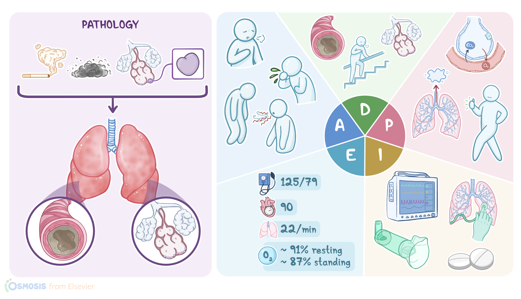
James Robyn is a 67-year-old male client who was brought to the emergency department, or ED, with a 3-day history of exertional breathlessness, wheezing, fatigue, and a worsening productive cough.

He states he has been having increased difficulty with normal day to day activities such as eating, talking and going up the stairs.

He has a history of cigarette smoking, two packs per day, since he was 25 years old.

However, he quit smoking a year ago after being diagnosed with chronic obstructive pulmonary disease, or COPD.

COPD, or chronic obstructive pulmonary disease, is a type of lung disease where chronic inflammation causes damage to the lungs and obstructs airflow.

It’s usually caused by inhalation of toxic substances, like tobacco smoke, or occupational pollutants like dust and silica.

In some people an autosomal dominant disorder called alpha-1 antitrypsin deficiency results in breakdown of the lung parenchyma by an enzyme called elastase.

COPD is characterized by long-term inflammation of the bronchial tubes, referred to as chronic bronchitis, and alveolar destruction, referred to as emphysema.

Most people diagnosed with COPD have elements of both chronic bronchitis and emphysema.

Chronic inflammation of the bronchial tubes in COPD causes a hypersecretion of mucus by the respiratory goblet cells.

The mucus then forms a plug that obstructs the airways causing air trapping, and it also causes chronic productive cough.

Obstruction of the bronchi can also cause exertional dyspnea, which can progress to resting dyspnea, fatigue, wheezing and chest tightness.

Destruction of the alveolar sacs impairs gas exchange, resulting in hypoxemia and hypercapnia.

Loss of elastic recoil causes collapse of the airways during exhalation, trapping the air and dilating the airspaces.

To make breathing easier, they often use the tripod position, where they sit up and lean forward with their hands on their knees.

They may use pursed lip breathing to prolong expiration and produce positive end-expiratory pressure, or PEEP, which allows them to breathe out as much air as possible.

Because this breathing technique requires use of accessory breathing muscles, they will expend a lot of energy just to breathe.

Air trapping also leads to an increased anteroposterior diameter of the chest, sometimes called a barrel chest.

Chronic hypoxemia can result in cyanosis, a bluish discoloration of the lips or fingertips.

It causes constriction of the pulmonary vessels and pulmonary hypertension.

This increases the workload on the right side of the heart, leading to right sided heart failure, or cor pulmonale.

Diagnosis of COPD is based on the client history, physical examination, and pulmonary function tests, or PFTs, to evaluate the degree of airway limitation.

The forced expiratory volume in one second, or FEV1, and the vital capacity, or FVC, are measured after the client is given a bronchodilator such as albuterol.

A FEV1/FVC ratio less than 70% indicates airway obstruction.

Since COPD is an irreversible disease, giving a bronchodilator does not change the person’s PFTs too much.

Finally, alpha-1 antitrypsin deficiency screening may also be done.

Long-term management for stable COPD includes smoking cessation and the avoidance of other triggers such as pollution and allergens.

Influenza and pneumococcal vaccines should be administered to decrease the risk of respiratory infections.

Although COPD is an irreversible disease, bronchodilators can help ease symptoms, and corticosteroids can decrease inflammation in the lungs.

Supplemental oxygen may be needed to maintain an oxygen saturation between 88 to 92 percent.

For these clients, the goal is not 100 percent saturation because hypoxemia is the main stimulus for their respiratory drive.

Finally, since these individuals expend much of their energy on simply breathing, dietary adjustments may be needed to maintain weight and muscle mass.

An exacerbation, or a sustained deterioration of their respiratory symptoms beyond their normal day to day variability, may manifest by increased dyspnea, cough, sputum production, and fatigue.

Increased wheezing may be noted and increased hypoxia may result in confusion or decreased level of consciousness.

Arterial blood gases, or ABGs, may show decreased PaO2, increased PaCO2, increased HCO3-, and decreased pH.

Okay, let’s get back to our client Mr. Robyn.

Since presenting to the ED, he has been admitted to the pulmonary ward for treatment of an acute exacerbation of COPD.

You have been assigned to his care.

After entering his room, you introduce yourself, wash your hands, and confirm his identity.

Mr. Robyn is sitting up on the side of his bed leaning over his bedside table in a tripod position.

You begin your assessment of Mr. Robyn by asking him how he is feeling today.

He states he is feeling short of breath and very tired.

You notice that he struggles to complete full sentences because he has to stop to breathe.

You notice nasal flaring and he is pursing his lips during expiration.

He states that he has had worsening respiratory symptoms for about a week including breathlessness when doing activities such as getting up to go to the bathroom, talking, and eating.

Sources

  1. "Ackley and Ladwig’s Nursing Diagnosis Handbook: An Evidence-Based Guide to Planning Care, 13th edition" Mosby (2022)
  2. "Update on Clinical Aspects of Chronic Obstructive Pulmonary Disease" N Engl J Med (2019)
  3. "The etiologic origins for chronic obstructive pulmonary disease" Int J Chron Obstruct Pulmon Dis (2019)
  4. "Harrison’s Principles of Internal Medicine, 21st edition" McGraw Hill / Medical (2022)
  5. "Definition, Causes, Pathogenesis, and Consequences of Chronic Obstructive Pulmonary Disease Exacerbations" Clin Chest Med (2020)
  6. "Critical Care Nursing: Diagnosis and Management, 9th edition" Elsevier (2021)