Esophageal cancer: Nursing
Introduction0:00–0:16
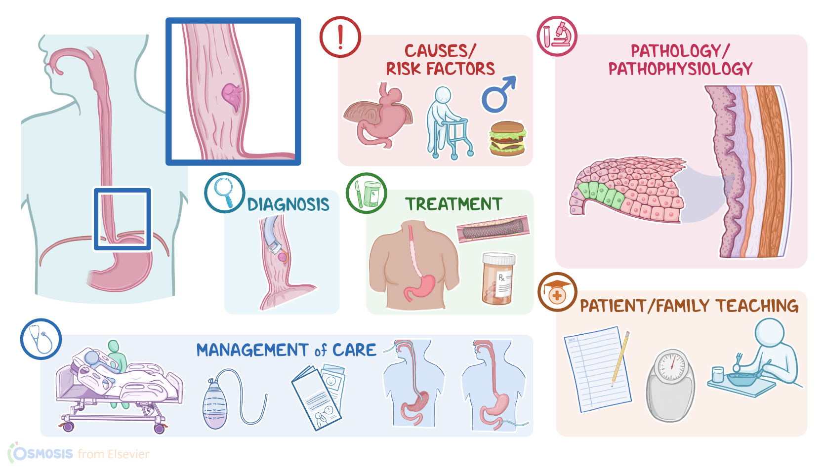
Esophageal cancer is a malignant tumor that originates in the mucosa of the esophagus. There are two types of esophageal cancer, squamous cell carcinomas and adenocarcinomas.Alright, now, the esophagus is a hollow tube that allows food to pass through from the pharynx to the stomach.
Physiology0:16–1:32
And at both ends is a sphincter or a bundle of muscles that can block off or open the esophagus. The upper esophageal sphincter relaxes to allow food to pass through.
The lower esophageal sphincter contracts to prevent acid reflux from the stomach.Let’s zoom into the wall of the esophagus, which is made up of four layers.
The outermost layer is a fibrous layer called the adventitia, and unlike the rest of the gastrointestinal tract, this layer does not contain a serosa.
Next is the muscular layer, which contracts to move food through the esophagus. After that is the submucosa, which consists of a dense layer of tissue that contains blood vessels, lymphatics, and nerves.
And then, there’s the innermost layer, called the mucosa, which comes into direct contact with food, and protects the esophageal wall from friction.
The mucosa also has three layers of its own: a layer made of stratified squamous epithelium; a layer of connective tissue called the lamina propria, and a layer of muscle cells called the muscularis mucosae.
Finally, at the lower esophageal sphincter, the esophageal mucosa joins the columnar gastric epithelium to form the gastroesophageal junction.Alright, now, esophageal cancer occurs when epithelial cells in the mucosa acquire mutations that let them grow and divide out of control.
Causes & risk factors1:32–3:27
Typically these are mutations in proto-oncogenes which result in a promotion of cell division, or mutations in tumor suppressor genes which result in a loss of inhibition of cell division.
Now, mutations may arise due to a variety of risk factors. Risk factors for both squamous cell carcinoma and adenocarcinoma include age over 60 years, being assigned male at birth, asbestos exposure, and a diet high in pickled and fermented foods, red meat, fat, and processed foods; and low in fruit, and vegetables.
Other risk factors include hiatal hernia, which is when part of the stomach bulges up into the chest through an opening in the diaphragm, and esophageal webs, or rings, which are concentric extensions of the normal esophageal wall into the esophageal lumen that can cause difficulty swallowing.
Specific risk factors for squamous cell carcinoma include smoking, excessive alcohol intake, excessive intake of very hot fluids, and caustic strictures, which is the narrowing of the esophagus following ingestion of a caustic substance, like household bleach.
Another predisposing condition is achalasia, which is when the smooth muscle of the lower portion of the esophagus doesn’t work well, making it difficult for food to pass towards the stomach.On the other hand, the strongest risk factor for adenocarcinoma is obesity, and gastroesophageal reflux disease, or GERD for short.
That’s because, with GERD, the lower esophageal sphincter is weaker than normal, and it allows acid from the stomach to go back up into the esophagus after meals.
The chronic presence of acid in the esophagus can lead to Barrett’s esophagus, where the distal part of the esophagus undergoes intestinal metaplasia.
This is where the normal squamous epithelium in the esophagus tries to adapt to the increased acidity by turning into columnar glandular epithelium that’s normally found in the intestine.
Moving on to pathology, a squamous cell carcinoma develops when the mutated cells originate in the squamous epithelium, typically in the proximal two-thirds of the esophagus.
Pathology3:27–4:32
In contrast, adenocarcinomas develop when the mutation occurs in the glandular epithelium, typically in the distal third of the esophagus.
Alright, now once an epithelial cell becomes mutated and cancerous, it starts dividing uncontrollably, forming a tumor mass.
This spread can be aggressive because the esophagus is not surrounded by a serosal layer. Esophageal cancer may also spread to nearby lymph nodes, or may metastasize to distant organs, such as the liver and lungs.
Complications of esophageal cancer include esophageal obstruction, hemorrhage, perforation, and tracheoesophageal fistula formation, which is an abnormal connection between the trachea, and the esophagus.Now, the clinical manifestations of esophageal cancer vary based on the size and location of the tumor.
Clinical manifestations4:32–5:38
Initially, clients can be completely asymptomatic. Over time, the body mounts an immune response to fight the tumor off, so clients may start experiencing unintentional weight loss, fever, and malaise.
If the tumor grows enough to physically obstruct the esophageal lumen, it can cause dysphagia or progressively difficult swallowing.
If the tumor surface bleeds, clients may also present with vomiting of blood, and iron deficiency anemia. If a tracheoesophageal fistula is present, it may lead to pulmonary aspiration of esophageal contents, which may cause symptoms like coughing and dyspnea.
Finally, if esophageal cancer spreads to the diaphragm, it can cause hiccups.Diagnosis of esophageal cancer starts with the client’s history and physical assessment, followed by endOscopy with biopsy.
Diagnosis5:38–6:04
Additional imaging via a CT, PET scan, or endoscopic ultrasound can be used to determine the stage of the tumor in the TNM system, where T stands for tumor location and local spread; N stands for lymph node involvement; and M for whether or not the tumor has metastasized.Treatment for esophageal cancer depends on its aggressiveness and extension.
Treatment6:04–7:06
Small, localized tumors can be treated with endoscopic mucosal ablation, which is when the tumor is removed with an endoscope.
On the other hand, for clients with larger tumors, the treatment of choice is esophagectomy, or removal of a part or the entire esophagus.
Also, any involved nearby lymph nodes should be resected. In addition to surgery, clients could be treated with chemotherapy, immunotherapy, or radiation therapy.
Targeted therapy, or medications that target specific molecules involved in the growth of cancer cells, has recently also been used to treat esophageal cancer.
For clients with dysphagia or esophageal obstruction, a stent can be placed to keep the lumen open so they can eat soft food or liquids.
If they can not eat, a feeding tube might be needed. Finally, clients may benefit from palliative care, which can include opioid analgesics and antiemetics, in order to decrease symptoms and improve their quality of life.
Alright, let’s look at the nursing care you’ll be providing for a client with esophageal cancer. Your priority goals are to promote nutrition, provide postoperative care, minimize the risk of complications related to treatment, and provide emotional support.Begin by assessing your client’s nutritional intake, looking for any changes in appetite, caloric intake, and weight, and the presence of dysphagia, gastric reflux, or regurgitation.
Management and care7:06–9:53
Consult the interdisciplinary care team to discuss nutritional status and assess the need for enteral nutrition. Remember to keep them in an upright position when eating and for at least one hour after eating to prevent aspiration.
Following an esophagectomy, provide routine postoperative care, and monitor for complications related to the procedure. Maintain NPO status, continue to provide hydration intravenously, and maintain their nasogastric tube, ensuring it is secured and attached to low intermittent suction.
Your client will usually have one or two chest tubes in place, as well as a Jackson-Pratt or JP drain. Monitor these closely according to your facility’s protocol, and immediately report a change in the amount or appearance of the drainage, or if the JP drain loses its suction, as these may indicate an anastomosis leak.
Your client may also have a jejunostomy or J-tube in place postoperatively in order to provide enteral nutrition. Maintain the tube by flushing it with 30 mL of water every 4 hours to promote patency.
Once bowel function has returned, begin small feedings as ordered, and advance feedings as tolerated. During care, remember to keep the head of the bed elevated to at least a 30 degree angle, and encourage your client to cough, deep breathe, and use the incentive spirometer every hour.
Also keep a close eye on their SpO2 and vital signs. Immediately report signs of pulmonary complications like crackles auscultated in the lungs, decreased SpO2, tachycardia, and fever.
Administer the prescribed antibiotics, and ensure your client’s pain is controlled to allow effective coughing, incentive spirometer use, and early ambulation.
Also be sure to assess your client for signs of laryngeal nerve injury, which can impair swallowing and lead to aspiration.
Assess their support system and provide information on local resources or counseling. Finally, begin planning for discharge by coordinating your client’s care with the case manager and clinical dietitian to ensure your client has the supplies they will need to take care of themselves at home.Okay, begin your client and family teaching by explaining that esophageal cancer is a malignant tumor that starts in the tissue cells of the esophagus, and that it can lead to difficulty swallowing, aspiration, and bleeding.
General client and family teaching9:53–12:15
Review the plan of care and treatments, including all medications to ensure they know how and when to take them at home.
Lastly, provide them with a schedule of their follow-up appointments and visits to the outpatient clinic for ongoing monitoring and care.Next, talk to them about how to maintain their nutritional intake and weight.
Teach them how to manage their tube and feeding pump, and reassure them that a home health nurse will also be visiting to assess the tube and assist them with feedings.
Then, explain how their diet will gradually progress to a soft oral diet and then to a normal diet, with the goal of being totally off tube feedings in a couple of months after discharge.
Instruct them to keep a log of all their dietary intake, and provide them with examples of nutritious liquids, foods, and liquid supplements.
Instruct them to contact their healthcare provider right away if they have trouble swallowing and are unable to consume liquids or foods orally.
Also remind them to avoid sugary foods and drinks such as fruit juices, ice cream, syrup, and jam, as this can cause food to pass through their stomach too quickly, causing a problem called dumping syndrome.
Teach them to recognize symptoms of dumping syndrome, and instruct them to let their healthcare provider know right away if they experience symptoms such as cramping, stomach pain, diarrhea, weakness or nausea after eating.
If your client needs help quitting, provide counseling and refer them to smoking cessation resources.Also teach them how to decrease the risk of gastric reflux and prevent aspiration.
Instruct them to drink and eat small amounts six times each day. Stress the importance of eating slowly and sitting upright while eating and for at least one hour after meals.
Instruct them to avoid eating or drinking 2 hours before bedtime, and to keep the head of the bed elevated to about 30 degrees.
Lastly, instruct them to monitor their weight daily, and to let their healthcare provider know if they experience weight loss.Alright, as a quick recap… Esophageal cancer is a malignancy that arises from the esophageal mucosa, classified as either squamous cell carcinoma or adenocarcinoma.
Review12:15–13:36
Common risk factors include age over 60, being assigned male at birth, and obesity. Smoking is a major risk factor for squamous cell carcinoma while GERD is a major risk factor for adenocarcinoma.
Complications of esophageal cancer can include esophageal obstruction, hemorrhage, fistula formation, or esophageal perforation.
Signs and symptoms of esophageal cancer include progressive dysphagia, odynophagia, weight loss, and pain. An esophagram, endoscopic examination, and biopsy are generally used to diagnose esophageal cancer.
Small localized tumors are treated with endoscopic mucosal ablation, while larger tumors are often removed by esophagectomy.
Chemotherapy and targeted therapy can also be used. Goals of nursing care include promoting nutrition, providing postoperative care, minimizing the risk of complications related to treatment, and providing emotional support.
Client and family teaching focuses on explaining the treatment plan, and how to maintain adequate
| ESOPHAGEAL CANCER | ||
| KEY POINTS | NOTES | |
| DEFINITION |
| |
| PHYSIOLOGY |
| |
| CAUSES AND RISK FACTORS |
| |
| PATHOPHYSIOLOGY |
| |
| SIGNS AND SYMPTOMS |
| |
| DIAGNOSIS |
| |
| TREATMENT |
| |
| MANAGEMENT OF CARE |
| |
| PATIENT AND FAMILY TEACHING |
| |
No notes for this video yet
Try adding a note below