Preeclampsia and eclampsia: Nursing

Last updated: February 15, 2022

Preeclampsia and eclampsia: Nursing

Acute Final

Acute Final

Endocrine system anatomy and physiology
Antepartum assessment - Fetus: Nursing
Assessment of gestational age: Nursing
Fetal circulation: Nursing
Fetal development: Nursing
Group B streptococcus (GBS) infection in pregnancy: Nursing
Hepatitis B virus (HBV) infection in pregnancy: Nursing
Hyperemesis gravidarum: Nursing
Large for gestational age (LGA) infant: Nursing
Preeclampsia and eclampsia: Nursing
Prenatal screening: Nursing
Placenta previa: Nursing process (ADPIE)
Placental abruption: Nursing process (ADPIE)
Birth-related procedures: Nursing
Cesarean birth: Nursing
Intrapartum assessment - Fetal heart rate patterns: Nursing
Intrapartum assessment - Uterine activity: Nursing
Premature rupture of membranes (PROM): Nursing
Shoulder dystocia: Nursing
Prolapsed umbilical cord: Nursing process (ADPIE)
Stages of labor: Nursing
Assessment - Postpartum: Nursing
Perinatal depression: Nursing
Physiology of lactation: Nursing
Postpartum infections: Nursing
Postpartum hemorrhage: Nursing
Biliary atresia: Nursing
Cleft lip and palate: Nursing
Congenital diaphragmatic hernia: Nursing
Congenital heart defects - Acyanotic: Nursing
Congenital heart defects - Cyanotic: Nursing
Esophageal atresia and tracheoesophageal fistula: Nursing
Craniosynostosis: Nursing
Hemolytic disease of the fetus and newborn: Nursing
Hyperbilirubinemia: Nursing process (ADPIE)
Infant of a diabetic mother (IDM): Nursing
Meconium aspiration syndrome: Nursing
Neonatal respiratory distress syndrome (NRDS): Nursing
Neonatal sepsis: Nursing
Neural tube defects: Nursing
Newborn adaptation to extrauterine life: Nursing
Persistent pulmonary hypertension of the newborn (PPHN): Nursing
Physical assessment - Neonate: Nursing
Small for gestational age (SGA) infant: Nursing
Postterm infant: Nursing
Thermoregulation - Neonate: Nursing
Arterial blood gas (ABG) - Overview: Nursing
Arterial blood gas (ABG) - Metabolic acidosis: Nursing
Arterial blood gas (ABG) - Metabolic alkalosis: Nursing
Arterial blood gas (ABG) - Respiratory acidosis: Nursing
Arterial blood gas (ABG) - Respiratory alkalosis: Nursing
Adrenal insufficiency (Addison disease): Nursing
Anemia - Iron-deficiency: Nursing
Anemia - Aplastic: Nursing
Anemia - Macrocytic: Nursing
Case study - Hypothyroidism: Nursing
Case study - Iron-deficiency anemia: Nursing
Case study - Sickle cell anemia: Nursing
Complete blood count (CBC) - Hemoglobin and hematocrit: Nursing
Complete blood count (CBC) - Red blood cells (RBC): Nursing
Complete blood count (CBC) - Platelets: Nursing
Complete metabolic panel (CMP) - Blood urea nitrogen (BUN) and creatinine (Cr): Nursing
Complete metabolic panel (CMP) - Estimated glomerular filtration rate (eGFR): Nursing
Complete metabolic panel (CMP) - Liver function tests (LFT): Nursing
Cushing syndrome and Cushing disease: Nursing
Hematopoietic growth factors: Nursing pharmacology
Hyperparathyroidism: Nursing
Hyperthyroidism: Nursing process (ADPIE)
Hypoparathyroidism: Nursing
Hyperpituitarism: Nursing
Hypopituitarism: Nursing
Hypothyroidism: Nursing process (ADPIE)
Medications affecting the parathyroid glands: Nursing pharmacology
Medications for growth hormone disorders: Nursing pharmacology
Medications for thyroid disorders: Nursing pharmacology
Neutropenia: Nursing
Polycythemia: Nursing
Thrombocytopenia: Nursing
Acute kidney injury (AKI): Nursing process (ADPIE)
Benign prostatic hyperplasia (BPH): Nursing process (ADPIE)
Case study - Cholecystitis: Nursing
Case study - Cirrhosis: Nursing
Case study - Chronic kidney disease (CKD): Nursing
Case study - Benign prostatic hyperplasia (BPH): Nursing
Case study - Gastroesophageal reflux disease (GERD): Nursing
Case study - Pediatric appendicitis: Nursing
Case study - Pyelonephritis: Nursing
Cholecystitis: Nursing
Cholelithiasis: Nursing
Chronic kidney disease (CKD): Nursing
Cirrhosis: Nursing process (ADPIE)
Diverticular disease: Nursing
Gastroesophageal reflux disease (GERD): Nursing process (ADPIE)
Hemolytic uremic syndrome: Nursing
Hirschsprung disease: Nursing
Intestinal obstruction: Nursing
Irritable bowel syndrome (IBS): Nursing
Nephrotic syndrome: Nursing
Pyloric stenosis: Nursing process (ADPIE)
Renal and urinary calculi: Nursing
Urinary incontinence - Stress: Nursing process (ADPIE)
Diabetes insipidus: Nursing process (ADPIE)
Dialysis care: Nursing
Case study - Diabetic ketoacidosis (DKA): Nursing
Case study - Pediatric diabetes mellitus type 1: Nursing
Diabetes mellitus (DM): Nursing process (ADPIE)
Hyperosmolar hyperglycemic state (HHS): Nursing process (ADPIE)
Diabetic ketoacidosis (DKA): Nursing process (ADPIE)
Case study - Epilepsy: Nursing
Case study - Head injury: Nursing
Epidural and subdural hematoma: Nursing
Case study - Stroke: Nursing
Hemorrhagic stroke - Intracranial hemorrhage (ICH) and subarachnoid hemorrhage (SAH): Nursing
Increased intracranial pressure (ICP): Nursing
Hydrocephalus: Nursing process (ADPIE)
Intracranial aneurysm: Nursing
Seizure disorder: Nursing process (ADPIE)
Stroke: Nursing process (ADPIE)
Jaundice: Nursing
Nutrition - Enteral: Nursing skills
Nutrition - Newborn: Nursing
Nutrition - Parenteral: Nursing skills
Phenylketonuria (PKU): Nursing
Arterial embolism: Nursing
Disseminated intravascular coagulation (DIC): Nursing
Hemophilia: Nursing process (ADPIE)
Acute respiratory distress syndrome (ARDS): Nursing
Asthma: Nursing process (ADPIE)
Atelectasis: Nursing
Bacterial pneumonia: Nursing process (ADPIE)
Bronchiolitis and respiratory syncytial virus (RSV): Nursing process (ADPIE)
Case study - Acute respiratory distress syndrome (ARDS): Nursing
Care of an intubated client: Nursing skills
Case study - Chronic obstructive pulmonary disease (COPD): Nursing
Case study - Impaired gas exchange: Nursing
Case study - Pediatric asthma: Nursing
Chest tube care: Nursing
Chronic obstructive pulmonary disease (COPD): Nursing process (ADPIE)
Cystic fibrosis: Nursing
Epiglottitis: Nursing process (ADPIE)
Flail chest: Nursing
Intraoperative care: Nursing
Pleural effusion: Nursing
Pneumothorax and hemothorax: Nursing
Pulmonary edema: Nursing
Smoke inhalation injury: Nursing process (ADPIE)
Tracheostomy: Nursing
Venous thromboembolism (VTE): Nursing process (ADPIE)
Arrhythmias - Asystole: Nursing
Arrhythmias - Atrial flutter (Aflutter): Nursing
Arrhythmias - Premature atrial contractions (PACs): Nursing
Arrhythmias - Heart blocks: Nursing
Arrhythmias - Atrial fibrillation (Afib): Nursing
Arrhythmias - Premature ventricular contractions (PVCs): Nursing
Arrhythmias - Sinus tachycardia and sinus bradycardia: Nursing
Arrhythmias - Supraventricular tachycardia (SVT): Nursing
Arrhythmias - Ventricular fibrillation (Vfib): Nursing
Arrhythmias - Ventricular tachycardia (Vtach): Nursing
Cardiac biomarkers - Troponin: Nursing
Case study - Acute coronary syndrome (ACS): Nursing
Case study - Atrial fibrillation (Afib): Nursing
Case study - Heart failure with reduced ejection fraction (HFrEF): Nursing
Case study - Deep vein thrombosis (DVT): Nursing
Case study - Hypertension: Nursing
Case study - Hypovolemic shock: Nursing
Coronary artery disease (CAD) and angina pectoris: Nursing process (ADPIE)
Electrocardiogram (ECG) - Normal sinus rhythm (NSR): Nursing
Heart defects that decrease pulmonary blood flow - Nursing considerations & client education: Nursing
Hypertension: Nursing process (ADPIE)
Left-sided heart failure: Nursing process (ADPIE)
Myocardial infarction (MI): Nursing process (ADPIE)
Pericardial effusion and cardiac tamponade: Nursing process (ADPIE)
Peripheral arterial disease (PAD): Nursing process (ADPIE)
Rheumatic heart disease: Nursing process (ADPIE)
Shock - Cardiogenic: Nursing
Shock - Neurogenic: Nursing
Shock - Obstructive: Nursing
Shock - Septic: Nursing
Sickle cell disease: Nursing process (ADPIE)
Valvular heart disease: Nursing

Notes

PREECLAMPSIA AND ECLAMPSIA

KEY POINTS
NOTES
DEFINITION
  • Preeclampsia 
    • New-onset hypertension and proteinuria after 20 weeks of gestation or during the postpartum period in a previously normotensive patient
  • Eclampsia 
    • Patient with preeclampsia develops generalized seizures

PHYSIOLOGY
  • Blood pressure regulation during pregnancy
    • Cardiac output increases
    • Intravascular volume increases
    • Peripheral vascular resistance decreases 
    • Decreased response to vasoconstrictor molecules
    • Higher levels of vasodilator molecules 
      • Blood vessels dilate more
      • Keeps </= BP 120/80 mm Hg
    • Placenta requires increased blood
      • Spiral arteries dilate and open into placenta
      • Umbilical vein gets nutrients and oxygen without contact with spiral arteries 
      • Deoxygenated blood and waste are carried to placenta by umbilical arteries
      • Eliminated by pregnant patient 

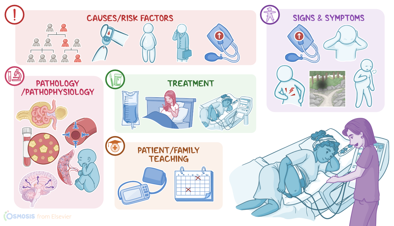
CAUSES AND RISK FACTORS
  • Causes
    • Unknown
  • Risk factors
    • Family history of preeclampsia
    • First pregnancy 
    • Age over 35 years 
    • Obesity
    • Chronic hypertension
    • Diabetes before pregnancy
    • Black descent

PATHOPHYSIOLOGY
  • Develop in patients that are sensitive to vasoconstrictor molecules 
  • Patients with lower levels of vasodilating molecules 
  • Patients have increased peripheral vascular resistance and low blood flow to target organs
  • Placenta 
    • Decreased perfusion causes release of proinflammatory cytokines
    • Aberrant development of spiral arteries
      • Systemic inflammation
    • Decreased blood flow to fetus
      • Intrauterine growth restriction
      • Low birth weight
      • Preterm birth 
      • Fetal death
    • Potential placental abruption
  • Kidneys
    • Decreased perfusion damage glomeruli
      • Proteinuria
      • Edema
      • Renal insufficiency 
  • Liver 
    • Decreased perfusion damages liver
    • Hyperbilirubinemia
    • Release enzymes into blood stream
  • Brain 
    • Vasoconstriction leads to cerebral blood vessel rupture
    • Risk for cerebral hemorrhage and stroke
    • Inflammation and high blood pressure causes cerebral edema and ischemia
    • Development of seizures 
  • Complication
    • HELLP syndrome


SIGNS AND SYMPTOMS
  • Hypertension
    • Systolic BP (SBP) > 140 mmHg
    • Diastolic BP (DBP) > 90 mmHg
    • Or both
  • Proteinuria
  • Dyspnea
  • Severe disease
    • SBP >160 mmHg or DBP >110 mmHg
    • Or S/S of end-organ damage
  • Eclampsia
    • Seizures before, during, or after labor

DIAGNOSIS
  • History 
  • Physical assessment 
  • Laboratory tests

TREATMENT
  • Deliver baby and placenta
  • For patients <37 weeks' gestation without severe disease 
    • Bed rest
    • Fetal monitoring
  • >37 weeks' gestation
    • Labor induction
  • Patients with features of severe disease regardless of gestational age
    • Labor induction 
    • IV antihypertensives 
    • IV magnesium sulfate 
    • Position patient in left lateral
  • <34 weeks' gestation
    • Antenatal corticosteroids

MANAGEMENT OF CARE
  • Goals of care 
    • Control blood pressure
    • Prevent complications
    • Provide emotional support
  • Establish IV access 
  • Administer IVFs, antihypertensives, and magnesium as prescribed 
  • Apply external fetal monitor
  • Insert urinary catheter
  • Seizure precautions
  • Apply oxygen
  • Emotional support
  • Monitor
    • Level of consciousness (LCO)
    • Vital signs
    • Lung sounds 
    • Oxygen saturation
    • Intake and output
    • Edema
    • Signs or symptoms of magnesium toxicity
    • Fetal heart rate (FHR)
  • Report to HCP 
    • Altered LOC
    • Decreased urine output
    • Severe headache
    • Epigastric pain
    • Visual changes 
    • Clonus 
    • Pulmonary edema 
    • Changes in FHR

PATIENT AND FAMILY TEACHING
  • Explain condition, plan of care, and safe medication administration
  • Report to HCP
    • Visual changes 
    • Headache
    • Shortness of breath
    • Extreme weakness
  • After delivery
    • Teach about medications
    • Self-monitor BP
    • Notify HCP
      • Increased BP
      • Sudden weight gain
      • Swelling of face, hands, or feet
      • Visual changes
      • Nausea 
      • Abdominal pain

Transcript

Watch video only

Preeclampsia is a pregnancy-specific condition characterized by new-onset hypertension and proteinuria after 20 weeks of gestation or during the postpartum period in a previously normotensive client; while eclampsia is when a client with preeclampsia develops generalized seizures. These two conditions differ from gestational hypertension, which simply refers to new onset of hypertension during pregnancy, without proteinuria or seizures.

Now, let’s quickly review the physiology of blood pressure regulation during pregnancy. There are three key factors to keep in mind: cardiac output, which is the amount of blood that the heart pumps out to the systemic circulation in a minute; intravascular volume, which is the amount of blood in the client’s circulation; and peripheral vascular resistance, which is the resistance of blood flow in peripheral arteries, and is regulated via vasoconstriction or vasodilation as needed. Remember that in pregnant clients, the cardiac output and intravascular volume increase, but the peripheral vascular resistance decreases.

That’s because during pregnancy, there’s a relative decrease in the response to vasoconstrictor molecules, like angiotensin II, and there are also higher levels of vasodilator molecules, like prostacyclin PGI2. As a result, blood vessels dilate more to accommodate the increased blood volume, and this keeps blood pressure in the normal range, below 120 over 80 mmHg.

This increased blood volume can come in pretty handy, especially since the placenta needs a lot of blood. Remember that the placenta is a temporary organ that develops in the uterus during pregnancy, and connects the pregnant client with the fetus. The way this works is that a type of endometrial arteries called the spiral arteries dilate during pregnancy, and open into the placenta to form little pools of blood. On the fetal end, the umbilical vein connects to the placenta and also opens into those pools. This way the umbilical vein can get nutrient and oxygen-rich blood for the fetus without it being in direct contact with the spiral arteries. In the same vein, pun intended, deoxygenated blood and waste products from the fetus are carried to the placenta by the umbilical arteries, and from there, they are picked up by the pregnant client’s veins to be eliminated.

That being said, while the exact cause of preeclampsia and eclampsia is unknown, some risk factors for developing these conditions have been identified. These include family history of preeclampsia, first pregnancy or being over 35 years of age, as well as obesity, chronic hypertension or diabetes before pregnancy, and Black race.

Now, the pathology of preeclampsia and eclampsia is thought to develop in clients that are particularly sensitive to vasoconstrictor molecules like angiotensin II, and they also seem to have lower levels of vasodilating molecules like prostacyclin PGI2. As a result, these clients have increased peripheral vascular resistance and low blood flow to target organs, like the placenta, as well as the kidneys, liver, and brain.

On the placenta end, decreased perfusion can cause the release of proinflammatory cytokines and, as a consequence, systemic inflammation. Another element that can play a role in decreased placental perfusion can be the aberrant development of spiral arteries, which also plays a role in systemic inflammation. Either way, low blood flow to the placenta means low blood flow to the developing fetus, which can cause intrauterine growth restriction, low birth weight, preterm birth or even fetal death. Additionally, with preeclampsia, there’s also an increased risk of placental abruption, meaning the placenta prematurely detaches from the uterine wall.

On the kidney end, low blood flow can damage the tiny glomeruli that normally work to keep blood proteins from making their way into the urine; so this results in proteinuria. As a consequence, less proteins in the blood can cause generalized or localized edema, like pulmonary edema. Additionally, over time, low renal blood flow can also lead to renal insufficiency.

On the liver end, decreased blood flow can cause liver damage and dysfunction. And because the liver is in charge of conjugating bilirubin to produce bile, some clients may develop hyperbilirubinemia. In addition, the liver is full of enzymes for various metabolic processes, the damaged liver may release these enzymes into the blood.

And let’s not forget about the brain! Now, the exact mechanism of brain damage during preeclampsia is not understood, but it appears to be twofold. On the one hand, the vasoconstriction of cerebral blood vessels means they can’t withstand the increased blood pressure, so the tiniest of cerebral blood vessels are at risk of rupturing and causing cerebral hemorrhage and stroke. On the other hand, inflammation and high blood pressure can alter the normal physiology of cerebral circulation, and cause cerebral edema and small areas of cerebral ischemia. This could play a role in the development of seizures and the progression of preeclampsia to eclampsia.

Okay, now, the clinical manifestations of preeclampsia and eclampsia can vary, but they include hypertension, defined as systolic blood pressure over 140 mmHg, diastolic blood pressure over 90 mmHg, or both. Most clients also develop proteinuria, and some might develop dyspnea as a result of pulmonary edema.

Now, features of severe disease are defined as having systolic blood pressure over 160 mmHg, or diastolic blood pressure over 110 mmHg; or signs of end-organ damage, such as decreased urinary output, which can indicate renal damage; jaundice and right upper quadrant or epigastric pain, which can indicate liver damage; and visual disturbances, such as blurry or double vision; scotomata, which means “seeing spots”, as well as headache, confusion, drowsiness, numbness and tingling, or hyperreflexia, which can indicate brain damage. Finally, if preeclampsia progresses to eclampsia, the client can develop seizures, which can occur before, during, or sometimes even after labor.

The diagnosis of preeclampsia and eclampsia starts with the client’s history and physical assessment confirming hypertension; followed by laboratory tests showing proteinuria. In some cases, preeclampsia can be diagnosed in the absence of proteinuria if there is evidence of end-organ damage. Some laboratory cues for end-organ damage can include elevated creatinine blood levels, hyperbilirubinemia, elevated liver enzymes, and thrombocytopenia or low platelets. When elevated liver enzymes and thrombocytopenia occur alongside hemolytic anemia, that could indicate a potentially life-threatening complication in pregnancy called HELLP syndrome, which stands for Hemolysis, Elevated Liver enzymes, and Low Platelets.