Renal cancer: Nursing
Notes
| RENAL CANCER | ||
| KEY POINTS | NOTES | |
| DEFINITION |
| |
| PHYSIOLOGY |
| |
| CAUSES AND RISK FACTORS |
| |
| PATHOPHYSIOLOGY |
| |
| SIGNS AND SYMPTOMS |
| |
| DIAGNOSIS |
| |
| TREATMENT |
| |
| MANAGEMENT OF CARE |
| |
| PATIENT AND FAMILY TEACHING |
| |
Transcript
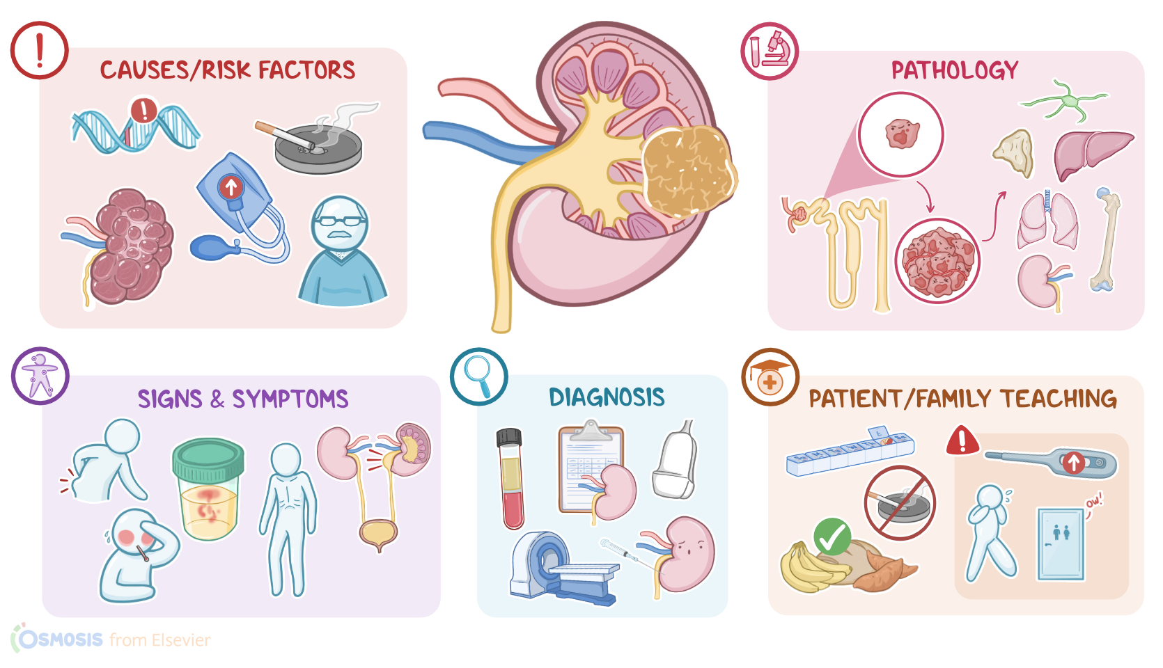
Renal cancer is a malignant tumor that arises from the cortex, pelvis, or the calyces of the kidneys. The most common type of renal cancer is renal cell carcinoma, which forms from the cells lining the proximal convoluted tubules of the kidney.
All right, let’s quickly review some kidney physiology! We can think of the kidneys as the body’s natural blood filter. Their main function is to clear blood of metabolic wasteful substances and toxins by excreting them through urine. In addition, they secrete important hormones, and are essential in regulating the acid-base balance, pH, blood pressure, and electrolyte levels in the body.
So, if we take a cross-section of the kidney, there is an outside rim, known as the renal cortex, and an inner portion, which is the renal medulla. The cortical tissue extends towards the medulla, forming renal columns that divide the medulla into pyramidal-shaped structures called the renal pyramids. The tips of the pyramids, called the renal papilla, project into minor calyces which join together to form major calyces which funnel into the renal pelvis. Urine collects in the renal pelvis and then heads out of the kidney through the ureter.
Now, within the cortex and the medulla there are millions of tiny functional units called nephrons, which consist of a renal corpuscle and a set of renal tubules. The renal corpuscle is made up of the glomerulus, a tiny bundle of capillaries, and the Bowman’s capsule, which is a cup-shaped structure that surrounds the glomerulus. So, blood gets filtered through the glomerulus, and then travels through the renal tubules, which are, in order: the proximal convoluted tubule, loop of Henle, distal convoluted tubule and finally, collecting ducts which drain urine into the renal papillae and eventually empty into the renal pelvis.
Now, the exact cause of renal cancer is often unknown, but there’s typically a genetic mutation in a cell of the cortex, pelvis or calyces, such as a mutation in the Von Hippel–Lindau or VHL gene. This is a tumor suppressor gene, so normally it suppresses the growth of tumor cells. And these mutations can be hereditary, meaning that the client inherits the mutation from one of their parents, or non-hereditary, also known as sporadic, which occur de novo or spontaneously.
Whatever the cause is, the chance of developing renal cancer increases with certain risk factors. Modifiable risk factors include exposure to toxins like tobacco smoke, asbestos, cadmium, and gasoline; as well as obesity, hypertension, unopposed estrogen use, and acquired cystic kidney disease. On the other hand, non-modifiable risk factors include age above 45 years, being assigned male at birth, and having a family history of renal cancer.
All right, now, the most common type of renal cancer is renal cell carcinoma, which occurs when an epithelial cell in the proximal convoluted tubule of the kidney becomes mutated and cancerous, and begins dividing uncontrollably, forming a tumor mass. As the tumor keeps growing, new blood vessels also develop via angiogenesis to supply it. Eventually, cancerous cells start invading neighboring tissues, and can spread to nearby lymph nodes or the adrenal gland on the same side; or even metastasize to other organs, like the liver, lungs, long bones, or to the other kidney.
Renal cell carcinoma is also frequently responsible for causing various paraneoplastic syndromes, which is where the tumor cells generate a hormone that causes its own set of symptoms. For example, these tumors can release the hormone erythropoietin which increases the production of new red blood cells, and this can lead to polycythemia or too many red blood cells, which can cause the blood to start sludging or slowing down its normal flow. Another paraneoplastic syndrome involves the release of renin, which causes hypertension.
Some other hormones that renal cell carcinomas are known for releasing include parathyroid hormone-related peptide or PTHrP and adrenocorticotropic hormone or ACTH. PTHrP mimics parathyroid hormone or PTH, causing hypercalcemia, fatigue, and muscle weakness while ACTH secretion results in Cushing syndrome, which can cause hyperglycemia, hypertension, skin hyperpigmentation, osteoporosis, weight gain, easy bruising, and frequent infections from a weakened immune system.
Finally, in rare cases, a large renal cell carcinoma affecting the left kidney can compress the left renal vein and impede normal venous drainage of the left testis. This leads to dilation of the testicular veins and formation of a varicocele.
Okay, so the clinical manifestations of renal cancer vary based on the size and location of the tumor. Initially, clients are typically asymptomatic. As the disease progresses over time, clients may develop a palpable mass in the abdomen or lower back, and can experience symptoms like unintentional weight loss, fever, malaise, nausea, and vomiting.
If the tumor grows enough to physically obstruct the urinary flow, it can cause urine to build up inside the ureter, called hydroureter. As the tumor invades the tissue and breaks through the basement membrane, clients can experience hematuria; while compression of nearby nerves can cause pain in the flank or near the hip bone. Finally, with paraneoplastic syndromes, clinical manifestations depend on the secreted hormone.
The diagnosis of renal cancer starts with the client's history and physical assessment. Additionally, laboratory studies include a complete blood count or CBC which may show anemia and increased erythrocyte sedimentation rate, or ESR. When the tumor secretes erythropoietin, clients can have an increased hematocrit and low ESR. If the tumor secretes PTHrP, calcium levels are also elevated. Kidney function tests can show elevated serum creatinine and blood urea nitrogen, or BUN for short. Urinalysis is typically also performed to look for red blood cells in the urine.
Imaging tests like a pelvic or abdominal CT scan can be used to stage the tumor using the TNM classification, by defining the location and looking for lymph node involvement or metastasis. Additional imaging tests include renal ultrasound, and a kidney, ureter, and bladder X-ray, or KUB. Once a suspicious lesion is found on imaging, a renal biopsy is performed, where multiple core specimens are obtained to confirm the diagnosis.
Treatment for renal cancer depends on its aggressiveness and extension. Small, localized tumors can be treated with partial nephrectomy, which is when the affected part of the kidney is surgically removed. On the other hand, for clients with larger tumors, the treatment of choice is radical nephrectomy, or removal of the entire kidney, along with the surrounding fat, the ipsilateral adrenal gland, and nearby lymph nodes. This surgical procedure is sometimes followed by chemotherapy and radiation therapy to kill the remaining cancer cells.
Now, for clients with unresectable metastatic tumors, as well as those who can’t have surgery, treatment can involve microwave ablation, or MWA, and cryoablation, which can slow tumor growth. Biologic response modifiers, or BRMs, including interleukin 2, interferon, and tumor necrosis factor, or TNF, have also been shown to increase survival time. Finally, targeted therapy or medications that target specific molecules involved in the growth of cancer cells has recently also been used to treat renal cancer.