Systemic lupus erythematosus (SLE): Nursing
1,337views
Systemic lupus erythematosus (SLE): Nursing
Exam 1
Exam 1
Notes
| SYSTEMIC LUPUS ERYTHEMATOSUS (SLE) | ||
| KEY POINTS | NOTES | |
| DEFINITION |
| |
| PHYSIOLOGY |
| |
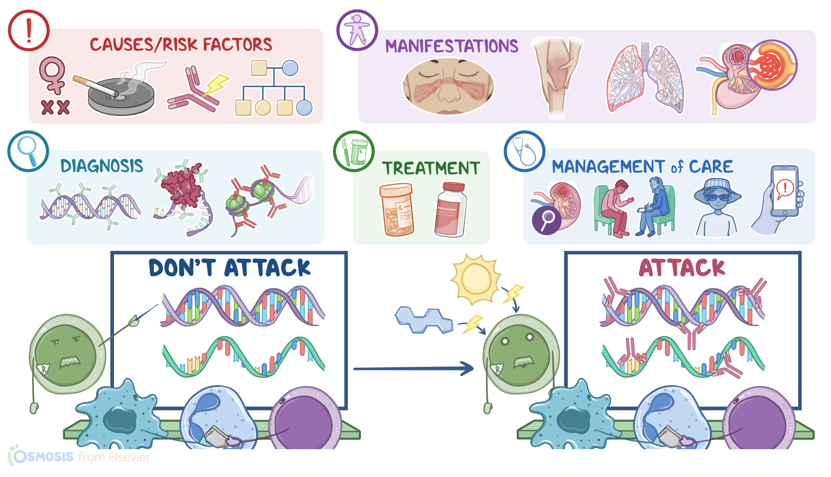
| CAUSES AND RISK FACTORS |
| |
| PATHOPHYSIOLOGY |
| |
| SIGNS AND SYMPTOMS |
| |
| DIAGNOSIS |
| |
| TREATMENT |
| |
| MANAGEMENT OF CARE |
| |
| PATIENT AND FAMILY TEACHING |
| |
Transcript
Content Reviewers
Systemic lupus erythematosus, or simply SLE or lupus, is a chronic autoimmune disease that affects multiple systems, mainly the skin, joints, and serous membranes.
SLE is progressive, meaning that it gets worse over time with alternating periods of remission and exacerbation. A less common type of lupus particularly affects the skin, called cutaneous lupus erythematosus, or CLE for short. Clients with this condition eventually develop a full-blown SLE over time.
Now, let’s quickly recap the physiology of the immune system, which protects the body from harmful agents. This can be done with the aid of immune cells, including macrophages and lymphocytes.
So, let’s imagine that a foreign particle enters the body, it will be faced by macrophages, which swallow this harmful agent and break it down, leaving a small particle called an antigen. The macrophage then presents this antigen on its surface for another type of immune cells to recognize, called T lymphocytes. Types of T lymphocytes include cytotoxic T lymphocytes, which fight against intracellular pathogens and cancer cells by initiating cell death; as well as T helper cells, which activate another type of lymphocytes, called B lymphocytes.
Once activated, B lymphocytes can differentiate into plasma cells or memory cells. Plasma cells produce antibodies. These bind to antigens found on the surface of the pathogen, producing immune complexes.
So, an antibody tags the pathogen for other immune cells to recognize. In contrast, memory cells monitor the body for the same antigen, and when the pathogen visits the body again, these cells convert to plasma cells, and start producing antibodies to fight this pathogen even faster than the first exposure.
During development, the immune cells learn how to skip the antigens found on the surface of body cells and not react to them, called self-tolerance. This is the main function of another type of T lymphocytes, called regulatory T cells, which suppress the immune system to maintain self-tolerance.
Now, the exact cause of systemic lupus erythematosus is unknown, but it’s thought to be multifactorial, involving genetic susceptibility, altered immune system function, and exposure to certain triggers such as cigarette smoke and ultraviolet light.
Lupus is also more common in individuals assigned female at birth, especially during childbearing years; those with a family history of Lupus; and Black, Hispanic, or Asian individuals.
Alright, now the pathology of systemic lupus erythematosus starts with a genetically susceptible client, meaning that they have less functional regulatory T cells. Add a hormonal or an environmental trigger to that, and self-tolerance is lost. This causes the immune system to form auto-antibodies that target nucleic acids, such as DNA or RNA.
Therefore, these auto-antibodies are called anti-nuclear antibodies, or ANA for short. Now, when these ANAs bind to nucleic acids, immune complexes are formed, and these immune complexes eventually precipitate in the walls of blood vessels supplying the brain, heart, skin, kidneys, and joints. This triggers an abnormal immune response, called type III hypersensitivity reaction, which leads to chronic inflammation of the affected blood vessels and reduced perfusion of the affected organs.
Complications of systemic lupus erythematosus typically arise from the organ damage caused by the disease, or as a side effect of the medications used to treat the disease. These complications include skin damage, alopecia, and increased risk of coronary artery disease, in addition to end-stage renal disease.
Complications can also arise in the nervous system, including visual changes, anxiety, and depression. Complications of the gastrointestinal system include intestinal obstruction and inflammatory bowel syndrome. Complications of the reproductive system include pregnancy loss, preeclampsia, and eclampsia. Finally, clients with lupus that receive long-term steroids can develop complications, such as osteoporosis and increased risk for infections.
In terms of clinical manifestations, there are general ones, like fever and weight loss, as well as specific manifestations depending on the organ system being affected. The skin is often affected, so clients with lupus classically present with a malar rash, or "butterfly rash," over the cheeks that spares the nasolabial folds. Additional skin manifestations include a discoid rash, which is a plaque-like chronic rash in sun-exposed areas, and a general photosensitivity of the skin.
Another type of tissue that can be damaged is the mucosa of various organs, so ulcers in the mucus membrane of the mouth or the nose might be present.
Lupus can also affect the serous, or outer membrane of an organ, so clients can get serositis, which could manifest as pleuritis, or inflammation of the lining around the lungs and chest cavity; as peritonitis, which is inflammation of the lining of the abdomen, or as pericarditis, which is inflammation of the lining of the heart.
In addition to pericarditis, lupus can also cause inflammation of the myocardium, leading to myocarditis, or the endocardium, leading to Libman-Sacks endocarditis, where clumps of fibrin and immune cells form vegetations on the mitral or aortic valve. The musculoskeletal system is also frequently involved, so clients can experience pain in multiple joints, and myalgia.
Next is the urinary system, where lupus can lead to glomerulonephritis, proteinuria, and hematuria. For neurological manifestations, these include seizures, psychosis, personality changes, and peripheral neuropathy.
Gastrointestinal manifestations, such as dysphagia, nausea and vomiting, abdominal pain, and diarrhea, are also common. Some clients may also experience reproductive manifestations like menstrual abnormalities. Finally, components of the blood can be affected , causing various hematologic disorders, anemia, thrombocytopenia, and leukopenia.
The diagnosis of systemic lupus erythematosus starts with the client’s history and physical examination, followed by diagnostic tests. The gold standard test for the diagnosis of lupus is the ANA test, which measures the amount of antinuclear antibodies in the blood. Now a large proportion of clients with lupus have these, meaning this test is very sensitive, but this test isn’t very specific, since these are seen in other autoimmune diseases.
Other autoantibodies can also be detected, including anti-dsDNA, anti-Smith, which are actually relatively specific for lupus, and antiphospholipid antibodies.
Additionally, clients with lupus that has developed as a reaction to certain medications typically have elevated auto-antibodies against histones, which are small proteins around which the DNA loops.
Additional diagnostic tests are sometimes also performed, including a complete blood count, serum complement levels, electrocardiography, urinalysis, as well as chest x-ray, and x-rays of the affected joints.