Hydrocephalus: Nursing process (ADPIE)
Client Report0:00–0:29
Ishan Mandal is a 5 week old male who presents with an increased head circumference, vomiting, and irritability. His mother Ziva reports that while Ishan appears hungry, he has difficulty suckling when attempting to breastfeed.
After an appointment with his pediatrician, Isha is directly admitted to the neonatal intensive care unit, or NICU for monitoring and treatment of hydrocephalus.
Pathology0:29–6:11
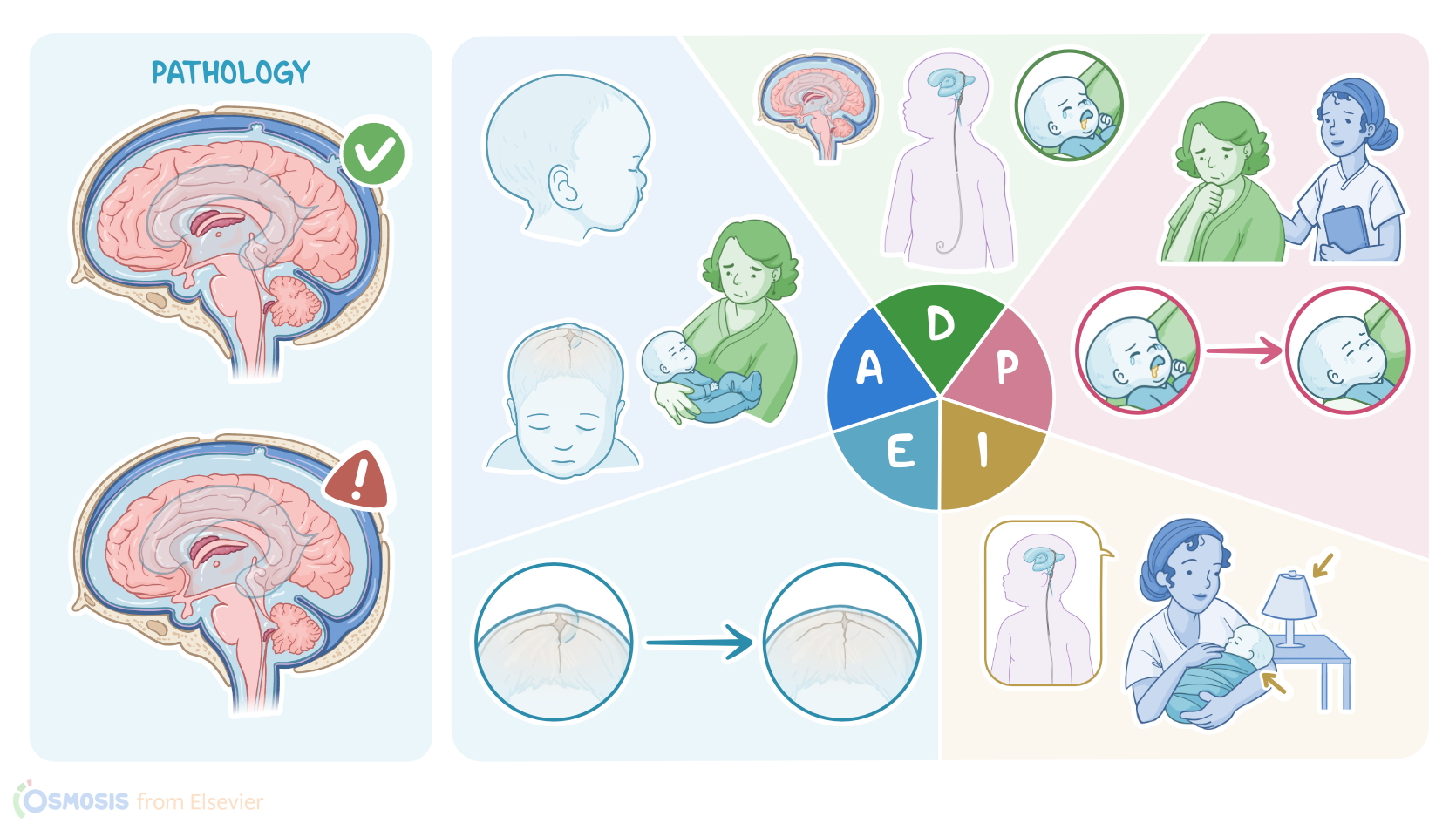
Hydrocephalus refers to the excessive buildup of cerebrospinal fluid or CSF within the brain. The CSF helps provide cushion and protection as well as metabolic fuel for the brain.
Now the brain has two lateral ventricles as well as the 3rd and 4th ventricles, which are all interconnected, and each one contains a structure called a choroid plexus.
The choroid plexus is made up of ependymal cells which produce the CSF that can drain down to the 4th ventricle. From there, the CSF enters the subarachnoid space and gets reabsorbed by the arachnoid granulations into the dural venous sinuses, which are pools of venous blood.
Finally, the CSF and venous blood are drained together out of the skull and into the internal jugular vein. Now since the skull is such a rigid structure, the volume of the brain, CSF, and blood must be constant and in balance.
So with hydrocephalus, the increased CSF volume causes the four ventricles to enlarge and intracranial pressure to rise, which can compress and damage brain structures.
Now there are two types of hydrocephalus. Non-communicating or obstructive hydrocephalus is caused by an obstruction of the CSF flow anywhere along its path.
This can be caused by a brain tumor, cyst, or by congenital causes like cerebral aqueduct stenosis. On the other hand, communicating or non-obstructive hydrocephalus is most often caused by decreased CSF reabsorption.
This occurs when there's inflammation or obstruction of the arachnoid granulations which can be caused by infections such as meningitis as well as subarachnoid hemorrhage.
Less frequently, communicating hydrocephalus can be caused by increased CSF production like by a choroid plexus tumor. Now there are some factors that can put the client at increased risk of hydrocephalus.
Non-bonofiable risk factors include male sex, having a family history of hydrocephalus, and some congenital malformations affecting the CSF flow.
On the other hand, modifiable risk factors include being unvaccinated against common bacteria that cause meningitis, as well as experiencing head trauma which may lead to subarachnoid hemorrhage.
Other possible modifiable risk factors include gestational diabetes mellitus, as well as the lack of prenatal care before pregnancy, which may increase the risk of congenital infections like congenital syphilis and rubella.
Now the main symptoms of hydrocephalus result from the increased intracranial pressure and include headache, nausea, vomiting, as well as seizures.
In addition, clients may have blurred vision, unsteady gait, and impaired consciousness which in severe cases may progress to coma.
In infants, additional symptoms can include irritability, high pitched cry, and a downward deviation of the eyes, which is called sunset eyes, as well as macrocephaly, which is an increase in head circumference.
Clients with hydrocephalus have a higher risk of developing long term complications such as permanent visual impairment, speech impairment, and learning difficulties affecting the client's attention, thinking, and memory formation.
In children this results in poor school performance. Other potential complications include recurrent seizures, as well as physical disabilities such as balance problems, poor motor coordination, and muscle weakness.
Diagnosis of hydrocephalus begins with history and clinical findings. In addition, fundoscopic examination may show papilloedema, which is the swelling of the optic nerve.
Next, in infants younger than 6 months of age, where fontanelles are still present, cranial ultrasonography is typically done to visualize the enlarged brain ventricles, while in older children and adults, MRI is preferred.
Finally, other tests can be performed based on the suspected cause, such as a lumbar puncture for meningitis or CT scan to rule out a subarachnoid hemorrhage.
Now, the main treatment of hydrocephalus focuses on draining the excess CSF from the brain to decrease the intracranial pressure.
If hydrocephalus develops over a short period of time, the intracranial pressure should be decreased with a lumbar puncture or by surgically placing a temporary shunt, such as an external ventricular drain into the brain ventricles.
This allows monitoring of the intracranial pressure while also draining the excess CSF into an external collection bag. In young infants, an anterior fontanelle ventricular tap can be done where a needle is inserted through the anterior fontanelle and into the ventricles, which allows the excess CSF to drip out.
Now, if hydrocephalus persists, the client may need surgical placement of a permanent shunt. Which drains the excess CFF from brain ventricles to somewhere else in the body where it can then be absorbed into the bloodstream.
The most commonly used shunt is a ventricular peritoneal shunt, or VP shunt for short, which drains the CSF into the peritoneal cavity in the abdomen.
Finally, keep in mind that clients with a shunt need to be followed up for complications like shunt obstruction, disconnection, and infection.
Eshan has just been admitted to the NICU, and you begin your assessment. Eshon appears listless as he rests in his mother's arms, and you instantly notice the macrocephaly.
Assessment6:11–8:27
You gently pick up Ishan and place him on the prepared radiant warmer bed and attach the cardio respiratory monitor and pulse oximeter.
Light palpation of his head reveals firm bulging fontanelles and widened cranial suture lines. Careful measurement of his skull reveals that his occipital frontal circumference or OFC is 15.6 inches or 39.6 centimeters.
Ishan's medical records indicate that his birth OFC measurement was within normal limits at 13.5 inches or 34.3 centimeters.
Next, you perform a focused neurological assessment and observe sluggish pupils, as well as delayed and decreased reflexes with mild lower extremity spasticity.
Throughout the physical assessment, you note that Ishaw has a shrill, high pitched cry. Ishan's vital signs are temperature 98.6 °F or 37 °C.
Apical heart rate is regular at 90 BPM. Respirations are regular at 42 breaths per minute.
BP of 78/42 millimeters of mercury, and oxygen saturation of 93% on room air. His pain level is 5 out of 10 on the FLC Behavioral pain assessment scale.
You administer 2 L of supplemental oxygen via nasal cannula. Next you review his recent lab results and take note of the following potassium 3.5 milliequivalents per liter, hematocrit 45%, hemoglobin 14.3 g per deciliter, ventriculomegaly is evident on anterior fontanelle ultrasound.
Ziva expresses fear about Ishaw not making a full recovery and how that would impact their family's future. You reassure Ziva that hydrocephalus is a treatable condition, and the healthcare team is here to help Ishaw.
After documenting your findings, you let Ziva know that the neurosurgeon will be in soon to obtain surgical consent for shunt placement.
With the assessment data you obtained, you identify these priority nursing diagnoses ineffective cerebral tissue profusion related to increased intracranial pressure, fluid and electrolyte imbalance related to vomiting and poor feeding, impaired comfort related to increased intracranial pressure, risk for infection related to surgical procedure, and compromised parental coping related to sudden onset of acute illness.
Diagnosis8:27–8:55
You next create a plan of care for Ishan while collaborating with his mother and the neurosurgical team. By discharge, Ishan will have stabilized intracranial pressure and an OFC measurement trending toward normal parameters, cessation of vomiting with adequate oral intake, increased consolability, and Ishan's mother will demonstrate adequate coping skills.
Planning8:55–9:19
Implementation9:19–10:04
It's now time to begin implementing the plan of care. You closely monitor intake, output, and laboratory values and perform routine vital signs and neurologic checks.
Next, you administer the ordered IV fluids and electrolytes, antibiotics, and antiemetic. You provide comfort by swaddling him and you teach Ziva how to position Ishan so his neck remains in neutral alignment while his head is elevated at 30 degrees.
You also keep environmental stimuli and handling to a minimum and keep the lights around Ishan's bed dimmed. Finally, you reinforce the neurosurgeon's explanation about Ishan's condition and the need for shunt placement while providing emotional support for Ziva.
Isan had a successful VP shunt placement and has been transferred back to the NICU for continued monitoring and evaluation.
Evaluation10:04–11:48
Intracranial pressure monitoring was initiated intraoperatively during the shunt placement and will be continued postoperatively.
You observe that his fontanels are soft and moderately rounded, and his cranial suture lines are closer together. The surgical sites on Ion's head and abdomen are intact with no signs of bleeding or infection.
He has had one wet diaper, and his laboratory values have normalized. Ishan's cry is now at normal pitch, and he is easily consoled.
His vital signs are temperature 98.4 °F or 36.8 °C, heart rate 120 BPM, respirations 56 breaths per minute, BP 80/44 millimeters of mercury, and oxygen saturation 96% on room air.
His pain level is now 3 out of 10 on the Flax scale. While teaching Ziva to monitor for signs and symptoms of shunt malfunction or infection such as fever, seizures, or other neurological signs.
She tells you she has been reading about Ishan's condition and proper home management and care of his shunt. You are happy that your interventions have been successful in supporting both Ishan and his mother.
As Ishan continues to recover, the health care team will continue to monitor Ishan's recovery and revise his plan of care as needed.
All right, as a quick recap, Ishan Mandal was admitted to the NICU with signs and symptoms of hydrocephalus, which is an excessive buildup of CSF within the brain.
Summary11:48–12:38
Your assessment revealed increased OFC, vomiting, suckling difficulty, and a shrill cry. The priority nursing diagnoses you established were ineffective cerebral tissue profusion, fluid and electrolyte imbalance, impaired comfort, risk for infection, and compromised parental coping.
Careful planning allowed you to create goals that focused on alleviating Ishan's symptoms and supporting Ziva's ability to cope.
Implementation of the plan of care involved decreasing intracranial pressure and providing information and emotional support to his mother.
Finally, you evaluated that Ishan's plan of care was effective in promoting his well-being.
| HYDROCEPHALUS | ||
| KEY POINTS | NOTES | |
| PATIENT REPORT |
| |
| PATHOPHYSIOLOGY |
| |
| DIAGNOSIS AND TREATMENT |
| |
| ASSESSMENT |
| |
| NURSING DIAGNOSES |
| |
| PLANNING |
| |
| IMPLEMENTATION |
| |
| EVALUATION |
| |
- "Ackley and Ladwig’s Nursing Diagnosis Handbook: An Evidence-Based Guide to Planning Care, 13th edition" Mosby (2022)
- "Medical-Surgical Nursing: Concepts for Interprofessional Collaborative Care, 10th Edition" Elsevier (2020)
- "Harrison’s Principles of Internal Medicine, 21st edition" McGraw Hill / Medical (2022)
- "Anaesthetic management for hiatal hernia repair in a child with Bartter's syndrome: A case report" J Pak Med Assoc (2020)
- "Critical Care Nursing: Diagnosis and Management, 9th edition" Elsevier (2021)
- "Syndromic Hydrocephalus" Neurosurg Clin N Am (2022)
No notes for this video yet
Try adding a note below