Increased intracranial pressure (ICP): Nursing
Introduction0:00–0:11
Increased intracranial pressure is a potentially life-threatening condition characterized by increased pressure within the skull.
First, let’s take a look at the physiology of intracranial pressure or ICP, which is how much pressure there is within the cranium and spinal column, and it plays an important role in how much oxygen gets to the brain.
Physiology0:11–2:05
ICP, which is normally between 7 and 15 mmHg, is determined by the cerebral perfusion pressure or CPP, which is the amount of force available to deliver blood to the brain.
CPP is defined as the difference between the mean arterial pressure, or MAP and the ICP.Now, when it comes to regulating ICP, remember that the mature skull is a rigid structure that can’t expand.
So, to maintain a normal ICP, there must be a balance between the volumes of its contents, including brain tissue, cerebrospinal fluid or CSF, and blood supplying the brain.
As a result, if there’s an increase in the volume of any one of these three, there should be a compensatory decrease in the other two, which is known as the Monroe-Kellie hypothesis.
Normally, the volume of the brain remains relatively stable, so ICP can be regulated by changes in CSF and blood volume.
When needed, CSF production can be decreased or reabsorption can be increased to help bring ICP back to normal. Similarly, cerebral blood volume can be decreased either by cerebral vasoconstriction or increasing the venous blood drainage out of the skull.
Finally, it’s worth noting that cerebral blood vessels are lined by highly selective endothelial cells that only allow certain substances to pass into the interstitial fluid surrounding the neurons.
This is commonly referred to as the blood-brain barrier, and it helps protect the neurons from pathogens, some immune cells, different products of metabolism as well as some medications that can be found in the systemic circulation.
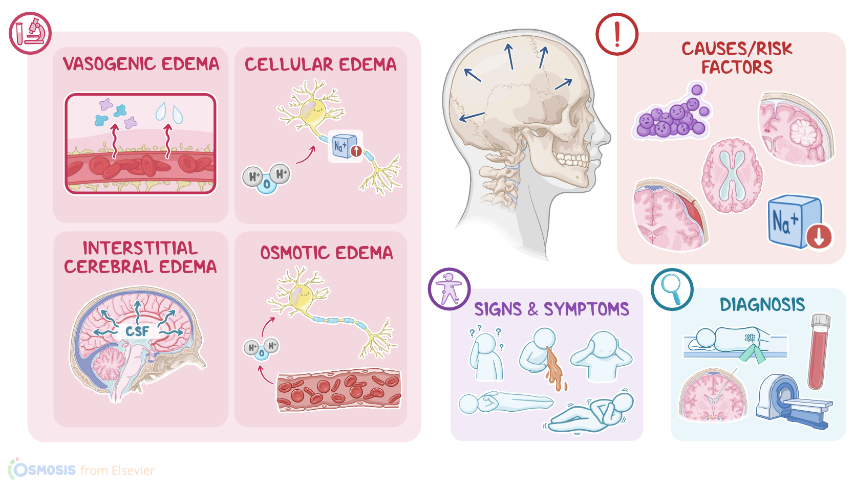
So, increased ICP can be caused by an increased volume within the skull. An increase in brain tissue volume might develop in the case of cerebral edema, which is an excessive buildup of fluid throughout the brain tissue.
Causes & risk factors2:05–2:53
Some causes of cerebral edema include encephalitis, hyponatremia, and a large ischemic stroke. In addition, space-occupying lesions such as brain tumors or abscesses, as well as intracranial hemorrhage, can also lead to increased ICP.
Finally, increased ICP may be caused by increased CSF production, like in a choroid plexus tumor; as well as impaired CSF circulation, like in obstructive hydrocephalus; or decreased CSF reabsorption, like in meningitis or a subarachnoid hemorrhage.
Now, when it comes to pathology, there are 4 major causes of cerebral edema. First up, there’s vasogenic edema, which is the most common form, and it’s when the blood-brain barrier is disrupted, allowing more molecules, like proteins, to pass out of the blood vessel and into the interstitium.
Pathology2:53–5:30
In turn, this creates an increased osmotic pressure that draws fluid into the brain interstitium, increasing the ICP. Second, there’s cellular edema, also known as cytotoxic edema.
In this case, injury to the brain cells causes an increase in intracellular sodium, which, in turn, makes more water enter the cells.
So increased ICP is on account of the cells swelling up with water. Third, there’s interstitial cerebral edema, which is when CSF flows from the brain ventricles into the interstitium.
And finally, there’s osmotic edema, which is when disturbances in the osmotic pressure in the blood, such as hyponatremia, makes brain cells pull in water from the blood, which increases the ICP.
Now, the compensatory mechanisms that keep ICP within a normal range have limits, and can be overwhelmed. When this happens, ICP starts to increase, which puts pressure on the ventricles and cerebral blood vessels.
In turn, this decreases cerebral blood flow and cerebral perfusion. Lower cerebral perfusion means that less oxygen is delivered to the brain tissue, which causes neurons to swell up and die, worsening the already increased ICP.
At the same time, carbon dioxide starts building up, causing vasodilation, which allows even more fluid to leak from the blood vessels into the brain tissue.
If not corrected, increased ICP can lead to complications, such as papilledema, which is the swelling of the optic disc, which is the point where the optic nerve leaves the retina.
The most severe complication, though, is brain herniation. Brain herniation occurs when a part of the brain is pushed into another space of the skull or even out of it, and this can affect other CNS structures, like the brainstem.
Brain herniation can either be supratentorial and infratentorial. Supratentorial herniation refers to displacement of the cerebrum above the tentorium, which is a meningeal fold located in the back of our skull that separates the cerebrum from the cerebellum.
Infratentorial herniation refers to herniation of the cerebellum, which is located below the tentorium. Now, clinical manifestations of increased ICP can be classified as early or late signs and symptoms.
Additionally, clients with papilledema may experience visual abnormalities, such as double vision or even visual loss. On the other hand, late signs and symptoms include hypertension, bradycardia, and irregular breathing; these signs are referred to as a Cushing’s triad, which indicate advanced brain stem dysfunction; as well as fixed or dilated pupils.
Clinical manifestations5:30–7:00
Finally, there could be loss of brainstem reflexes such as the gag reflex, the swallowing reflex or the pupillary and corneal reflexes.
There’s also progressive deterioration of the client’s level of consciousness, and if not promptly treated, clients may fall into a deep state of unconsciousness, or coma.
Lastly, with severe brain injury, the client may show abnormal posturing, such as decerebrate or decorticate posturing. Decerebrate posturing is where the arms are stiffly extended and abducted, and the wrists are pronated, with flexed fingers; while the legs are extended, with the feet in plantar flexion.
With decorticate posturing, on the other hand, the arms are adducted and flexed on the chest, with flexed wrists and fingers, while the legs are extended and internally rotated, with plantar flexion of the feet.
Diagnosis of increased ICP starts with the client’s history and physical assessment. Then, diagnostic tests can be divided into tests that confirm the increased ICP, and tests that look for the underlying cause.
A lumbar puncture can confirm increased ICP and once the needle is inserted into the spinal canal, it’s attached to a manometer that measures CSF pressure.
However, a non-contrast CT should always be done before preforming a lumbar puncture when increased ICP is suspected. This is to rule out mass occupying lesions like a tumor or hematoma that increase the risk of cerebral herniation during the procedure, which is often fatal.Alternatively, ICP can be measured by inserting an intraventricular catheter directly in CSF within the lateral ventricle of the brain.
Diagnosis7:00–8:20
A fundoscopic examination of the eye can also be done, which can identify papilledema as an indirect sign of increased ICP.
Tests to look for the underlying cause include imaging techniques, such as a CT scan or MRI, to look for lesions within the skull; as well as blood tests, drug and toxicology screens, and CSF analysis, which may reveal signs of infection or inflammation.Treatment of increased ICP is aimed at reducing the pressure within the skull.
This can be done by keeping the client’s head in a neutral position and elevating the head of the bed at 30 degrees to increase venous blood outflow.
Also ensuring adequate respirations is needed to maintain cerebral oxygenation, since increased PaCO2 and hypoxemia tend to increase ICP.
In addition, analgesics and anxiolytics can be given to decrease agitation, which can increase ICP, and osmotic diuretics, such as mannitol, can be used to help draw fluid from the brain tissue into the intravascular space.
Treatment8:20–9:27
If these measures cannot lower ICP, an intraventricular catheter can be placed to drain excess CSF from the ventricles into an external system.
Another way of treating increased ICP involves doing a decompressive craniectomy, where a part of the skull is temporarily removed to help relieve the pressure.
Finally, definitive treatment may involve treating the underlying cause, such as surgical removal of a tumor, abscess, or hematoma, when possible.Alright, as a quick recap… Increased intracranial pressure or ICP, is when ICP is higher than 15mmHg.
To maintain a normal ICP, there must be a balance between the volumes of its contents, including brain tissue, cerebrospinal fluid or CSF, and blood supplying the brain.
One of the major causes of increased ICP is cerebral edema, and there are 4 types: vasogenic, cytotoxic, interstitial, and osmotic edema.
Causes of cerebral edema include encephalitis, hyponatremia, and a large ischemic stroke. In addition, increased ICP can also be caused by space-occupying lesions such as brain tumors or abscesses, intracranial hemorrhage, or imbalances in CSF production, reabsorption or circulation.
Review9:27–11:20
Late signs and symptoms include hypertension, bradycardia, and irregular breathing; these signs are referred to as a Cushing’s triad, which indicate advanced brain stem dysfunction; as well as fixed or dilated pupils.
Diagnostic tests used to confirm increased ICP include a lumbar puncture or placing an intraventricular catheter, while tests used to look for the underlying cause include imaging techniques like a CT or MRI scan, as well as blood tests, drug and toxicology screens, and CSF analysis.
Treatment can be done by elevating the client’s head, using analgesics, anxiolytics and osmotic diuretics, placing an intraventricular catheter to drain the excess CSF, or doing a craniotomy to relieve the pressure.
breathing. The signs are referred to as a Cushing's Triad, which indicate Advanced brain stem dysfunction as well as fixed or dilated pupils.
Diagnostic test, used to confirm increased ICP include a lumbar puncture or placing an introvert. Regular catheter will test used to look for the underlying cause include Imaging techniques like a CT or MRI scan as well as blood test drug toxicology screens, and CSF analysis.
Treatment can be done. By elevating the clients head using analgesics anxiolytics and osmotic diuretics.
Placing an intervention with a catheter to drain the excess USF, or doing a craniotomy to relieve the pressure.
| INCREASED INTRACRANIAL PRESSURE (ICP) | ||
| KEY POINTS | NOTES | |
| DEFINITION |
| |
| PHYSIOLOGY |
| |
| CAUSES AND RISK FACTORS |
| |
| PATHOPHYSIOLOGY |
| |
| SIGNS AND SYMPTOMS |
| |
| DIAGNOSIS |
| |
| TREATMENT |
| |
No notes for this video yet
Try adding a note below