Myasthenia gravis: Nursing

1,314views

Myasthenia gravis: Nursing

Watch later

Watch later

Hypokalemia: Clinical
Movement of water between body compartments
The role of the kidney in acid-base balance
Blood histology
Blood components
Respiratory acidosis
Metabolic acidosis
Respiratory alkalosis
Metabolic alkalosis
Metabolic and respiratory alkalosis: Clinical
Metabolic and respiratory acidosis: Clinical
Bronchodilators: Beta 2-agonists and muscarinic antagonists
Chronic obstructive pulmonary disease (COPD): Clinical
Chronic obstructive pulmonary disease (COPD): Nursing process (ADPIE)
Adrenergic antagonists: Alpha blockers
Sympatholytics: Alpha-2 agonists
Alpha-2 adrenergic agonists: Nursing pharmacology
Adrenergic receptors
Adrenergic antagonists: Beta blockers
Diabetes mellitus: Clinical
Diabetes mellitus (DM): Nursing process (ADPIE)
Diabetes mellitus
Diabetes mellitus: Pathology review
Stroke: Nursing process (ADPIE)
Stroke: Clinical
Ischemic stroke
Intracerebral hemorrhage
Peptic ulcers and stomach cancer: Clinical
GERD, peptic ulcers, gastritis, and stomach cancer: Pathology review
Sickle cell disease (NORD)
Sickle cell disease: Nursing process (ADPIE)
Sickle cell disease: Clinical
Blood groups and transfusions
Erythropoietin
Blood products: Nursing pharmacology
Oxygen binding capacity and oxygen content
Blood products and transfusion: Clinical
Hemophilia
Hemophilia: Nursing process (ADPIE)
Leukemia: Nursing process (ADPIE)
Chronic leukemia
Leukemias: Pathology review
Acute leukemia
Leukemia: Clinical
Lymphomas: Pathology review
Lymphoma: Clinical
Non-Hodgkin lymphoma
Hodgkin lymphoma
Lymphatic system anatomy and physiology
Anatomy of the abdominal viscera: Liver, biliary ducts and gallbladder
Liver anatomy and physiology
Cirrhosis
Cirrhosis: Pathology review
Cirrhosis: Clinical
Cirrhosis: Nursing process (ADPIE)
Hepatitis C virus
Hepatitis A and Hepatitis E virus
Liver cancer: Nursing
Cholestatic liver disease
Non-alcoholic fatty liver disease
HIV (AIDS)
HIV and AIDS: Pathology review
Antiretrovirals for HIV/AIDS - Protease inhibitors: Nursing pharmacology
Antiretrovirals for HIV/AIDS - NRTIs and NNRTIs: Nursing pharmacology
Antiretrovirals for HIV/AIDS - Integrase strand transfer inhibitors: Nursing pharmacology
Anatomy of the abdominal viscera: Pancreas and spleen
Pancreatitis: Nursing process (ADPIE)
Glaucoma: Nursing process (ADPIE)
Glaucoma
Eye conditions: Refractive errors, lens disorders and glaucoma: Pathology review
Acute respiratory distress syndrome (ARDS): Nursing
Anatomy of the coronary circulation
Coronary artery disease: Pathology review
Coronary artery disease: Clinical
ECG basics
ECG normal sinus rhythm
ECG rate and rhythm
ECG axis
ECG intervals
ECG QRS transition
ECG cardiac infarction and ischemia
Cardiac cycle
Arterial disease
Delirium
Dementia and delirium: Clinical
Vascular dementia
Frontotemporal dementia
Dementia with Lewy bodies
Dementia: Pathology review
Tumor lysis syndrome (TLS): Nursing Process (ADPIE)
Cholelithiasis: Nursing
Coronary artery disease (CAD) and angina pectoris: Nursing process (ADPIE)
Breast cancer: Nursing process (ADPIE)
Ovarian cancer: Nursing
Cervical cancer: Nursing
Hormones and hormone modulators for cancer: Nursing pharmacology
Endometriosis: Nursing
Heart failure
Left-sided heart failure: Nursing process (ADPIE)
Heart failure: Pathology review
Pneumonia: Pathology review
Pneumonia
Bacterial pneumonia: Nursing process (ADPIE)
Respiratory stimulants: Nursing pharmacology
Corticosteroids - Inhaled: Nursing pharmacology
Pneumothorax and hemothorax: Nursing
Chest trauma: Clinical
Pleural effusion: Nursing
Tuberculosis (TB): Nursing
Parkinson disease: Nursing process (ADPIE)
Huntington disease
Multiple sclerosis (MS): Nursing
Multiple sclerosis
Guillain-Barré syndrome: Nursing
Guillain-Barre syndrome
Myasthenia gravis: Nursing
Acute kidney injury (AKI): Nursing process (ADPIE)
Acute kidney injury: Clinical
Chronic kidney disease (CKD): Nursing
Chronic kidney disease
Chronic kidney disease: Clinical
Polycystic kidney disease (PKD): Nursing
Renin-angiotensin-aldosterone system
Osteoarthritis: Nursing
Osteoarthritis
Rheumatoid arthritis (RA): Nursing process (ADPIE)
Rheumatoid arthritis: Clinical
Rheumatoid arthritis
Rheumatoid arthritis and osteoarthritis: Pathology review
Systemic lupus erythematosus
Systemic lupus erythematosus (SLE): Pathology review
Systemic lupus erythematosus (SLE): Clinical
Systemic lupus erythematosus (SLE): Nursing
Mycobacterium tuberculosis (Tuberculosis)
Peripheral venous disease (PVD): Nursing process (ADPIE)
Peripheral arterial disease (PAD): Nursing process (ADPIE)
Buerger disease: Nursing
Raynaud phenomenon: Nursing
Aortic dissections and aneurysms: Pathology review
Aortic aneurysms and dissections: Clinical
Aortic dissection
Aortic aneurysm: Nursing process (ADPIE)
Venous thromboembolism (VTE): Nursing process (ADPIE)
Benign prostatic hyperplasia (BPH): Nursing process (ADPIE)
Benign prostatic hyperplasia
Prostate cancer: Nursing
Prostate cancer
Testicular cancer: Nursing
Testicular cancer
Diabetic ketoacidosis (DKA): Nursing process (ADPIE)
Endocrine system anatomy and physiology
Vitamin B12 deficiency
Thalassemia: Nursing
Anemia - Iron-deficiency: Nursing
Anemia - Macrocytic: Nursing
Anemia - Aplastic: Nursing
Thyroid hormones
Medications for thyroid disorders: Nursing pharmacology
Hyperthyroidism: Nursing process (ADPIE)
Hypothyroidism: Nursing process (ADPIE)
Hypoparathyroidism: Nursing
Hyperparathyroidism: Nursing
Anxiety disorders: Nursing process (ADPIE)
Spinal cord injury (SCI): Nursing
Cluster A personality disorders
Smoke inhalation injury: Nursing process (ADPIE)
Shock - Anaphylactic: Nursing
Shock - Obstructive: Nursing
Shock - Neurogenic: Nursing
Shock - Hypovolemic: Nursing
Shock - Cardiogenic: Nursing
Shock - Septic: Nursing
Pulmonary edema: Nursing
Burn injury: Nursing

Notes

MYASTHENIA GRAVIS

KEY POINTS
NOTES
DEFINITION
  • Chronic, progressive autoimmune disease with disrupted neuromuscular communication

PHYSIOLOGY
  • Muscle contraction
    • Initiated at neuromuscular junction
    • Acetylcholine (ACh) released
    • Binds nicotinic receptors
    • Voluntary muscle contraction

CAUSES AND RISK FACTORS
  • Causes
    • Autoantibodies that target the neuromuscular junction
  • Risk factors
    • Assigned female at birth in their 20s-30s
    • Assigned male at birth in their 60s-70s

PATHOPHYSIOLOGY
  • Autoantibodies bind ACh receptor
  • Loss of ACh receptor function and inflammation triggered
  • Number of ACh receptors decreases
  • Muscle contraction less effective over time

SIGNS AND SYMPTOMS
  • Fatigue
  • Skeletal muscle weakness
  • Diplopia
  • Ptosis
  • Impaired facial expressions
  • Difficulty chewing or swallowing
  • Slurred speech
  • Difficult walking or climbing stairs
  • Abnormal posture
  • Difficult breathing
  • Myasthenic crisis
    • Generalized muscle weakness
    • Respiratory failure

DIAGNOSIS
  • History
  • Physical assessment
  • Laboratory tests
  • Electromyography (EMG)
  • Tensilon test
  • X-ray
  • CT

TREATMENT
  • Acetylcholinesterase inhibitors
  • Immunosuppressive medications
  • Immunoglobulins
  • Plasmapheresis
  • Surgery

MANAGEMENT OF CARE
  • Goals of care
    • Prevent and manage complications
    • Promote muscle strength and quality of life
  • Assess weakness 
  • Assess lungs and ability to cough and swallow
    • Notify HCP
      • Respiratory rate below 12 or above 20
      • Oxygen saturation below 92%
      • Abnormal chest movement
      • Reduced or absent breath sounds
  • Administer medications as prescribed
  • Assist to high Fowler position
  • Assess gag reflex
  • Encourage soft or non-solid foods
  • Consider referral to speech language pathologist
  • Keep items within reach
  • Institute fall precautions
  • Notify HCP
    • Signs of deterioration
    • Signs of cholinergic crisis
  • Provide encouragement and psychosocial support

PATIENT AND FAMILY TEACHING
  • Explain condition, plan of care, and how to safely self-administer medications
  • Emphasize consultation with HCP before beginning new medications
  • Organize daily activities 
  • Safety precautions
  • Eat small, frequent meals
  • Rest between bites
  • Choose foods that are easy to chew
  • Infection prevention
  • Recognize symptoms of myasthenia crisis
  • Provide information on local support groups
  • Wear medical identification bracelet

Transcript

Watch video only

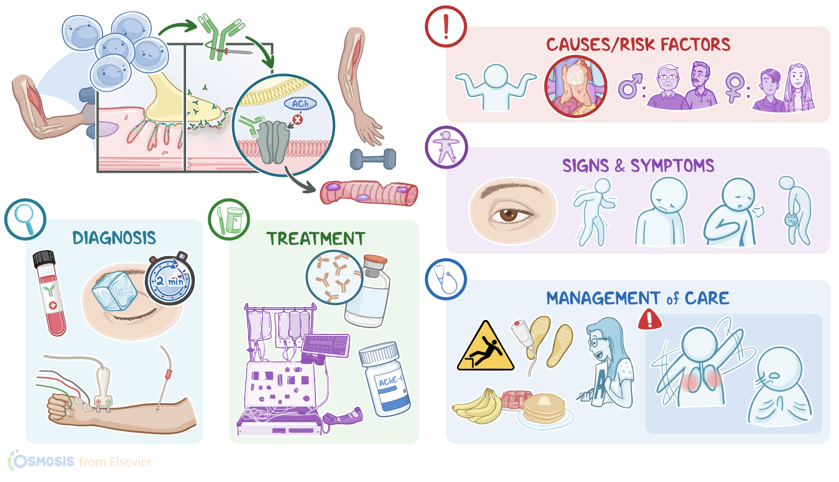
Myasthenia gravis is a chronic, progressive autoimmune disease caused by antibodies that bind to and destroy neuromuscular communication, where nerves send impulses to trigger skeletal muscle contraction. Over time, this results in fatigue and muscle weakness due to impaired muscle contractions.

Now, to better understand myasthenia gravis, let's review the normal physiological process of muscle contraction that occurs at cellular level. Muscle contraction is initiated at the neuromuscular junction, which is basically a connection between the axonal end of a motor neuron and a skeletal muscle fiber. So, the end of the neuron is called the presynaptic membrane, while the muscle cell is called the postsynaptic membrane.

Now, to trigger muscle contraction, the axonal end of the motor neuron releases the neurotransmitter acetylcholine, also called ACh, at the neuromuscular junction. Acetylcholine then binds to the nicotinic acetylcholine receptors present on the muscle cell membrane. The binding of acetylcholine to its receptor activates a chain reaction within the muscle cell, which ultimately results in voluntary muscle contraction.

Now, myasthenia gravis is caused by autoantibodies that target the neuromuscular junction. In a minority of cases, this occurs as a paraneoplastic syndrome, where an underlying cancer like thymoma or bronchogenic carcinoma can result in the production of autoantibodies. However, in the majority of cases, myasthenia gravis does not have a clear underlying cause. Clients at risk are those who are assigned female at birth in their 20s and 30s, or clients assigned male at birth in their 60s and 70s.

So, in myasthenia gravis, the client's own B cells generate autoantibodies, setting in a type II hypersensitivity reaction. These autoantibodies specifically bind with the postsynaptic acetylcholine receptor, or AChR for short, in 90% of clients, or sometimes target the muscle-specific tyrosine kinase receptors, or MuSK for short, in 10% of clients, both of which ultimately lead to loss of acetylcholine receptor function. In addition, binding of autoantibodies to acetylcholine receptors activates the classical pathway of the complement, which triggers inflammation and damage of the postsynaptic membrane of muscle cells, ultimately decreasing the number of available acetylcholine receptors on the muscle membrane. As a result of all these, initiation of a muscle contraction becomes less effective over time.

Typically, myasthenia gravis presents with fatigue and skeletal muscle weakness that fluctuates throughout the day. Clients with myasthenia gravis might wake up feeling fine, but get progressively weaker as their daily activities go on, and by the end of the day they might feel very weak. This weakness usually increases when performing repetitive movements, and improves with rest. Moreover, myasthenia gravis is a chronic and progressive disease that worsens over time.

The initial symptoms of myasthenia gravis often involve the extraocular muscles, which control the movement of the eyes and eyelids, so clients might experience diplopia or ptosis. If facial muscles are involved, clients may present with impaired facial expressions, difficulty in chewing or swallowing, slurred speech, and fading out voice along the conversation. Neck muscles can become weak too, and these clients might feel their head is heavy and it’s hard to keep upright. As the disease progresses, arm and leg muscles are also involved, so clients might experience difficulty with walking, climbing stairs, and even performing simple tasks such as hair combing and brushing their teeth. Clients may also develop an abnormal posture due to weakness of the hip muscles.

Myasthenia gravis can also affect the respiratory muscles, so the diaphragm and the intercostal muscles, leading to difficulty breathing. Finally, a potentially life-threatening acute exacerbation of the disease is a myasthenic crisis, which can present with a worsening generalized muscle weakness, and even lead to respiratory failure. Triggers of myasthenic crisis include stressful events like an infection, surgery, emotional trauma, temperature extremes, or pregnancy. Lastly, certain medications can make myasthenia gravis worse, and should be avoided or used with caution, including certain antibiotics like fluoroquinolones, macrolides, and aminoglycosides; as well as beta-blockers like propranolol; and antiarrhythmic medications like procainamide and quinidine.

Now, the diagnosis of myasthenia gravis starts with a history and physical assessment, as well as laboratory tests to detect AChR or MuSK antibodies in blood. Additional diagnostic tests include electromyography, or EMG for short, which uses repetitive electrical stimuli to assess muscle contraction. A Tensilon test can be performed by intravenously injecting edrophonium chloride, which is an acetylcholinesterase inhibitor that briefly blocks the breakdown of acetylcholine and temporarily increases its levels at the neuromuscular junction; in myasthenia gravis, this is enough to overcome the symptoms caused by the decreased number of available receptors, so the weakness is improved. Lastly, chest X-ray and CT scan can be performed to rule out an underlying cause like a thymoma.

Although there’s no cure for myasthenia gravis, certain medications can be used to help mitigate some of the symptoms and improve the client’s quality of life. Initial treatment consists of acetylcholinesterase or AChE inhibitors, such as neostigmine or pyridostigmine. These medications prevent the enzyme acetylcholinesterase from degrading acetylcholine, which increases its concentration around muscle cells and, subsequently, its effects are increased and prolonged. Severe cases of myasthenia gravis can also be treated with immunosuppressive medications, such as corticosteroids like prednisone, which reduce the production of autoantibodies.

Clients experiencing an acute exacerbation or myasthenic crisis can be treated with intravenous immune globulins or IVIGs, in order to quickly neutralize the autoantibodies and stop the acute exacerbation; or with plasmapheresis, or plasma exchange to filter out the circulating autoantibodies from the client’s blood; and some clients may require respiratory support. Finally, clients with a thymoma can undergo surgical removal of the thymus, known as thymectomy.

All right, now let’s look at nursing care for a client with myasthenia gravis. The main goals of nursing care are to prevent and manage complications, as well as promote optimal muscle strength and improved quality of life. Begin assessing your client’s muscle weakness, including the onset, duration, and frequency of muscle weakness. Assess your client’s respiratory rate, depth, effort, breathing pattern, breath sounds, and oxygen saturation.

Report concerning assessment findings, like a respiratory rate below 12 or above 20 breaths per minute, an oxygen saturation below 92%, abnormal chest movement, or reduced or absent breath sounds. If this occurs, ensure oxygen, suction equipment, and a bag valve mask are readily available. Be sure to also assess your client’s ability to cough and swallow, and implement aspiration precautions.