Myocardial infarction (MI): Nursing process (ADPIE)
Client Report0:00–0:38
Miguel Garcia is a 60-year-old Hispanic male with a history of hypertension who was brought to the emergency department, or ED, for chest pain.
The pain began two hours ago and he states it feels like an elephant is sitting on his chest. The physician just confirmed with an electrocardiogram, or ECG, that Mr.
Garcia is experiencing a STEMI, which stands for ST segment elevation myocardial infarction. The team is acting swiftly to prepare him for an emergent percutaneous coronary intervention, known as a PCI.
Pathology0:38–9:30
A myocardial infarction, also known as an MI or heart attack, refers to the death of cardiac muscle tissue from prolonged ischemia.
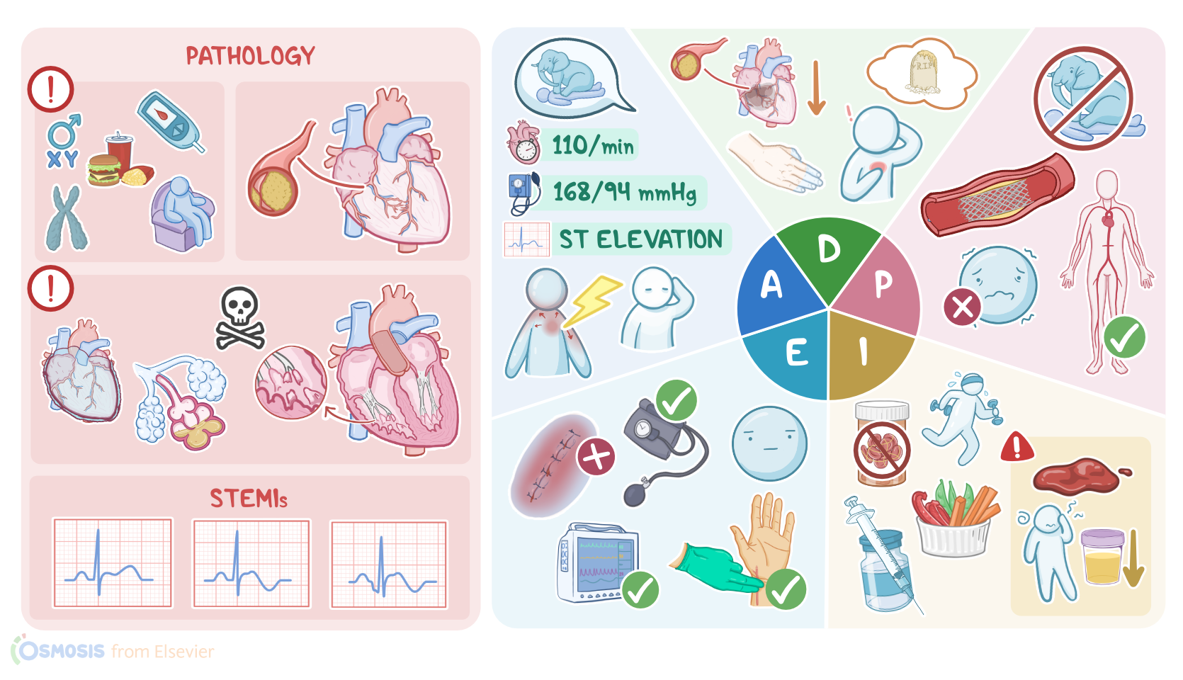
Risk factors include being over 40 years old or male; having diabetes mellitus, dyslipidemia, hypertension, or a family history of MIs; smoking tobacco; obesity; a sedentary lifestyle; and eating a high fat diet.
The most common cause of MI is atherosclerosis. This is when lipid containing plaque builds up in the coronary arteries.When the coronary artery is occluded, blood and oxygen supply cannot meet the demands of the myocardium, causing ischemia.
This leads to angina, or a type of chest pain. Angina can either be stable or unstable.
Stable angina is provoked by activities that increase oxygen demand, such as exercise, lasts up to 15 minutes, and is relieved by rest or vasodilators like nitroglycerin.
This is because the occlusion limits blood flow while the oxygen demand of the myocardium increases, but when demand decreases or if the artery becomes dilated, the pain resolvesOn the other hand, unstable angina can occur without exertion or even at rest, persists longer than 15 minutes, is a more severe pain than stable angina, and is not relieved by rest or vasodilators.
This is often due to the plaque rupturing which causes platelets to attach to the plaque, forming a thrombus. The result is a more severe reduction in coronary blood flow than what occurs in stable angina, so it doesn’t resolve with rest or vasodilators.Initially, ischemic damage is potentially reversible, but after about 20 to 40 minutes the damage is irreversible and myocardial tissue starts to necrose, causing an MI.
During an MI, the lack of blood supply to the myocardium can cause severe crushing substernal chest pain. The pain is unprovoked, which means it occurs spontaneously regardless of activity, persists over thirty minutes, is unrelieved by rest or vasodilators like nitroglycerin, and can radiate to the shoulders, arms, jaw, neck, or back.
The myocardial oxygen supply and demand imbalance during an MI can also lead to blood pressure and heart rate changes, dysrhythmias, dyspnea, nausea, vomiting, fever, diaphoresis, a feeling of impending doom, and coolness or cyanosis of the extremities.
However, symptoms can vary greatly. Women and elderly clients are more likely to experience atypical symptoms, and about 20 percent of MIs are silent, meaning the client was asymptomatic or attributed symptoms to another condition, such as heartburn.The damage caused to the myocardium during an MI can lead to several complications, including arrhythmias, cardiogenic shock, pericarditis, heart failure, papillary muscle rupture, pulmonary edema, and sudden cardiac death.
If an MI is suspected based on symptoms, the next step is to diagnose it as soon as possible. When it comes to diagnosing MIs, there are two types to keep in mind based on the ECG findings: NSTEMIs, or non ST segment elevation myocardial infarction, and STEMIs.
During an MI, the inner third of the myocardium, called the subendocardium, is affected first because it’s farthest from the coronary arteries making it more vulnerable to changes in coronary perfusion.
If the infarct is limited to the subendocardium, this is known as a subendocardial infarction and shows on an ECG with a depressed or non-elevated ST segment.
If the necrosis spreads beyond the endocardium, it’s called a transmural infarction and will have ST segment elevation on an ECG.
For a client with unstable angina, the ECG findings are typically normal. A clear way to differentiate between angina and a MI is with cardiac biomarkers.
Cardiac biomarkers are only present in the case of a MI because they indicate there has been irreversible damage to the myocardium.
These include troponin I, troponin T, and creatinine kinase isoenzyme MB, called CK-MB. Troponin I and T, which are specific markers of cardiac injury, are elevated 2 to 4 hours after an infarction, peak around 48 hours, and remain elevated for 7 to 10 days.
CK-MB levels also start to rise 2 to 4 hours after an infarction, peak around 24 hours, and remain elevated for 48 hours.
Although CK-MB is not specific to cardiac injury, it is useful for identifying a reinfarction before troponin levels have returned to normal.
Because cardiac biomarkers can take several hours to be detected, they should be checked initially upon client arrival and again 6 hours later.Now, treatment should be initiated as soon as possible when a STEMI is diagnosed to restore perfusion to the myocardium.
Oxygen should be administered to clients who are breathless, have an oxygen saturation less than 90 percent, or who have heart failure.
Chewable aspirin should be administered to slow clotting. Vasodilators, like nitroglycerin, should be administered to foster coronary vasodilation but are contraindicated in clients with hypotension, bradycardia, or who have taken phosphodiesterase inhibitors in the past 72 hours.
Pain control increases myocardial oxygenation, so an analgesic such as morphine should be administered too. Within 30 minutes of client arrival, a chest x-ray should be performed to detect cardiomegaly, pulmonary edema, pleural effusion, aortic dissection, or heart failure.
A complete blood count should be performed to assess for anemia and to obtain a baseline platelet count prior to initiating thrombolytic therapy.
A complete metabolic panel can reveal abnormalities in electrolytes and kidney function. Also, clotting times may be ordered to establish baseline levels for anticoagulation therapy.Once a STEMI is confirmed, reperfusion is needed immediately and can be accomplished through PCI; thrombolytic therapy, also called fibrinolytic therapy; or a coronary artery bypass graft, referred to as CABG.
Emergent PCI is the preferred reperfusion strategy and should be performed within 90 minutes of client arrival to the ED.
PCI involves inserting a catheter into the femoral or radial artery, injecting contrast to locate the occlusion, inflating a tiny balloon to compress the obstructing plaque, and inserting a stent to keep the artery patent.
If PCI is unavailable, thrombolytic therapy should be administered within 30 minutes of client arrival to the ED. Thrombolytic therapy involves IV administration of a thrombolytic medication, such as reteplase, to dissolve the occluding clot.
CABG, which involves grafting a vein or artery from elsewhere in the body onto the coronary artery to bypass the blockage.
It’s not used as frequently as PCI or thrombolytic therapy but is indicated when PCI is unsuccessful. If more than 12 hours have passed since symptom onset, reperfusion strategies are generally not performed, but PCI may be considered for clients with ongoing ischemia.After reperfusion or if reperfusion couldn’t be done, anticoagulation agents, such as heparin, are administered, in addition to dual antiplatelet therapy using aspirin and a PY12 inhibitor like clopidogrel.
Cardioprotective medications, including angiotensin converting enzyme, or ACE inhibitors; angiotensin receptor blockers, or ARBs, for clients intolerant of ACE inhibitors; beta blockers; and statins are recommended as long term therapy after a STEMI.
Additionally, lifestyle modifications, like following a low fat and low salt diet, exercising, and smoking cessation, as well as enrolling in a cardiac rehabilitation program are important aspects of post STEMI care.All right, let’s get back to Mr.
Assessment9:30–11:47
Garcia and begin his assessment. Upon entering his exam room, you introduce yourself, confirm his identity, and perform hand hygiene.
Mr. Garcia appears pale, diaphoretic, and anxious.
When asking him to describe his pain, he states about 2 hours ago he was sitting at his desk at work and suddenly felt like his chest was being crushed by an elephant.
He rates the pain as a 10 out of 10 and says it radiates down his left arm. His second dose of nitroglycerin was administered 5 minutes ago, so you administer his third dose, along with the ordered IV analgesic morphine and chewable aspirin.
His blood pressure is 168/94 mmHg, heart rate is 110 beats per minute, and an S4 heart sound is auscultated. He was connected to a 12 lead ECG immediately on arrival and it currently shows ST segment elevation.
Lung sounds are clear, respirations are 20 breaths per minute, and oxygen saturation is 95% on 4 liters per nasal cannula.
Bowel sounds are active and Mr. Garcia reports feeling nauseated.
His extremities are cool and capillary refill is delayed. His temporal artery temperature is 99.2°F or 37.3°C.
Throughout your assessment, Mr. Garcia remains alert and oriented but anxious stating “My dad died from a heart attack when he was around my age and I don’t want that to happen to me.” You reassure Mr.
Garcia that he is in good hands and the PCI will help him get on the road to recovery. After completing your documentation, you note the following results from his STAT labs and note his troponin I, troponin T, CK-MB have all increased.
Other results include platelets 254,000/mm3, hemoglobin 16 g/dL, leukocytes 13,000/mm3, potassium 3.5 mEq/L, creatinine 1.0 mg/dL.
As you leave the room, you update the physician of Mr. Garcia’s continued chest pain and the interventions you provided.Priority nursing diagnoses you choose for Mr.
Garcia’s care include: decreased cardiac output related to coronary artery occlusion, ineffective cardiac and peripheral tissue perfusion related to reduced coronary blood flow, acute pain related to myocardial ischemia, anxiety related to a new situational crisis and family history of fatal MI, and deficient knowledge related to post MI and PCI self care.Next, you collaborate with Mr.
Diagnosis11:47–12:14
Garcia and the healthcare team to plan goals for his care. Within 24 hours after his emergent PCI, Mr.
Garcia’s cardiac output will effectively maintain myocardial and peripheral tissue perfusion; he will have no chest pain, and will achieve a tolerable level of pain at the PCI catheter insertion site; and his anxiety will be decreased.
Planning12:14–12:47
At the time of hospital discharge, Mr. Garcia will verbalize understanding of post MI and PCI care instructions.OK, now it’s time to implement your plan for Mr.
Garcia. You review the physician’s orders and delegate vital sign collection to the patient care technician, requesting that you be notified immediately of any changes.
Meanwhile, you focus on continued monitoring of Mr. Garcia and preparing him for the PCI.
Implementation12:47–16:15
Mr. Garcia reports his chest pain is 9/10, so you administer another dose of IV morphine, which decreases his pain to a 7/10.
When the cardiac catheterization lab calls to say they are ready for your client, you transport Mr. Garcia and give a report to the cardiac catheterization team.The next day, you are assigned to care for Mr.
Garcia in the cardiovascular intensive care unit. Mr.
Garcia successfully underwent a PCI with stent placement to open his left anterior descending artery, which was 100% occluded.
Throughout your shift, you monitor his vital signs and ECG; heart and lung sounds; pain; level of consciousness; anxiety level; urine output; extremities for color and changes in temperature; peripheral pulses; and the PCI catheter insertion site in his right femoral artery.
You are prepared to notify the physician immediately if Mr. Garcia develops abnormal vital signs or heart rhythms; has decreased urine output; develops chest pain or shortness of breath; exhibits changes in level of consciousness; or has signs of bleeding or hematoma formation at the PCI insertion site.You administer the ordered anticoagulant heparin, and antiplatelet medications including aspirin and the PY12 inhibitor clopidogrel.
Mr. Garcia is also prescribed the ACE inhibitor lisinopril, the beta blocker carvedilol, and the statin atorvastatin.
You teach Mr. Garcia that these medications are needed to prevent clotting, to control his blood pressure, and reduce plaque formation, all of which helps him avoid a future MI.For the first 24 hours after his PCI, Mr.
Garcia will be on bed rest to reduce oxygen demand and prevent bleeding at the femoral catheter insertion site with his leg immobilized and the head of his bed elevated no more than 45 degrees.
With the help of another nurse, Mr. Garcia is carefully repositioned every 2 hours to prevent pressure injury.
The physical therapy assistant is consulted to help Mr. Garcia safely perform range of motion exercises, and a referral to cardiac rehabilitation has been placed for when Mr.
Garcia is discharged. You start preparing Mr.
Garcia for his discharge home by teaching him about post MI care, which includes recognizing symptoms that warrant medical attention such as chest pain or shortness of breath.
Next, you review lifestyle modifications, like following a heart healthy diet and increasing physical activity; taking his prescribed medications, and the importance of following up with cardiac rehabilitation.
Discharge instructions for his PCI include activity restrictions, keeping the incision site dry, and symptoms that warrant medical attention, such as redness, bleeding, or other discharge from the incision site; development of a fever, or his leg becoming numb or cool to the touch.
During your shift, you document nursing interventions performed and assessment findings.Now that it’s the end of your shift and it’s time to evaluate Mr.
Garcia’s care. Throughout your shift, his vital signs have remained stable with the most recent readings being: oral temperature 98.9°F or 37.1°C, heart rate 75 beats per minute, respirations 14 per minute and lung sounds are clear bilaterally, blood pressure 128/70 mmHg, oxygen saturation 98% on room air.
Chest pain is 0/10 and PCI insertion site pain is 2/10, which is Mr. Garcia’s stated level of tolerance.
Evaluation16:15–17:58
The 12 lead ECG is reading normal sinus rhythm with no signs of a new infarction. His skin is warm and dry, and his capillary refill is less than 3 seconds.
He voided 900 mL of pale yellow urine. Mr.
Garcia denies numbness in his extremities, and his peripheral pulses have been equal and normal. The PCI insertion site in his right femoral artery has had no signs of bleeding, hematoma, or infection.
When he hasn’t been sleeping, Mr. Garcia has remained alert and oriented and states he feels much less anxious than when he arrived in the ED.
He tells you he understands the lifestyle modifications he will adopt after discharge and that he understands how to monitor his insertion site.
He says he is looking forward to starting cardiac rehabilitation sessions. Finally, you note his latest lab results show his troponin I, troponin T, CK-MB have peaked.
Through the work of the interdisciplinary team, Mr. Garcia will continue to have a smooth recovery during the rest of his hospital stay.All right, as a quick recap….
Mr. Garcia came to the ED with chest pain and was diagnosed with a STEMI, which most commonly occurs after plaque in the coronary arteries ruptures, forms a clot, and occludes blood flow to the myocardium.
This leads to myocardial ischemia and necrosis, along with assessment findings like chest pain, elevated ST segments on ECG and elevated cardiac biomarkers.
An emergent PCI with stent placement was performed to restore coronary blood flow. Your priority nursing diagnoses included decreased cardiac output, ineffective cardiac and peripheral tissue perfusion, acute pain, anxiety, and deficient knowledge.
Summary17:58–18:58
Care planned for Mr. Garcia included preparing him for PCI, monitoring for complications, and education for self-care after discharge.
Your plan was implemented, and along with the healthcare team, you continue to evaluate and adjust the plan of care as needed to achieve the best outcome possible.
with assessment findings, like chest pain, elevated ST, segment on ECG and elevated. Cardiac biomarkers, an emergent PCI with stent.
Placement was performed to restore coronary blood flow, your priority nursing diagnosis include decreased. Cardiac output ineffective cardiac and peripheral tissue perfusion acute pain anxiety and deficient knowledge care plan for mr.
Garcia included, preparing him for PCI monitoring for complications and education for self care after discharge your plan was implemented and along with the healthcare team, you continue to evaluate and adjust the plan of care as needed to achieve the best outcome possible.
| MYOCARDIAL INFARCTION (MI) | ||
| KEY POINTS | NOTES | |
| PATIENT REPORT |
| |
| PATHOPHYSIOLOGY |
| |
| ASSESSMENT |
| |
| NURSING DIAGNOSES |
| |
| PLANNING |
| |
| IMPLEMENTATION |
| |
| EVALUATION |
| |
No notes for this video yet
Try adding a note below